OCD - its development and effect on Elbow Dysplasia
←
→
Page content transcription
If your browser does not render page correctly, please read the page content below
OCD – its development and effect on Elbow Dysplasia
                                                                       Karen Hedberg BVSc 2006
Definitions of terms:-
Osteochondritis (OC) arises from an error in conversion of cartilage to bone in the rapidly growing dog, resulting
in the development of thickened and/or necrotic cartilage within joints and growth plates.
Osteochondritis Dessicans (OCD) is a progression of OC resulting in deep cracks and fissures within the
cartilage, as well as joint swelling and lameness. OCD results from a combination of genetic, nutritional and rapid
growth factors.
Osteoarthritis (OA) develops over time if there are remnants of cartilage flaps retained within joints and where
incongruent joint surfaces have developed subsequent to the OCD lesions.

Osteochondritis is a developmental bone disease characterised by defective cartilage in various to multiple joints.
The main feature of OC is a failure of the cartilage to properly convert to bone during the growth process. Due to
rapid growth, the cartilage cells divide at a normal or increased rate, but the cells do not fully mature. This can
result in thickening of the cartilage in various areas of the joint and a delay in bone formation.

As the cartilage continues to grow without being normally converted to bone, it loses part of its nutrition because
of its increased thickness. Cartilage has no direct blood supply and relies on nutrition from the joint fluid and the
underlying developing bone. The thicker cartilage results in loss of blood supply, in turn causing necrosis or death
of the lower layer of cartilage.
Once this happens, the overlying cartilage separates from the underlying bone (which can also occur as a result of
trauma) causing small cartilage “flaps” to lift. Such splits allow synovial (joint) fluid in direct contact with
underlying bone, creating pain and an inflammatory response. This in turn causes the joint capsule to react and
become inflamed as well, creating bursitis (joint swelling).
Cartilage flaps or loose sections that break away (often called “joint mice”) results in continuing pain and
inflammation. Small loose fragments can attach to the joint capsule or migrate into a connecting tendon sheath
connected to the joint capsule, causing chronic inflammation around the tendon sheath (most commonly seen in
shoulder OCD).
Diagrams- Development of OCD

Development of OCD
1.Normal Development of a joint surface
2. Failure of developing cartilage to convert to bone results in a delay in bone formation and a flattening effect
and a thickening of the cartilage. Changes can be due to reduced blood supply, genetic and environmental factors.
3. Loss of nutrition to the lower sections of thickened cartilage results in necrosis and splits in the cartilage.
Lifting of the cartilage exposes bone tissue to joint fluid, resulting in an inflammatory response and joint pain.
One of the important features of OC is to remember that on X-ray, cartilage is radiolucent. Therefore, early
changes to cartilage are not readily detectable until there is a delay in the bone formation underneath the
actual cartilage defect, creating a “flattening affect” of the underlying developing bone.

Early lesions of OC can go three ways:
1. Heals through rest and appropriate treatment such that there is a return to normal bone and cartilage
development and maturation
2. Remains unchanged
3. Progress to OCD, the development of arthritis and of clinical lameness

OCD can occur in many different joints, most commonly in the elbows, followed by shoulders, and less
commonly in the hocks. The affected joints can be single or bilateral. Different breeds have higher incidences or
predilections for various joints to be affected.

Osteoarthritis (OA) or chronic joint disease develops further over time.

Diagram - OCD of the Shoulder

Joint Disease and Rapid Growth
Many of the joint diseases discussed in this chapter occur in the younger dog and can arise as a consequence of
rapid growth in an increasingly heavy breed of dog (over time).

Osteochondrosis and joint dysplasias have been studied in many species, in particular in pigs. In pigs, where the
animals were selected for an increasingly heavy end weight and for rapidity of weight gain, the incidence of
symmetrical lesions in joints and many growth plates. Experimentally in pigs, the incidence and severity of OCD
was directly related to rapid growth ie. rate of weight gain. When the diet was restricted and they were grown at a
slow growth rate, the incidence of OCD was dramatically reduced (almost to zero).

All dog studies in this area support the concept that high caloric intake rather, than the specific intake of protein,
minerals or vitamins, influences the frequency and severity of osteochondrosis and HD. The causes of ED while
not as thoroughly studied, show similarities and probably similar outcomes.
The common conclusion from studies on dogs is that excessive calcium, phosphorus and vitamin D along with a
high energy diet and rapid weight gain causing rapid growth, are almost a sure fire recipe for pushing the
parameters for normal structural growth and joint soundness well beyond their normal limits, resulting in joint
disorders.

The higher incidence of osteochondrosis in males versus females is probably a direct reflection of this as males
can be anywhere between 15-25% heavier than females at any one time (and certainly by full maturity), despite
being born at a comparable weight.

Equally, this is not to say that genetics does not pay a very important part in the body’s structural soundness.
Excessive rates of weight gain and thus rapid growth result in pushing the body’s parameters beyond which they
can cope, particularly if they were not the most structurally stable to start with. For example, an excessive rate of
growth and weight will not create severe HD in itself but it can make an existing problem considerably worse.

Rate of Weight Gain
Rapid weight gain and rate of growth through excessive nutritional intake may cause a disparity of
development of supporting tissues.

Factors affecting cartilage integrity (thickness and stability) and joint fluid composition, such as repeated
trauma from excessive looseness of the joint, can increase joint fluid production, thickening of the joint
capsule, resulting in both joint pain and reduction in joint stability. These factors contribute to the
development of joint looseness and subsequent subluxation, resulting in early clinical signs and joint changes.

Elbow Dysplasia (ED)
The elbow is quite a complicated joint, as there are 3 bones that must line up precisely in order to form a stable
working joint.
Any deviation from proper congruity between the various bones, either in length between the weight bearing
surfaces of the radius and ulna, or a reduced ulna notch where the distal end of the humerus sits to form the
top half of the joint, will result in arthritis.
Diagram Normal elbow

a) normal elbow, lateral view
b) lateral view showing good open joint space
c) Front view, left radius and ulna
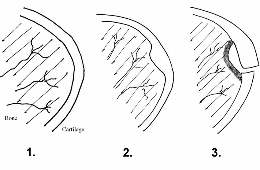
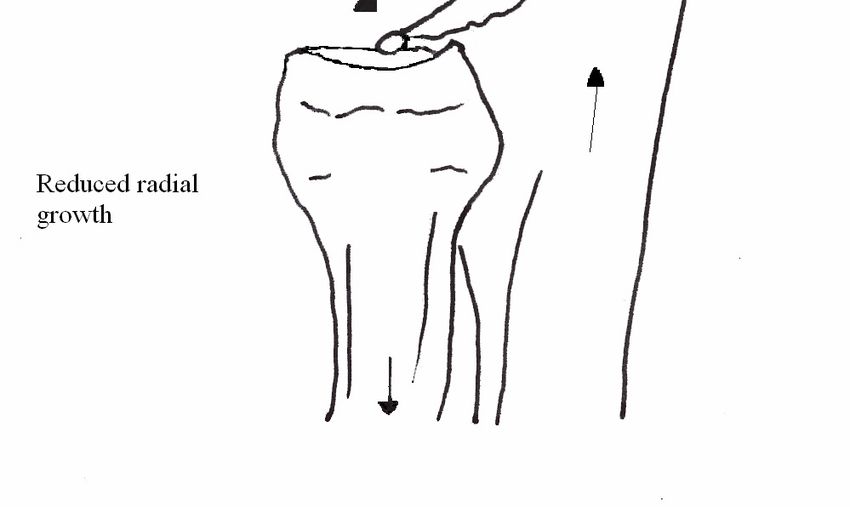
ED results from an incongruity of the joint, mainly as a result of under development of the ulna trochlear notch
(the inside surface of the joint at the top of the ulna). If the incongruity is minor, there may be minimal changes
with no obvious clinical problems. The incongruity results in a step down effect from a poorly developed ulna
notch in relation to the weight bearing surface of the top of the radius. If the incongruity is large, a combination of
the following 3 major conditions may be seen – UAP, OCD, FCP.

Another form of elbow dysplasia can be seen from radial over growth (relative to the ulna) due to early closure of
the distal growth plate of the ulna. Again this is where the cartilage core in this growth plate fails to get adequate
nutrition causing a delay in growth and resulting in a shortened ulna and overgrowth of the radius. This causes
incongruity in the elbow and in severe cases may result in bowing of the front legs, lateral luxation of the proximal
end (near the elbow) of the radius. The resulting incongruity can lead to a combination of the above conditions.
FCP (medial Fragmented Coronoid Process) Occasionally, after the anconeal process has united, there can be later
development of joint incongruity. The mico-movement and stresses placed within the joint can result in a
fragmented coronoid process, which nearly always affects the medial process.

There are 4 main subgroups of elbow dysplasia which may occur alone or in combination. Generally
they are considered inherited in most breeds and the incidence may be adversely affected by incorrect diets in
some breeds.
Excessive rate of weight gain can affect the incidence of many elbow conditions, often leading to joint incongruity
(uneven rate of growth between the radius and ulna), and OCD, particularly in males.

i.UAP – Ununited anconeal process
The anconeal process is a large triangular shaped piece of bone situated at the back of the elbow joint. This has a
separate ossification centre in a handful of breeds mainly larger, heavier breeds notably the GSD, Labrador, Great
Dane, Irish Wolfhound, Newfoundlands, Bloodhound, Basset and Dogge de Bordeaux.This process normally is
fully ossified (sealed) by 16-18 weeks of age. This condition is considered inherited with a possible 3 different
genes being involved.
Diagram - UAP due to radial overgrowth
The anconeal process forms the hinge at the back of the elbow. When the anconeal process has a separate
ossification (growth area) in breeds such as in the GSD, this area becomes a target for poor conversion of cartilage
to bone. The combination of the separate ossification centre and a too small circumference of the ulna trochlear
notch (inside edge of the back of the joint) results in small movements along the cartilaginous bridge between the
ossification centre of the anconeal process and the ulna. This results in failure of the anconeal process to unite.
Chronic movement of the UAP results in ongoing development of joint arthritis and further wear and tear of the
cartilage surfaces of the joint.
Closure of the ossification centre -The anconeal process in the GSD should be closed by 16-18 weeks of age,
definitely by 20 weeks of age. If screening for UAP, X rays taken at 20 weeks should identify a problem if it is
present.
Symptoms - Clinically signs are seen from as young as 5-6 months with intermittent lameness, which is
exaggerated by exercise. Full flexion and extension of the elbow will elicit pain. There is often an accompanying
lateral bursitis (fluid swelling).
Diagnosis - X rays of the elbow in the flexed lateral position will readily show whether the process has unified or
not. Long standing cases often have boney arthritic changes as well.
Treatment (depending on other conditions/joint incongruities present) is either:
a) removal of the process which, if done while young, usually has a reasonable prognosis with some arthritis
later.
b) fixation of the loose fragment by a lag screw will also give a reasonable prognosis in early cases.
As the anconeal process is not a weight bearing surface, early surgery gives better long term prospects than in the
FCP cases (as loss of that process affects the weight bearing surfaces of the joint).
The majority of UAP cases in the GSD have good congruity of the head of the radius relative to the ulna. Where
there is poor congruity, ie. the radius is too long for the ulna, an osteotomy (cutting) of the ulna to allow it to
lengthen naturally is another option.
Affected dogs should not be breed with. UAP is generally considered to be inherited as a separate condition to
OCD and FCP in the GSD and this may be true for other breeds as well.
ii. OCD – osteochondritis dessicans (see above as well)
OCD occurs in many larger breeds and can affect almost any breed greater than 18-20kg at adulthood. There is a
higher incidence in males versus females. This can affect many joints, the commonest being the elbow. Breeds
that see with a reasonably high incidence of elbow OCD would include the following: Rottweiler (high incidence),
GSD, Golden Retriever, St Bernard, Great Dane, Border Collie, Rhodesian Ridgeback and Labrador.
Symptoms – OCD is generally seen as a shifting lameness in the forequarter from around 5-8 months of age, some
joint capsule swelling and usually a turning out of the front legs at stance as the inner edges of the elbow are most
commonly affected. The cause of the problem is considered to be due to a faulty blood supply to the joint cartilage
secondary to very rapid growth.
Diagnosis - On X ray the signs are often quite subtle in mild cases with minor “fluffiness” and/or flattening of the
joint surfaces to the more distinct pot holes of larger lesions. It is generally diagnosed by X rays of a straight
extended and slightly medially rotated view of the elbow.
Treatment - If the condition is mild, treatment with drugs such as Cartrophen* which increases the blood supply to
cartilage can be very effective along with complete rest, slowing down of the rate of weight gain and low doses of
anti-inflammatories. Repair and recovery can take up to 6-8 weeks, depending on the severity of the condition.
Very heavy puppies may have to be kept reasonably restricted until 9 months of age by which stage all rapid
growth has slowed dramatically. Severe cases of OCD are often found in conjunction with FCP, and may require
surgical intervention. Most cases >80% show good responses to medical, dietary and exercise management.
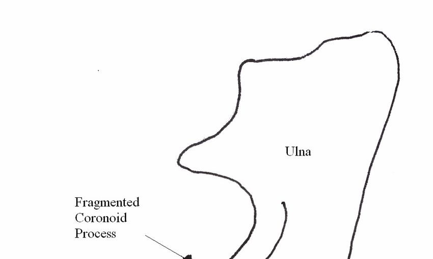
iii. FCP – fragmented coronoid process
FCP of the ulna generally refers to the medial coronoid process, a process that stabilises the medial edge of the
joint. Fragmentation of this process means that the inside edge of the elbow is not stable, hence the very typical
lateral rotation of the leg away from the pain. It affects the same age group as above.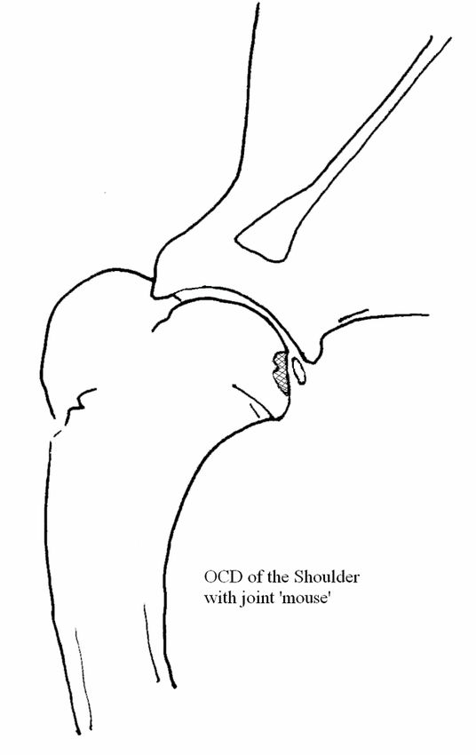
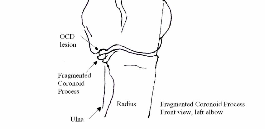
Diagram - FCP/OCD lesions – medial view, left elbow

Diagram - FCP lesion - reduced growth of radius leading to FCP
Diagnosis - On X ray, with the elbow extended straight out and a second view with slight medial rotation the
process can be visualised and assessed. The coronoid process can be seen as being separated from the main section
of the ulna.
Treatment - If these are diagnosed early, surgical intervention can give reasonable results. In the older dog, where
there are considerable secondary arthritic changes, medical management with tablets is probably preferred.
Outcome - Regardless of the treatment, the resulting joint incongruity (unevenness) will lead to osteoarthritis over
time.

iv.Joint Incongruity
 While most forms of elbow dysplasia can, by their development, result in joint incongruity, what we are looking
at here is where there has been a possible early closure of a distal growth plate in the foreleg resulting in uneven
growth of the radius (usually slightly shorter) in relation to the ulna. The resulting uneven ends of the bones within
the joint can cause excessive wear on cartilages and in the worst cases, force the anconeal process distally (ie.
create a UAP).

Other forms of elbow “dysplasia” exist which generally involve the lateral displacement of the top of the radius in
relation to the ulna (giving a cabriole effect) but these are uncommon.

General Treatment of Elbow Dysplasia
As with any painful bone disorder, regardless of the age presented, common treatment is aimed at pain
management, sensible diet and weight control and a restricted, suitable exercise regime. Where there are only
minor changes in joint surfaces, medical management and conservative treatment with anti-inflammatory agents
and rest is generally all that is needed.
Those animals with UAP require surgical intervention to minimise future arthritis. Younger dogs diagnosed with a
FCP or loose cartilaginous flaps, should have these removed surgically in order to minimise future damage to the
joint. Due to the incongruity of the joint, there will be ongoing arthritic changes. In the older dog with advanced
arthritic changes, medical management with conservative exercise regimes is, generally, the preferred method of
treatment.

Diet and Elbow Dysplasia
 While diet may not of itself create elbow dysplasia, it can affect the severity of OCD seen. Rapid weight gain will
push factors such as joint congruity, as well as affecting the blood supply to the cartilage within the joints. Reduce
the energy levels in the diets, particularly for dogs that need to be confined, and ideally, change puppies onto an
adult maintenance diet. Affected puppies should have their weight at or below the normal for their breed, sex and
age.

Rule outs (differential diagnoses)
 Not all forelimb lameness is due to elbow dysplasia! Panosteitis and simple injuries should be checked for,
particularly where there is a very sudden onset of lameness.

Controlling the incidence of Elbow Dysplasia –
The major task in reducing the severity and or incidence of any breed inherited disorder, is establishing the mode
of inheritance. The inheritance of problems such as hip and elbow dysplasia where there are two or more genes
affecting the inheritance pattern, one can only, at this stage use quantifying tests such as X rays and sires progeny
results to assist in our control schemes. Some genes can behave incompletely, ie. blend effects with a normal
gene and not be expressed, as the normal gene carries sufficient enzyme making ability to hide the effects of a
defective gene. The animal will appear normal but is in fact a "carrier" of the abnormal gene.
The more genes affecting a characteristic, the harder and slower it is to eradicate or affect the characteristic and
the more environmental effects may come into play (weight, diet, rate of growth etc). Where there are ways to
measure the effect of the characteristic, then progress can be made in controlling the effect of the polygenes in
the overall population eg. X raying of individuals and their progeny.

The schemes currently in use for control/reduction in severity of HD and ED aim to reduce the incidence and
overall severity of these conditions across a breed a) as a whole and b) over time. Trying to shift the genetic
structure of polygentic conditions within a breed is a long term goal and cannot be pushed rapidly without
severe consequences in other areas (eg. type, temperament etc).

. As this is a group of highly inherited conditions, fairly rapid improvements can be made over reasonably short
periods of time. Breeding from severely affected dogs should be heavily discouraged. Generally dogs with a UAP,
FCP and arthritis of greater than 4mm should not be used for breeding.

Grading of Elbow X rays
When reading or scoring elbow X rays, the major measurement is done on the caudal (back) edge of the anconeal
process. When this process is clean and shows no changes, then the elbow is graded normal. Grade 1 dogs are
allowed up to just under 2mm of arthritic changes, Grade 2 dogs are allowed between 2mm to just under 4mm.
UAP, 4mm of arthritic change and greater are automatic fails.

Occasionally a dog may have no changes on the anconeal process, but have minor changes on the ulna around the
joint or the space between the top of the radius and ulna. While these dogs may receive a 0 from the anconeal
process, they may then receive a Grade 1 due to minor arthritic changes elsewhere around the edges of the joint.

Breeding from dogs with significant arthritic changes (Grade 2) in the elbows should only be with care and ideally
always be to normal partners and preferably to those lines with low incidences of elbow problems. Grade 1
animals can be bred to a wider range of animals, but, again, ideally to normal partners.

With the elbow results, as with the hips, the average score of the sire’s progeny (where more than 20 progeny
are scored) is of greater benefit in predicting the genetic worth of the dog than it is the score of the sire
himself. To try and give an extra loading on results, we can loosely grade sires by the % of normal elbows
being produced. Slight variations due to state averages (local population genetics) can effect these results.

Sires can further be loosely grouped on their progeny results and the % of normals. These sires can then be
graded as to their effectiveness as sire for producing good elbows. I would suggest the following loose
designation in assessing sires for the soundness of their elbow producing.

Sires that produce greater than:-
>85% normal elbows should be considered excellent producers of good elbows
>70% normal elbows should be considered very good producers of elbows
>60% normal elbows should be considered good producers of elbows
Sires that produce less than :-
< 60 % normal elbows should be considered poor producers of elbows

The whole idea of these schemes is to give breeders information so as to give them knowledge prior to
breeding an animal as to what one can afford to do.

 The conclusion one gets from these schemes, is that the more information one has for both of the parents and
of their close relatives, particularly offspring of the sire, the better one can plan and get successful results
across a litter. In breeds where such information is limited and/or sire statistics are not available, breeders
have a much harder time selecting good sires and good breeding combinations.
You can also read