Original Article A Case Study on Biomechanical Analysis of Kneeling and Squatting Methods while Manual Lifting Using Motion Capture Analysis
←
→
Page content transcription
If your browser does not render page correctly, please read the page content below
June 2021, Vol 18 No. 1

                                                                                 Original Article

A Case Study on Biomechanical Analysis of Kneeling and
Squatting Methods while Manual Lifting Using Motion Capture
Analysis
Tam Jenn Zhueng,a,* Yeoh Wen Liang,b Choi Jeewon,b Zuraida Mohamed,a Loh Ping Yeapb

a
    Negeri Sembilan State Health Department, Ministry of Health, Malaysia
b
    Department of Human Science, Faculty of Design, Kyushu University, Japan

*Corresponding author: jztam79@gmail.com

ABSTRACT: Weight lifting techniques had been long discussed and debated between squatting and semi-
squatting (kneeling) techniques. Using motion analysis and force plates, the weight lifting tasks were measured
for comparison. Participant was required to lift different weights of 5kg, 10kg and 15kg using squat and semi-
squat techniques. The tasks were randomized and the performance was measured using Cortex 7.0 and Mokka
6.0. Then kneeling technique was indeed a more efficient and safe posture as compare to the squatting technique
that would expose an acute onset of spinal injury. The case study illustrated that kneeling technique was the
better technique in protecting the spinal health from any chronic musculoskeletal disorders. Therefore, the study
recommended that weight lifting training exercises to be promoted for kneeling postures and provide core
muscle in strengthening exercises as an important intervention and training program at workplace.

Keywords: Force, Intervertebral Angles, Motion Capture, Technique, Weight Lifting
                                                                                                  All rights reserved.

1.0 INTRODUCTION
Supervisors need to be cautious when managing his co- workers’ physical activities. Training had always been
limited with regards to multitasking. In many instances, proper engineering innovation and controls could not be
allocated due to various financial and commitment constraints. Therefore, the cost- efficient control strategies
would be adopting hazard isolation and administration control strategies. In the service sector, (e.g.: nurses,
stewardess or receptionists) workers would be exposed to various multitasking including lifting objects at
various degrees and frequencies in a day. The musculoskeletal system would be chronically affected if the job
task required high repetitive movement (even contact stress). Night shifts, improper sleeping posture, inadequate
recovery time and poor quality of sleep were all indirectly associated with low back pain with time. Procedures
such as venipunctures, dialysis, dressing and nursing care were commonly done under non-ergonomic
conditions (Theodora et al., 2010).

                                                                   35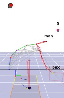
Journal of Occupational Safety and Health

                  Papi et al. (2017) and Tam et al. (2019) correctly pointed out that there were ergonomic and
         psychological factors related to low back pain especially at work. Numerous efforts have been spent on
         developing tools to prevent chronic back pain, which include questionnaires (Traeger et al., 2014) and simple
         field- assessments especially addressing sports related injuries. Surprisingly, objective and standardized
         anthropometric and ergonomic assessments at workplace settings were not easy to come by. Most physical
         assessment of the human body catered for human efficiency enhancement for development of sports. Studies
         have shown that the intervertebral discs of running athletes were reported to be healthier than individuals who
         were not active in sports (Belavy et al., 2017) while Yoo & An (2009) correlated musculoskeletal pain with poor
         physical posture. Therefore, the human posture and anthropometric knowledge could answer the chronic
         presentations of musculoskeletal diseases.
                  Other sports that emphasized on the technique would be the weight lifting category. Weight lifting
         techniques were catered for competitive performances and prevention of long-term injuries and disabilities. It
         would be interesting to assess the movement of the human body when challenge with weight lifting under
         working environment or requirements. Pourahmadi et al. (2019) concluded that kinematic/ motion analyses were
         very different assessments that medical science assessments and that there were not many motions analysis
         reviews (even assessments among healthy population were limited) in the literature. Meanwhile, Yamamoto et
         al. (2017) and van der Have et al. (2019) mentioned of low back pain association with forward bending or
         stooping posture movements. Besides the movement and forces, the angle movement could also be observed
         using the motion analysis (Suter et al., 2019); squatting compared with kneeling (semi-squat) techniques.
         Nevertheless, there are limited documented visuals or figures on how the vertebrae, typically the lumbar angles
         changes and its relationship to low back exposures pain while lifting in squatting versus kneeling (semi-
         squatting) positions.
                  This case study aims to present the relationship of forces created via different lifting postures (kneeling
         versus squat postures) while lifting various different weights from the ground to the standing posture. The study
         would identify the movement of the thoracolumbar spine (thoracic kyphosis) and lumbosacral spine (lumbar
         lordosis) with the spinal angle changes in relation to the forces during lifting. By comparing the two postures,
         the benefits and limitations on the two lifting postures can provide preventive knowledge for ergonomic health
         purposes.

         2.0 METHOD
         This study was approved by the Ethics Committee of the Faculty of Design at Kyushu University, Japan. A male
         participant (35 years old, 178cm, 78.3kg and BMI of 24.62kgm -2) had volunteered and consented to participate
         in this study. The participant’s kinematic data were collected using a 3D motion- capture system (Cortex 7.0;
         Motion Analysis Corporation, Santa Rosa, CA, USA) while two force platforms (9286A; Kistler, Winterthur,
         Switzerland) were sampled at 1000 Hz in sync with the kinematic data through the 3D motion- capture systems.
         There were 11 infrareds high- speed cameras that would be recording movements with a sampling rate of 120
         Hz.
                   The task conditions were box lifting at weight 5kg, 10kg, and 15kg with kneeling or squatting method
         in randomized order. The weight was based from the Ergonomic Risk Assessment at Workplace 2017 of
         Malaysia (DOSH, 2017). Subsequently, the participant was asked to rate his perceived exertion based on the
         Borg’s Rating of Perceived Exertion (RPE) scale (Borg, 1990) after every trial. The kneeling method in this
         study refers to a singular kneeling of either side of the human body while the task was randomized as with the
         weighs mentioned above and as shown in Fig. 1. The squatting method was referred as crouch posture with his
         knees bent and the heels were adjacent to the hamstring muscles. At force plates, the measured forces were
         identified as X (left to right), Y (back to forward) and Z (top to bottom) vectors. Finally, the total resultant
         forces (in Newton- N) were used in this report.

                                                                 36
June 2021, Vol 18 No. 1
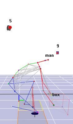
A total 22 reflective markers were placed on the participant’s body surfaces to collect the kinematic
data; from the anterior trageus, distal acronium process to the 5 th little toe, 8 markers over the box and 8 markers
were placed on the two tables placed above one another. Each dot on Fig. 1 represents the refractory markers.
Before the initiation of the study, the participant was allowed to change and prepare himself. Pre-assessment
medical screening such as weight and height was also taken. The 22 markers were placed on the anatomical
surfaces of the important structures of the body off-lined and the movements of the markers were recorded and
calculated by the Cortex 7.0. The participant was told to begin the trial by standing at rest. Then, the participant
would have to pick up the weighted box and finally to stand straight (Fig. 1) with the weighted box either using
kneeling or squatting according to the randomized protocol. The task conditions (two postures and three
weights) were randomized for the participant, and each condition was repeated for three repetitions.
Randomization is needed to prevent the participants to be able to prepare himself on the various different trials
that would need to be performed.
A single task would take 30 seconds (maximum). In between each task, the participant was allowed 60
seconds of recovery time. The entire motion capture would require approximately an hour to complete. For the
purpose of the study, the focus was on the spinal movements of C7, T6, T12, L5 and S1 vertebraes in relation to
each other as they move on their respective vertebral planes. The lumbar lordosis angle was calculated from the
thoracolumbar and thoracosacral segments. The thoracic kyphosis angle was calculated from the upper thoracic
segments (the C7-T6-T12 vertebral angle changes) and the lumbosacral segments (the T12-L5-S1 vertebral
angle changes) were also measured. For each task, the participant would have to repeat the task 3 times. All the
randomization effect, data, weight, movement, graphic videos and forces produced and captured by the cameras
and force plates were recorded into the Cortex 7.0 software.
OR
Free Standing Before Lifting Lifting Object Standing with Object
and Foot Off the
e.g. Kneeling e.g. Squatting
Force Plate
Phase 1 Phase 2
Figure 1 Difference Postures and Phases during Lifting
37Journal of Occupational Safety and Health
In this case study, the single lifting motion cycle was divided into Stand to Lift (Phase 1) and Box
Lifting (Phase 2) (Fig. 1). The measured forces were identified as X (left to right), Y (back to forward) and Z
(top to bottom) vectors. Lastly, the total resultant forces were used in this report. The maximal forces produced
over the left foot during the movements were recorded as the right knee would be used during the kneeling
posture that would be less consistent compared to the left foot. The Borg’s RPE was also obtained from the
participant after each task was performed. Besides the experimental movement of the tasks that were captured,
the changes in the management of the thoracic and the lumbar curvatures were also observed and measured.
Finally, the reporting method of Institute of Biomechanic Standards were used as reference (Wu et al., 2005 and
Hasegawa et al., 2018) when the angle changes were recorded by the Cortex 7.0 and Mokka 6.0 system.
3.0 RESULTS & DISCUSSIONS
Fig. 2 showed the difference values of the Borg’s Rapid Perceived Exertion (RPE) between kneeling (semi-
squatting) and squatting postures with increased weight. Using Borg’s RPE, we could conclude that the tasks
were not too physically strenuous and should not increase the cardiovascular demand (Max RPE: 6 units) on
both occasions. Findings suggested that the participant felt less amount of effort was needed when lifting objects
above 10kg when kneeling as compared to squatting postures. Although very efficient, the squatting technique
did increase perceived exertion to the participant as compared to kneeling technique.
Borg’s RPE
(unit)
Weight
Figure 2 Documented RPE when Kneeling or Squatting
38June 2021, Vol 18 No. 1
However, the study managed to record higher forces that were generated when the participant was
adopting the squatting technique as compared to kneeling technique to lift the similar 5kg and 15kg from the
ground (Fig. 3). Phase 1 (Blue Segment) referred to the participant standing to box lifting position. In phase 1,
the participant would need to bend his body and move nearer to the box. Phase 2 (Red Segment) referred to the
position from lifting the box to standing position with more movement and force generated. The squatting
technique produced a localised movement force (Fm) as compared to the kneeling techniques (Lifting hump-
LH). The squatting technique would expose a smaller number of muscles in a short period of time as compared
to the kneeling technique. Therefore, the intensity of the ergonomic risk would be much higher in the squatting
technique as compared to kneeling technique. The kneeling posture with 15kg object was noted to be using
larger number of muscles of the body as they body adjust, prepare and accommodate to the increased physical
activity that needed to be performed. With additional energy or force generated, the angle range on both
kneeling and squatting was relatively similar.
a) b)
LH LH
d)
c)
LH LH
Figure 3: Forces Generated with various Posture and Weighted Box.
LH- Lifting Hump
a) Kneeling Posture with 5kg box
b) Kneeling Posture with 15kg box
c) Squatting Posture with 5kg box
d) Squatting Posture with 15kg box.
Figure 3 Forces Generated with Various Posture and Weighted Box
39Journal of Occupational Safety and Health
Table 1 Resultant Force Measured
Standing with Standing with box Standing with box Standing with box
Level Rest empty box plus 5kg plus 10kg plus 15kg
(95% CI) (95%CI) (95%CI) (95%CI)
Force 781.02 808.31 853.43 877.50 927.93
(N) ±7.37 (805.53 to 811.10) (849.95 to 856.91) (874.71 to 877.52) (923.78 to 930.03)
Table 2 Maximal Forces during Lifting Objects from Floor
Weight Stand-to-Lift (Phase 1) Box Lifting (Phase 2)
(kg)
Knee Squat Knee Squat
5 665.57 ± 26.59 510.17 ± 71.0 878.84 ± 14.42 862.64 ± 5.53
10 662.03 ± 14.05 521.66 ± 75.40 869.17 ± 48.10 918.00 ± 6.24
15 672.60 ± 9.15 522.39 ± 38.61 955.60 ± 0.85 959.31 ± 22.32
Table 3 Angle Changes from Neutral Positions
Stand to Lift (Phase 1) Box Lifting (Phase 2)
Weight Posture
Thoracic Lumbar Thoracic Lumbar
Knee 4.57 ± 0.96 14.72 ± 0.07 4.01 ± 0.48 14.04 ± 0.51
5kg
Squat 10.70 ± 1.10 12.95 ± 0.22 6.89 ± 0.91 13.34 ± 0.44
Knee 6.47 ± 2.19 13.67 ± 0.19 5.33 ± 2.31 13.46 ± 0.13
10kg
Squat 7.62 ± 0.93 14.37 ± 0.41 6.44 ± 0.77 13.90 ± 0.36
Knee 8.29 ± 1.00 14.16 ± 0.32 8.31 ± 1.30 14.39 ± 0.37
15kg
Squat 10.70 ± 6.81 13.00 ± 1.29 6.89 ± 0.38 13.34 ± 0.38
The results are presented in mean±SD, unless otherwise specified. Table 1 showed the resultant forces
at rest from no object to exposure when lifting objects with 15kg. During the participant’s movement from
standing to kneeling or squatting to pick up the object that was on the floor, the force plates recorded forces that
were changing during movements (Table 2). Table 3 illustrated the forces that were documented during the
initiation stages as the participant kneeled or squatted to lift an object during Phase 1 to Phase 2. The forces
along with the movements were dynamic and similar forces were generated between both postures in Phase 2.
40June 2021, Vol 18 No. 1
a) b)
MLS MLS
c) d)
MLS
MLS
MLS- Maximal Lumbar Stability
a) Kneeling Posture with 5kg box
b) Kneeling Posture with 15kg box
c) Squatting Posture with 5kg box
d) Squatting Posture with 15kg box.
Figure 4 Lumbar Angle Changes
Figure 5 Thoracic Angles Lifting 5kg Objects (Kneeling and Squatting Postures)
41Journal of Occupational Safety and Health
However, the squatting technique exhibited relatively erratic and unstable angle movements to recovery as
compared to kneeling technique (Fig. 4). In fact, the squatting technique transfers some of the physical force to the
upper back (thoracic region) as the body entered Phase 2. Such thoracic angles were relatively protected when using
the kneeling technique as compared to the squatting technique (Fig. 5). Therefore, there would be stronger and
forceful impact of the vertebral during the squatting technique as compared to kneeling technique even at 5kg.
Fig. 4 illustrated in detail the different physical (muscle) hazards that the body was exposed when different
techniques were used. As similar angles were required, the final postures of lifting were similar (Maximal Lumbar
Stability- MLS). Our motion findings were consistent with van der Have et al. (2019). The kneeling technique
provided a much stable and gradual spinal angle changes. Once MLS was achieved, the core muscles would enter
the predominant isometric phase which would allow the lumbar spine to return to its anatomical angle to reduce
injury.
There were several advantages in using motion capture to document spinal movements during object
lifting. Although, motion capture along with force plates would be able to document movements and the forces that
interplay during the physical activities, it should be important to note that other modalities could be added or
incorporate to increase the precision and to detail out other important parameters such as muscle contractility,
muscle strength, oxygen- ventilation perfusion, neural impulses, spinal kinematics (Papi et al., 2017) and many
others anthropometric assessments. This study was designed to document inter-vertebral movements during object
lifting. With surface markers, the angle movements of the vital skeletal and muscle points were objectively reported
and represented (Stinton, 2011).
From literatures, kneeling technique was ergonomically efficient and would prevent back injuries (Vecchio,
2017), but relatively less power. Both techniques had similar loss of lumbar lordosis giving added pressure and
compression to the disc. However, the stability of the kneeling posture was affected when the objects were 15kg.
Our findings were consistent with Wang et al. (2012). Without adequate recovery and rest, chronic fatigue would
further injure the affected workers (Vecchio 2017 and Patel 2020). Therefore, untrained or unprepared workers
should be allowed to decline such tasks if the activity was done without supervision by a trained senior colleague or
health professionals (e.g., fitness trainer or physiotherapists). In the study, the untrained participant had consistently
described difficulty in lifting objects via squatting compared to kneeling (semi-squat) technique. As a case study,
this participant was a healthy, a 30-year-old Asian male that reported healthy lumbosacral or lumbar lordosis angles.
The participant was a teaching staff of the university as all students in Japan was told to return home due to the
Covid-19 pandemic. It was intended to have more than 20 participants form the university (majority Asians) to
participate in this study. With large number of participants and data, we could compare and measure the range of
muscle movement among the individuals. Therefore, the level of Asian referential and representation of this study is
limited.
The lumbar angle changes increased with additional weight during both squatting and semi- squatting
(kneeling) techniques. Caglayan et al. (2014) managed to report that an increased lumbar lordosis angle was
associated with onset of low back pain. A key important discussion that squatting lifts (trained appropriately
especially in sport performance) generate more efficient and effective competitive heavy lifting, particularly with a
straightened back and training (Wang et al., 2012, Kim 2014 and Cho et al., 2017). The motion capture had
successfully provided that insight and would recommend the importance of training and physiotherapy and core
exercises for workers to build a strong back.
42June 2021, Vol 18 No. 1
Postures of lifting in competitive weight lifting sports applied weights to be supported on shoulders/ upper
trunk in order to exceed daily lifting capacity (Myer et al., 2014 and Kushner et al., 2015). Although acceptable and
practiced in the past among sea-farers unloading shipments at seaports or docks, these techniques had not been
commonly used in this modern era. Kneeling technique would be a rather appropriate and safer technique of lifting
objects less than 10kg. Even procured tools to assist lifting had to be introduced and taught to all workers
effectively, especially in the service industries like banking, corporate business and the healthcare industry.
Secondly, perhaps a paradigm shifts especially for nursing staff or discipline to incorporate a healthier practice to
work on standing computer designs along with serving of medication and attending to patients. This would allow
nurses to really enjoy their rest and breaks sitting down as compared to repetitive or prolonged sitting with limited
movement which would be less healthy to the human body.
As recommendation, this study proposes the need for employers to designate dedicated trained personnel to
be in- charge of the back care treatments aimed to prevent musculoskeletal injuries with proper lifting technique
training, nutrition and medical care to strengthen the back muscles typically the multifidus muscle. Interestingly,
most literatures suggested that body- building squatting exercises were good core exercises to strengthen the back
structures (Al-Otaibi, 2015, Cho et al., 2017 and Lorenzetti et al., 2018). The multifidus muscle thickness was noted
to have a very close relationship with low back pain (Wallwork et al., 2009). ‘BackCare’ in the form of preventive
back strengthening programmes and workplace ergonomic investments would delay or prevent an early onset of
degenerative changes to the spine of all workers at work. Medical screening recommendation such as the Unilateral
Hip Bridge Endurance Test (UHBET) would be a good physical assessment of the strength a worker’s core muscles
(Butowicz et al., 2016). The idea would be to prevent occurrence of micro-injuries over the vertebral disc that would
be dehydrated and replaced by fibrous tissues (Ract et al., 2015). Such un-prevented pathogenesis would promote
back pain and prolapsed or herniated intervertebral discs. As such, core muscle strengthening and squatting
exercises (Lorenzetti et al., 2018) would improve performance and reduce dependency on the spine/ back to lift
objects from the ground (Patel, 2020).
Besides that, this study also proposes that employers from all sectors to designate a ‘Team Lifting’
personnel who has knowledge in ergonomics awareness and weight lifting training at work particularly in the
service and administrative industries (Theodora et al., 2010). These individuals should be upskilled further with the
knowledge on muscle strengthening and fitness benefits, covered in the scheduled nutritionist and physiotherapist
sessions. Our study clearly showed that structured physical training activities would be required to be implemented
for manufacturing workers or industries that involved extensive manual lifting work (Cho et al., 2017 and Patel,
2020).
However, there are limitations in this study. While testing out the kneeling posture, the knee was used to
balance the human body to lift the box. Therefore, the recording or motion captured between the lower limbs were
relatively inconsistent. Although the systemic bias had been addressed, the quality of knee posture information
would be limited as compared to the squat posture data. On the other hand, the participant successfully demonstrated
that the squat technique provided a powerful and sustained force during lifting (Fig. 3). It is advised that it would be
important to maintain a straight posture of the back to prevent possible injuries during the squat posture (Kim,
2014).
At the muscle level, Hemming et al. (2018) studied the kinematics movement of the spine among
individuals with non- specific chronic low back pain. These people specifically had more markers (13 markers)
along the spine as compared to our study (6 markers). As the spine moved, the spinal changes involved flexion and
all active extension pattern motor control impairment should be captured accurately. The benefits of this study
would be enhanced further with a larger recruitment of participants in safer conditions, post pandemic.
43Journal of Occupational Safety and Health
5.0 CONCLUSION
Both techniques of kneeling and squatting while lifting had different advantages with regards to lifting the objects.
Lifting heavy objects using the kneeling technique may have illustrated lower ergonomic risk as compared to the
squatting technique. Based on the law of energy conversion, additional pressure/ force would receive or be absorbed
by the joints, discs and soft tissues of the human body during the squatting technique. Although the squatting
technique could achieve a lifting task faster and easier, the squatting could promote more micro- injuries to the spine
as compared to the kneeling technique. Therefore, all manual workers and relevant stakeholders need to be trained
with Back Care awareness and prevention programme to prevent prolapsed intervertebral disc at work.
ACKNOWLEDGEMENTS
This work was part of the sub-specialty international training of Dr Tam Jenn Zhueng and supported by the Public Health
Development Division, Ministry of Health, Malaysia.
REFERENCES
Al-Otaibi, S.T. (2015). Prevention of Occupational Back Pain. Journal of Family & Community Medicines, 22(2),
73–77. doi: 10.4103/2230-8229.155370.
Borg, G. (1990). Psychophysical Scaling with Applications in Physical Work and The Perception of Exertion.
Scandinavian Journal of Work Environment & Health, 16(1), 55-58.
Butowicz, C.M., Ebaugh, D., Noehren, B. & Silfies, S.P. (2016). Validation of Two Clinical Measures of Core
Stability. The International Journal of Sports Physical Therapy, 11(1), 15-23.
Caglayan, M., Tacar, O., Demirant, A., Oktayoglu, P., Karakoc, M., Cetin, A., Em, S., Bozkurt, M., Ucar, D. & Nas,
K. (2014). Effects of Lumbosacral Angles on Development of Low Back Pain. Journal of Musculoskeletal
Pain, 22(3), 251–255.
Chaudhry, H., Bukiet, B., Ji, Z. & Findley, T. (2011). Measurement of Balance in Computer Posturography:
Comparison of Methods- A Brief Review. Journal of Bodywork & Movement Therapies, 15, 82- 91.
Cho, M.K., Kang, J.Y, Oh, J.H, Wu, J.G, Choi E.B, Park, S.E & Choi, M. (2017). The Effects of Performing Squats
on an Inclined Board on Thigh Muscle Activation. Physical Therapy Rehabilitation Science, 6, 39-44.
https://doi.org/10.14474/ptrs.2017.6.1.39 [30 September 2020]
DOSH. (2017). Ergonomic Risk Assessment Guidelines at Workplace 2017. Department of Occupational Safety and
Health, Ministry of Human Resource, Malaysia.
Hasegawa, T., Katsuhira, J., Oka, H., Fujii, T. & Matsudaira, K. (2018). Association of Low Back Load with Low
Back Pain during Static Standing. PLoS ONE, 13(12). e0208877.
https://doi.org/10.1371/journal.pone.0208877
Hemming, R., Sheeran, L., Van Deursen, R. & Sparkes, V. (2018). Non-specific Chronic Low Back Pain:
Differences in Spinal Kinematics in Subgroups during Functional Tasks. European Spine Journal, 27, 163–
170. https://doi.org/10.1007/s00586-017-5217-1.
Kim, J.S. (2014). Lower Body Kinematic Comparisons between Front and Back Squats in Response to Load.
College of Graduate Studies. Bridgewater State University.
44June 2021, Vol 18 No. 1
Kushner, A.M., Brent, J.L., Schoenfeld, B.J., Hugentobler, J., Lloyd, R.S., Vermeil, A., Chu, D.A., Harbin, J.,
McGill, S.M. & Myer, G.D. (2015). The Back Squat Part 2: Targeted Training Techniques to Correct
Functional Deficits and Technical Factors that Limit Performance. Journal of Strength and Conditioning
Research, 37(2), 13–60. doi:10.1519/SSC.0000000000000130.
Lorenzetti, S., Ostermann, M., Zeidler, F., Zimmer, P., Jentsch, L., List, R., Taylor, W.R. & Schellenberg, F. (2018).
How to Squat? Effects of Various Stance Widths, Foot Placement Angles and Level of Experience on
Knee, Hip and Trunk Motion and Loading. BMC Sports Science Medicine and Rehabilitation, 10, 14.
doi:10.1186/s13102-018-0103-7.
Myer, G.D., Kushner, A.M., Brent, J.L., Schoenfeld, B.J., Hugentobler, J., Lloyd, R.S., Vermeil, A., Chu, D.A.,
Harbin, J. & McGill, S.M. (2014). The Back Squat: A Proposed Assessment of Functional Deficits and
Technical Factors that Limit Performance. Strength & Conditioning Journal, 36(6), 4–27.
doi:10.1519/SSC.0000000000000103.
Papi, E., Koh, W.S. & McGregor, A.H. (2017). Wearable Technology for Spine Movement Assessment: A
Systematic Review. Journal of Biomechanics, 64, 186–197.
Patel, N. (2020). What to Do When You Get a Sore Back After Squats and Deadlifts. Fitbod.
https://www.fitbod.me/blog/sore-back-after-squats-and-deadlifts [28th September 2020]
Pourahmadi, M.R., Takamjani, I.E., Jaberzadeh, S., Sarrafzadeh,J., Sanjari, M.A., Bagheri, R. & Taghipour, M.
(2019). Kinematics of The Spine During Sit-To-Stand Movement Using Motion Analysis Systems: A
Systematic Review of Literature. Journal of Sport Rehabilitation, 28, 77-93.
Ract, I., Meadeb, J.M., Mercy, G., Cueff, F., Hussond, J.L. & Guillin, R. (2015). A Review of the Value of MRI
Signs in Low Back Pain. Diagnostic and Interventional Imaging, 96, 239-249.
Suter, M., Eichelberger P., Frang, J., Simonet, E., Baur, H. & Schmid, S. (2019). Measuring Lumbar Back Motion
during Functional Activities Using a Portable Strain Gauge Sensor-Based System: A Comparative
Evaluation and Reliability Study. Journal of Biomechanics. doi.org/10.1016/j.jbiomech.2019.109593.
Tam, J.Z., Mohamed, Z., Wan Puteh, S.E. & Ismail, N.H. (2019). A Systematic Review on Identifying Associated
Factors in Deciding Work- relatedness of Chronic Back Pain among Employees at Work. Malaysian
Journal of Public Health Medicine, Vol. 19 (1), 1-14.
Theodora, K., Dimosthenis, Z., Michael, K., Athanasios, K. & Evaggelos, S. (2010). Looking into the Factors
Affecting Low Back Pain Incidents in General Hospital Nurses: A Questionnaire Research. Hellenic
Journal of Nursing Science, 36-42.
Traeger, A.C., Moseley, G.L., Hübscher, M., Lee, H., Skinner, I.W., Nicholas, M.K., Henschke, N., Refshauge,
K.M., Blyth, F.M., Main, C.J., Hush, J.M., Pearce, G. & McAuley, J.H. (2014). Pain Education to Prevent
Chronic Low Back Pain: A Study Protocol for a Randomised Controlled Trial. BMJ Open, 4. e005505.
doi:10.1136/bmjopen-2014-005505.
Van der Have, A., Van Rossom, S. & Jonkers, I. (2019). Squat Lifting Imposes Higher Peak Joint and Muscle
Loading Compared to Stoop Lifting. Applied Sciences. doi:10.3390/app9183794.
Vecchio, L.D. (2017). Choosing A Lifting Posture: Squat, Semi-Squat or Stoop. MOJ Yoga & Physical Therapy 7,
2(2), 56‒62.
Wang, Z.L., Wu, L., Sun, J.Z., He, L.H., Wang, S. & Yang, L. (2012). Squat, Stoop, or Semi-squat: A Comparative
Experiment on Lifting Technique. Journal of Huazhong University of Science and Technology, 32(4), 630-
636.
45Journal of Occupational Safety and Health
Wu, G., van der Helm, FCT., Veeger, D. & Makhsons, M. (2005). ISB Recommendation on Definitions of Joint
Coordinate Systems of Various Joints for the Reporting of Human Joint Motion - Part II: Shoulder, Elbow,
Wrist and Hand. Journal of Biomechanics, 38(5), 981-992. DOI:10.1016/j.jbiomech.2004.05.042.
Yoo, WG & An, DH. (2009). The Relationship between the Active Cervical Range of Motion and Changes in Head
and Neck Posture after Continuous VDT Work. Industrial Health, 47, 183–188.
46You can also read