Partial Distal Biceps Avulsion Results in a Significant Loss of Supination Force
←
→
Page content transcription
If your browser does not render page correctly, please read the page content below
812
C OPYRIGHT Ó 2021 BY T HE J OURNAL OF B ONE AND J OINT S URGERY, I NCORPORATED
Partial Distal Biceps Avulsion Results in a Significant
Loss of Supination Force
Yoshiaki Tomizuka, MD, Christopher C. Schmidt, MD, Anthony J. Davidson, BS, Christopher S. Spicer, BS,
Michael P. Smolinski, BS, Ryan J. Mauro, BS, Sean M. Delserro, MME, Linsey H. Szabo, Patrick J. Smolinski, PhD, and
Mark Carl Miller, PhD
Investigation performed at the Shoulder and Elbow Mechanical Research Laboratory, Department of Orthopaedic Surgery, University of Pittsburgh
Downloaded from https://journals.lww.com/jbjsjournal by BhDMf5ePHKav1zEoum1tQfN4a+kJLhEZgbsIHo4XMi0hCywCX1AWnYQp/IlQrHD3i3D0OdRyi7TvSFl4Cf3VC1y0abggQZXdtwnfKZBYtws= on 05/05/2021
Medical Center, Pittsburgh, Pennsylvania
Background: Partial avulsions of the short and/or long head of the distal biceps tendon cause pain and loss of strength.
The goal of the present study was to quantify the loss of supination and flexion strength following a series of surgical
releases designed to simulate partial and complete short and long head traumatic avulsions.
Methods: Mechanical testing was performed to measure supination moment arms and flexion force efficiency on 18
adult fresh-frozen specimens in pronation, neutral, and supination. The distal biceps footprint length was divided into 4
equal segments. In 9 specimens (the distal-first group), the tendon was partially cut starting distally by releasing 25%,
50%, and 75% of the insertion site. In the other 9 specimens (the proximal-first group), the releases started proximally.
Mechanical testing was performed before and after each release.
Results: Significant decreases in the supination moment arm occurred after a 75% release in the distal-first release
group; the decrease was 24% in pronation (p = 0.003) and 10% in neutral (p = 0.043). No significant differences in the
supination moment arm (p ‡ 0.079) or in flexion force efficiency (p ‡ 0.058) occurred in the proximal-first group.
Conclusions: A simulated complete short head avulsion significantly decreased the supination moment arm and
therefore supination strength.
Clinical Relevance: A mechanical case can be made for repair of partial distal biceps tendon avulsions when the rupture
involves ‡75% of the distal insertion site.
T
raumatic partial avulsion of the distal biceps tendon can whereas the long head is a more powerful supinator in the
cause activity-related pain and substantial loss of supi- supinated forearm21. Furthermore, the distal insertion of the
nation and flexion strength1-14. A majority of patients short head allows it to generate more elbow flexion force21.
with this type of lesion require surgical treatment for the reso- Partial distal avulsions of the short and/or long head can
lution of symptoms1-15. The precise anatomical lesion requiring occur as a result of both traumatic and atraumatic etiologies9-14,23,24.
surgical reattachment is unknown. Traumatic avulsions commonly propagate in a distal-to-proximal
A number of investigators have pointed out the dramatic direction, whereas in atraumatic tears the degeneration starts on
variation in biceps structure, ranging from a fused muscle with a the deep surface of the tendon and progresses superficially9-14,23,24.
highly interdigitated distal tendon to 2 distinct muscles with A large clinical series showed that a magnetic resonance imaging
separate short and long-head tendons—that is, a bifurcated (MRI)-diagnosed partial tear of >50% was a predictor of the need
muscle3,9-12,16-22. In most cadaveric dissections, the distal biceps for surgery to resolve the symptoms (odds ratio, 3.0; p = 0.006);
tendon is easily separated into its short and long-head compo- however, the study failed to describe the tear location5. Tear
nents, with the short head attaching distal to the long head on location and size may be important factors in determining the
the radial tuberosity (Fig. 1) and occupying 60% of the footprint need for surgery. The effects of partial short and long-head
area (Fig. 2)16,17,19-22. The short head generates a greater supina- avulsions on forearm supination and elbow flexion strength are
tion moment arm in neutral and pronated forearm positions, not known.
Disclosure: The authors indicated that no external funding was received for any aspect of this work. On the Disclosure of Potential Conflicts of Interest
forms, which are provided with the online version of the article, one or more of the authors checked “yes” to indicate that the author had a relevant financial
relationship in the biomedical arena outside the submitted work (http://links.lww.com/JBJS/G324).
J Bone Joint Surg Am. 2021;103:812-9 d http://dx.doi.org/10.2106/JBJS.20.00445
813
                 TH E JO U R NA L O F B O N E & JO I N T SU RG E RY J B J S . O RG
                                                                      d
                                                                                           P A R T I A L D I S TA L B I C E P S AV U L S I O N R E S U LT S   IN A   S I G N I F I C A N T LO S S
                             V O LU M E 103 -A N U M B E R 9 M AY 5, 2 021
                                                 d              d
                                                                                           O F S U P I N AT I O N F O R C E

                                                                                           cadaveric specimen was inspected for abnormalities and
                                                                                           bifurcation.
                                                                                                  The specimens were randomly assigned to either distal-first
                                                                                           or proximal-first releases. The cut sequence (distal-first or
                                                                                           proximal-first) defined the 2 conditions of the study. The humeri
                                                                                           were osteotomized and the wrists were disarticulated to permit
                                                                                           bolt fixation to the simulator. The soft tissue between the oste-
                                                                                           otomy and disarticulation sites, the interosseous membrane, and
                                                                                           the distal radioulnar joint were preserved. A dorsal skin incision
                                                                                           was made over the radial tuberosity, and the extensor carpi ulnaris
                                                                                           and supinator muscles were split to expose the insertion of the
                                                                                           distal biceps on the radial tuberosity. The biceps footprint length
                                                                                           was measured with a scientific caliper (L.S. Starrett) with an
                                                                                           accuracy of 0.01 mm and was divided into 4 equal segments
Fig. 1
                                                                                           (Figs. 3-A and 3-C). In 9 specimens (the distal-first group), the
Cadaveric dissection showing the distal attachment sites of the short and
                                                                                           biceps tendon was partially cut from its attachment site starting
long heads on the posterior aspect of the radial tuberosity. The short head
                                                                                           distally by sequentially releasing 25%, 50%, and 75% of the
inserts distal to the long head. (Reprinted from J Shoulder Elbow Surg.
                                                                                           insertion site (Fig. 3-B, 50% distal-first release). In the other 9
2012;21:942-8, Jarrett CD, Weir DM, Stuffmann ES, Jain S, Miller MC,
                                                                                           specimens (the proximal-first group), the biceps tendon was
Schmidt CC. Anatomic and biomechanical analysis of the short and long                      released starting proximally (Fig. 3-D, 50% proximal-first release).
head components of the distal biceps tendon. With permission of Elsevier.)                 The releases were done under loupe magnification, with the
                                                                                           specimens secured in the simulator, after native mechanical testing.
        The goal of the present study was to simulate partial and
complete traumatic avulsions of the short and long heads with                              Elbow Simulator
use of sequential surgical releases in a distal-to-proximal and a                          The elbow simulator, forearm supination moment arm
proximal-to-distal direction and to simultaneously measure the                             test, and elbow flexion force test have been previously vali-
resultant forearm supination moment arm and elbow flexion force                             dated21,25-27. The testing apparatus comprised an aluminum
efficiency. Supination moment arm and elbow flexion force effi-                               frame mounted to a material testing system (MTS) (MTS
ciency directly determine supination and flexion strength, respec-                          Systems) (Figs. 4 and 5). The humerus was bolted to the vertical
tively. We hypothesized that short-head releases would lead to a                           limb of the frame. The elbow was placed in 90° of flexion, and
decrease in the supination moment arm in 60° of pronation and
neutral forearm rotation and a reduction in elbow flexion force
efficiency whereas partial long-head releases would result in a
decrease in the supination moment arm in 60° of supination.

Materials and Methods

E    ighteen fresh-frozen human shoulder-to-hand cadaveric
     specimens from male donors with an average age (and
standard deviation) of 56.6 ± 13.6 years were used. All specimens
had preservation of the proximal and distal biceps insertions and
full forearm and elbow range of motion.
       Mechanical tests were performed to measure isometric
forearm supination moment arms in 60° of pronation, neutral,
and 60° of forearm supination and elbow flexion force at 90° as
previously reported21,25-27. After baseline testing, 9 specimens (the
distal-first group) underwent a serial 25%, 50%, and 75% release
of the biceps tendon from its insertion, starting distally (Figs. 3-A                      Fig. 2

and 3-B). In the other 9 specimens (the proximal-first group),                              Three-dimensional reconstruction of the radial tuberosity (green) with super-
the biceps tendon was released in a similar manner, but starting                           imposed short (red) and long (blue) head footprints. The radial protuberance is
proximally (Figs. 3-C and 3-D). The sequence of release (distal                            marked by the asterisk. The forearm is pronated, and the tuberosity is visu-
or proximal first) was randomized with use of a number gen-                                 alized from a posterior approach. The short head occupies 60% of the total
erator. Mechanical testing was performed after each release.                               footprint area, whereas the long head occupies 40%. Note that the tendon
                                                                                           attaches on the posterior aspect of the tuberosity. (Reprinted from J Shoulder
Specimen Preparation                                                                       Elbow Surg. 2012;21:942-8, Jarrett CD, Weir DM, Stuffmann ES, Jain S, Miller
The lines of pull for the proximal short and long-head biceps                              MC, Schmidt CC. Anatomic and biomechanical analysis of the short and long
tendons were anatomically replicated for each specimen26. Each                             head components of the distal biceps tendon. With permission of Elsevier.)
814
TH E JO U R NA L O F B O N E & JO I N T SU RG E RY J B J S . O RG
d
P A R T I A L D I S TA L B I C E P S AV U L S I O N R E S U LT S IN A S I G N I F I C A N T LO S S
V O LU M E 103 -A N U M B E R 9 M AY 5, 2 021
d d
O F S U P I N AT I O N F O R C E
Fig. 3-A Fig. 3-B
Fig. 3-C Fig. 3-D
Figs. 3-A through 3-D The biceps tendon was approached through the extensor carpi ulnaris/supinator window with the forearm in pronation. The
length of the footprint was measured and divided into 4 equal segments. The biceps tendon was partially cut from its attachment starting
distally (distal-first) or proximally (proximal-first) by sequentially releasing 25%, 50%, and 75% of the insertion site. Fig. 3-A Distal-first release
specimen prior to release. Fig. 3-B A 50% distal-first release. Fig. 3-C Proximal-first release specimen prior to release. Fig. 3-D A 50% proximal-
first release.
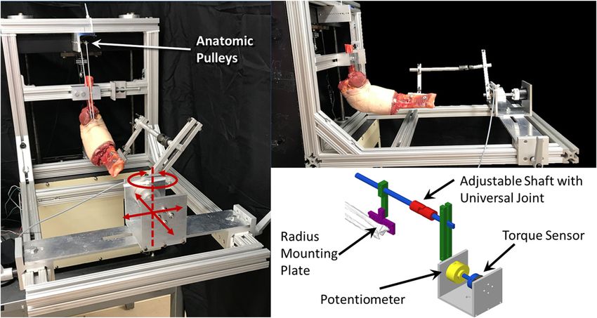
the radius was fixed to a mounting plate, which was attached Mechanical Testing
to the torque sensor (Transducer Techniques) housed within Forearm supination moment arm and elbow flexion force tests
the adjustable carriage. The torque sensor data were recorded were completed before and after each release for each specimen
with use of a data acquisition system (National Instruments). in a single session with use of a standard protocol21,25-27. To test
The adjustable shaft with universal joint and carriage were the forearm supination moment arm, the specimens were
adjusted to recreate the anatomical axis of rotation for each mounted on the elbow simulator with the humerus and ulna
specimen. The axis of rotation was found to be anatomical fixed firmly to the frame at 90° of flexion (Fig. 4). The position
when the forearm easily rotated, without binding, from full was chosen on the basis of a previous report indicating that the
pronation to supination. The anatomical lines of pull of the maximum biceps supination moment arm occurs at 90° of
proximal short and long-head tendons were connected to a elbow flexion28. The forearm was then rotated and locked into 3
single actuator. positions: 60° of pronation, neutral, and 60° of supination. The
815
                   TH E JO U R NA L O F B O N E & JO I N T SU RG E RY J B J S . O RG
                                                                        d
                                                                                             P A R T I A L D I S TA L B I C E P S AV U L S I O N R E S U LT S   IN A   S I G N I F I C A N T LO S S
                               V O LU M E 103 -A N U M B E R 9 M AY 5, 2 021
                                                   d              d
                                                                                             O F S U P I N AT I O N F O R C E

Fig. 4
Photographs and illustration showing an anatomical elbow simulator with a cadaveric elbow at 90° of flexion and the forearm in neutral position. This setup was utilized to
conduct the forearm supination moment arm testing protocol. (The schematic is reprinted from JSES Open Access. 2019;3:225-231, Schmidt CC, Madonna TJ, Vandreuil
N, Brown BT, Liu SY, Delserro S, Smolinski MP, Styron J, Smolinski PJ, Miller MC, et al. The effect of tendon rotation on distal biceps repair. With permission of Elsevier.)

neutral position was defined by a reference line drawn on the                                 least-squares regression line; the slope of this regression line was
radius that bisected the scaphoid and lunate fossae by con-                                  the elbow flexion force efficiency. The elbow flexion test was
necting the midpoints of the radial styloid and sigmoid notch27.                             repeated 3 times, and the recorded values were averaged.
The reference line was aligned vertically according to mea-
surements read from a digital goniometer. The proximal short                                 Statistical Analysis
and long heads of the tendon were attached to the actuator with                              A 1-factor repeated-measures analysis of variance (ANOVA)
use of low-friction cables. The proximal biceps tendon was                                   with partial tear size as the factor followed by post hoc analysis
preloaded in each of the 3 forearm positions up to 10 N for 10                               with use of Bonferroni correction was used for each forearm
cycles and then was loaded up to 67 N at a rate of 1 cm/sec. The                             position to evaluate the effects of partial releases on the biceps
resulting supination torque was measured with the torque                                     tendon moment arm and elbow flexion force efficiency. For all
sensor. A least squares regression line was fitted to the curve of                            statistical analyses, the level of significance was set at p < 0.05.
supination torque versus biceps load, and the slope of this                                         An a priori power analysis was performed to determine
regression line represented the supination moment arm. The                                   the minimum number of specimens that would be required to
test was repeated 3 times at each forearm position, and the                                  detect a 15% difference in the supination moment arm and
values were averaged for each position tested. The sequence of                               flexion force. On the basis of the study data, a sample size of
the forearm positions was randomly assigned.                                                 8 specimens in each group would provide adequate power
       The elbow flexion force test measured the biceps flexion                                (a = 0.05 and b = 0.80) to detect a 15% difference.
force efficiency, which is the ratio of flexion load to biceps load
(Fig. 5)21,25,26. The torque device was removed while maintaining                            Results
humeral fixation to the simulator. The radius and ulna were
pinned in 60° of forearm supination, and the biceps was loaded
until the elbow reached 90° of flexion. A cord attached to the distal
                                                                                             N    o bifurcated biceps muscles were observed. Seventeen
                                                                                                  specimens were free of tendon abnormality, and 1 con-
                                                                                             tained gouty crystals. The latter specimen was randomized into
part of the forearm was connected to a force sensor (Transducer                              the proximal-first group and ruptured after a 75% release
Techniques), allowing a counterforce to maintain the elbow at 90°                            during force supination testing. The specimen was removed
of flexion. The flexion force was recorded by preloading and                                   from mechanical analysis, leaving 9 specimens in the distal-first
loading the biceps as previously described for the supination test.                          group and 8 specimens in the proximal-first group. The average
Recorded flexion load versus applied load was used to calculate a                             footprint length as measured with the caliper was 22.1 ± 2.3 mm.
816
                 TH E JO U R NA L O F B O N E & JO I N T SU RG E RY J B J S . O RG
                                                                      d
                                                                                     P A R T I A L D I S TA L B I C E P S AV U L S I O N R E S U LT S   IN A   S I G N I F I C A N T LO S S
                             V O LU M E 103 -A N U M B E R 9 M AY 5, 2 021
                                                 d              d
                                                                                     O F S U P I N AT I O N F O R C E

                                                                                           In the distal-first group, the only significant decreases in
                                                                                     the supination moment arm occurred in association with a
                                                                                     75% release (p £ 0.043) (Table I). In pronation, the moment
                                                                                     arm was 24% less than that in the intact state (p = 0.003),
                                                                                     whereas in neutral, the moment arm was 10% less than that in
                                                                                     the intact state (p = 0.043). Additionally, a 29% decrease
                                                                                     occurred in supination (p = 0.056).
                                                                                           In the proximal-first group, there was no significant dif-
                                                                                     ference in the supination moment arm in association with 25%,
                                                                                     50%, and 75% releases in pronation, neutral, or supination (p ‡
                                                                                     0.079) (Table II). However, proximal-first releases of 50% and
                                                                                     75% reduced the supination moment arm in supination by 35%
                                                                                     (p = 0.079) and 37% (p = 0.131), respectively.
                                                                                           The flexion force efficiency results are shown in Table III.
                                                                                     Elbow flexion force did not significantly change relative to the
                                                                                     intact state in any of the distal-first or proximal-first release

                                                                                          TABLE I Distal-First Release Supination Moment Arms

                                                                                              Release Comparison                     Percent Change                   P Value

                                                                                              60° pronation
                                                                                                 Native versus 25%                         210%                        0.158
                                                                                                 Native versus 50%                         212%                        0.122
                                                                                                 Native versus 75%                         224%                       0.003*
                                                                                              Neutral
                                                                                                 Native versus 25%                          22%                        0.999
                                                                                                 Native versus 50%                         210%                        0.377
                                                                                                 Native versus 75%                         210%                       0.043*
                                                                                              60° supination
                                                                                                Native versus 25%                           27%                        0.996
                                                                                                 Native versus 50%                         218%                        0.999
                                                                                                 Native versus 75%                         229%                        0.056

                                                                                         *Significant (p < 0.05).

                                                                                          TABLE II Proximal-First Release Supination Moment Arms

                                                                                              Release Comparison                     Percent Change                   P Value

                                                                                              60° pronation
                                                                                                 Native versus 25%                          24%                        0.999
Fig. 5                                                                                           Native versus 50%                         213%                        0.971
Photograph showing an anatomical elbow simulator with corresponding                              Native versus 75%                          21%                        0.999
force sensor. With use of this setup, a counterforce was applied to maintain                  Neutral
the elbow at 90° of flexion, and the subsequent force generated during the                        Native versus 25%                           0%                        0.999
flexion force efficiency test was recorded.                                                        Native versus 50%                          24%                        0.999
                                                                                                 Native versus 75%                          27%                        0.999
      The supination moment arm results are summarized in                                     60° supination
Tables I and II (with the raw data shown in the Appendix). The                                   Native versus 25%                          22%                        0.999
native moment arm values were not significantly different                                         Native versus 50%                         235%                        0.079
from those in our previously published work (p ‡ 0.131)                                          Native versus 75%                         237%                        0.131
(Appendix)21,25-27.
817
                TH E JO U R NA L O F B O N E & JO I N T SU RG E RY J B J S . O RG
                                                                      d
                                                                                    P A R T I A L D I S TA L B I C E P S AV U L S I O N R E S U LT S   IN A   S I G N I F I C A N T LO S S
                            V O LU M E 103 -A N U M B E R 9 M AY 5, 2 021
                                                 d             d
                                                                                    O F S U P I N AT I O N F O R C E

                                                                                    (p = 0.32) and showed that 20 (44%) of 45 patients with a
   TABLE III Flexion Force Efficiency
                                                                                    traumatic partial distal biceps injury presented with a complete
      Release Comparison             Percent Change            P Value              short-head avulsion (Fig. 6)9-13,18,24,31. The present study did not
                                                                                    investigate atraumatic partial ruptures, which are believed to
      Proximal-first                                                                 occur as a result of radial tuberosity impingement4,14,23,24,29,30. In
         Native versus 25%                  1%                     0.999            degenerative tears, it is believed that the tendon first fails on its
         Native versus 50%                  4%                     0.482            deep surface adjacent to osseous irregularities on the tuberosity
         Native versus 75%                  5%                     0.058            and then progresses superficially14,23. However, we speculate
      Distal-first                                                                   that the mechanics would not be different from the uninjured
         Native versus 25%                  3%                     0.716            state as long as the superficial fibers of both the short and long
         Native versus 50%                  2%                     0.999            heads remain intact, given that force-transmission studies have
         Native versus 75%                 22%                     0.999            shown that the supination moment arm does not change from
                                                                                    the native state as long as the tendon is reattached posterior to
                                                                                    its radial protuberance27,32-34.
groups following 25%, 50%, or 75% sectioning (p ‡ 0.058).                                  In a previous mechanical study, the short and long-head
The proximal-first 75% release increased the flexion force                            components were separated, tested individually, and compared
efficiency by 5% (p = 0.058).                                                        with each other21. In that study, the short head was found to be a
                                                                                    better supinator in pronation and neutral, whereas the long head
Discussion                                                                          was a better supinator in a supinated position because the shape of

T    raumatic avulsion of the short head of the distal biceps
     tendon can result in a substantial loss of supination
strength. In the present study, a 75% distal-first release, which
                                                                                    the radial protuberance maximized the moment arms for each
                                                                                    head in their respective positions. The present study was different
                                                                                    because the tendons were not separated but rather were released
simulates a complete short-head avulsion, decreased the supi-                       from the radius to simulate partial avulsions and subsequently
nation moment arm by 24% in pronation (p = 0.003), 10% in                           were compared with the intact tendons. The significant decreases
neutral (p = 0.043), and 29% in supination (p = 0.056). The                         in the supination moment arm that were observed in association
supination moment arm can be thought of as the efficiency of                         with a complete (75% distal-first) short-head release in pronation
the biceps muscle to rotate the forearm; that is, the greater the
moment arm, the greater the supination strength that can be
generated by a given biceps contraction27. A drop in the
moment arm value reduces the effectiveness of biceps force
transmission, which could result in a clinically meaningful loss
of supination strength, endurance, and/or power.
       A recent clinical series showed that an MRI-diagnosed
partial tear of >50% was a predictor of the need for surgery to
resolve the symptoms (odds ratio, 3.0; p = 0.006)5. The
mechanical results of the present study supported those clinical
findings when the tear was distal and involved ‡75% of the
insertion site. A simulated complete short-head avulsion was
associated with a significant (p £ 0.043) decrease in the supi-
nation moment arm in the pronated and neutral forearm
positions. The authors of the clinical series did not address the
location of the >50% tear5. In the future, using preoperative
advanced imaging to clarify tear size and location may help to
further define appropriate surgical indications.
       The pathoanatomy of partial distal biceps avulsions can
be divided into traumatic and atraumatic etiologies, and the                        Fig. 6

understanding of this subject is still evolving9-14,23,24. The cutting              Clinical photograph showing a short head tendon avulsion, after the entire
sequence of distal-to-proximal (distal-first) or proximal-to-                        tendon was detached to aid in repair. Prior to the photograph, a posterior
                                                                                                                                                                         32
distal (proximal-first) instead of deep-to-superficial was de-                        extensor carpi ulnaris-slitting approach was used for exposure . The short
signed to model a traumatic distal partial avulsion and not a                       head was found avulsed, and the long head was found intact. The long head
chronic degenerative tear4,14,23,24,29,30. The merit of our cutting                 was then temporarily sutured for future traction. The long head was sur-
sequence is supported by the following clinical findings: (1) the                    gically detached at the insertion site. The entire tendon was retracted to
most common mode of traumatic failure of bifurcated distal                          gain exposure for permanent short and long-head suture placement. In this
biceps tendons is a complete short-head rupture and (2) a                           photograph, the avulsed short head is held by 2 forceps. The short and long
recent MRI study demonstrated no significant difference in tear                      heads were then reattached to their respective insertion sites with 2 single
morphology between bifurcated and non-bifurcated tendons                            cortical buttons (not shown).
818
                   TH E JO U R NA L O F B O N E & JO I N T SU RG E RY J B J S . O RG
                                                                        d
                                                                                       P A R T I A L D I S TA L B I C E P S AV U L S I O N R E S U LT S   IN A   S I G N I F I C A N T LO S S
                               V O LU M E 103 -A N U M B E R 9 M AY 5, 2 021
                                                   d              d
                                                                                       O F S U P I N AT I O N F O R C E

(24%; p = 0.003) and neutral (10%; p = 0.043) and the substantial                      ability of the human body to adapt to a partial distal biceps
decrease in the supination moment arm in association with a                            avulsion. However, other mechanical studies on reattach-
complete (50% proximal-first) long-head release in supination                           ment position have correlated with subsequent clinical
(35%; p = 0.079) are in agreement with the findings of the above-                       findings27,32-34. In conclusion, the size and location of the
mentioned mechanical study21 (Tables I and II). However, one                           partial distal biceps tear affect the supination moment arm.
would expect that proximal-first release would increase the                             A partial distal biceps avulsion involving ‡75% of the distal
supination moment in pronated and neutral positions because of                         footprint substantially decreases the supination moment
increasing force transmission through the short head, and like-                        arm, and therefore strength; a strong mechanical case can be
wise, that distal-first release would increase the supination                           made for surgical repair with the goal to restore full clinical
moment in a supinated position because of increasing force                             function.
transmission through the long head. These findings were not
observed in the present study as none of the supination moment                         Appendix
arms increased after sectioning. Perhaps a proximal-first release of                           Supporting material provided by the authors is posted
25% shifts the long-head load to the short head, but in an                                    with the online version of this article as a data supplement
asymmetrical manner, whereby the tendon fibers next to the                              at jbjs.org (http://links.lww.com/JBJS/G325). n
release receive most of the load; this asymmetrical transmission
shifts the resultant force vector off its apex on the protuberance
toward the release and thereby fails to increase its expected
moment arm. Proximal-first releases of >40% cut into the short-
head tendon and also shift the resultant vector off its apex, away
                                                                                       Yoshiaki Tomizuka, MD1,2
from the release. The overall moment arm fails to increase because                     Christopher C. Schmidt, MD2,3
the resultant force fails to act on the protuberance at its maximum                    Anthony J. Davidson, BS2,3
point away from the forearm axis of rotation20,25. The above                           Christopher S. Spicer, BS2,4
rationale also can be applied to distal-first release, but it is                        Michael P. Smolinski, BS2,4
important to remember that the long head occupies 40% of the                           Ryan J. Mauro, BS2
footprint21.                                                                           Sean M. Delserro, MME2,4
       It is surprising that partial releases, either distal-first or                   Linsey H. Szabo2
                                                                                       Patrick J. Smolinski, PhD2,4
proximal-first, did not significantly affect the native flexion                           Mark Carl Miller, PhD2,4
force efficiency (p ‡ 0.058). Anatomical studies have clearly
shown that the short head inserts further than the long head                           1Department  of Orthopaedic Surgery, Nihon University School of
from the center of rotation of the elbow joint16,19,21,22. Intuition                   Medicine, Tokyo, Japan
would suggest a complete short-head avulsion (distal-first
                                                                                       2Shoulder
75% release) would substantially reduce the flexion force                                         and Elbow Mechanical Research Laboratory, Department of
                                                                                       Orthopaedic Surgery, University of Pittsburgh Medical Center, Pittsburgh,
efficiency. Furthermore, a previous mechanical study dem-
                                                                                       Pennsylvania
onstrated that the ratio of flexion load to biceps load was 15%
higher (p = 0.001) in the short head as compared with the                              3Department   of Orthopaedic Surgery, University of Pittsburgh Medical
long head21. In that study, the short and long-head muscles                            Center, Pittsburgh, Pennsylvania
and tendons were completely separated and any interconnec-
                                                                                       4Department of  Mechanical Engineering and Materials Science, University
tions were released. In the present study, the interconnections
between the short and long-head muscles and tendons were                               of Pittsburgh, Pittsburgh, Pennsylvania
preserved, and these bridging structures could redistribute the                        Email address for C.C. Schmidt: schmidtc@upmc.edu
flexion force after a partial release and thereby mechanically
compensate for the loss of the distal attachment site20. Another                       ORCID iD for Y. Tomizuka: 0000-0001-8528-8696
explanation for the lack of significant findings could be the                            ORCID iD for C.C. Schmidt: 0000-0002-0363-4282
limited number of specimens that completed the mechanical                              ORCID iD for A.J. Davidson: 0000-0002-0223-4252
analysis (n = 17, including 9 in the distal-first group and 8 in the                    ORCID iD for C.S. Spicer: 0000-0001-7978-8387
proximal-first group). An a priori power analysis showed that 8                         ORCID iD for M.P. Smolinski: 0000-0002-6112-3034
                                                                                       ORCID iD for R.J. Mauro: 0000-0002-5588-489X
specimens were needed to detect a 15% difference in supination                         ORCID iD for S.M. Delserro: 0000-0001-7778-7575
moment arm and flexion force efficiency.                                                 ORCID iD for L.H. Szabo: 0000-0001-6281-6898
       The findings of the current study may not be clinically                          ORCID iD for P.J. Smolinski: 0000-0003-3795-8565
applicable, given the limitations of our injury model and the                          ORCID iD for M.C. Miller: 0000-0002-4902-3452

                                                                              References
1. Bourne MH, Morrey BF. Partial rupture of the distal biceps tendon. Clin Orthop      2. Dellaero DT, Mallon WJ. Surgical treatment of partial biceps tendon ruptures at
Relat Res. 1991 Oct;271:143-8.                                                         the elbow. J Shoulder Elbow Surg. 2006 Mar-Apr;15(2):215-7.
819
                    TH E JO U R NA L O F B O N E & JO I N T SU RG E RY J B J S . O RG
                                                                          d
                                                                                          P A R T I A L D I S TA L B I C E P S AV U L S I O N R E S U LT S   IN A   S I G N I F I C A N T LO S S
                                V O LU M E 103 -A N U M B E R 9 M AY 5, 2 021
                                                     d               d
                                                                                          O F S U P I N AT I O N F O R C E

3. Rokito AS, McLaughlin JA, Gallagher MA, Zuckerman JD. Partial rupture of the           20. Fogg QA, Hess BR, Rodgers KG, Ashwood N. Distal biceps brachii tendon
distal biceps tendon. J Shoulder Elbow Surg. 1996 Jan-Feb;5(1):73-5.                      anatomy revisited from a surgical perspective. Clin Anat. 2009 Apr;22(3):346-51.
4. Vardakas DG, Musgrave DS, Varitimidis SE, Goebel F, Sotereanos DG. Partial rupture     21. Jarrett CD, Weir DM, Stuffmann ES, Jain S, Miller MC, Schmidt CC. Ana-
of the distal biceps tendon. J Shoulder Elbow Surg. 2001 Jul-Aug;10(4):377-9.             tomic and biomechanical analysis of the short and long head components of the
5. Bauer TM, Wong JC, Lazarus MD. Is nonoperative management of partial distal            distal biceps tendon. J Shoulder Elbow Surg. 2012 Jul;21(7):942-8. Epub 2011
biceps tears really successful? J Shoulder Elbow Surg. 2018 Apr;27(4):720-5. Epub         Aug 3.
2018 Feb 1.                                                                               22. Kulshreshtha R, Singh R, Sinha J, Hall S. Anatomy of the distal biceps brachii
6. Frazier MS, Boardman MJ, Westland M, Imbriglia JE. Surgical treatment of partial       tendon and its clinical relevance. Clin Orthop Relat Res. 2007 Mar;456:117-20.
distal biceps tendon ruptures. J Hand Surg Am. 2010 Jul;35(7):1111-4.                     23. Davis WM, Yassine Z. An etiological factor in tear of the distal tendon of the
7. Jockel CR, Mulieri PJ, Belsky MR, Leslie BM. Distal biceps tendon tears in             biceps brachii; report of two cases. J Bone Joint Surg Am. 1956 Dec;38(6):
women. J Shoulder Elbow Surg. 2010 Jul;19(5):645-50. Epub 2010 Apr 18.                    1365-8.
8. Kelly EW, Steinmann S, O’Driscoll SW. Surgical treatment of partial distal biceps      24. Nicolay RW, Lawton CD, Selley RS, Johnson DJ, Vassa RR, Prescott AE, Omar
tendon ruptures through a single posterior incision. J Shoulder Elbow Surg. 2003          IM, Marra G. Partial rupture of the distal biceps brachii tendon: a magnetic reso-
Sep-Oct;12(5):456-61.                                                                     nance imaging analysis. J Shoulder Elbow Surg. 2020 Sep;29(9):1859-68. Epub
9. Cho CH, Song KS, Lee SM. Isolated short head component rupture of a bifurcated         2020 Jun 9.
distal biceps tendon mimicking as a complete rupture. J Hand Surg EurVol. Vol 2011        25. Schmidt CC, Brown BT, Williams BG, Rubright JH, Schmidt DL, Pic AC, Naka-
May;36(4):333-4. Epub 2011 Mar 3.                                                         shian MR, Schimoler PJ, Miller MC. The importance of preserving the radial tuber-
10. Koulouris G, Malone W, Omar IM, Gopez AG, Wright W, Kavanagh EC. Bifid                 osity during distal biceps repair. J Bone Joint Surg Am. 2015 Dec 16;97(24):
insertion of the distal biceps brachii tendon with isolated rupture: magnetic resonance   2014-23.
findings. J Shoulder Elbow Surg. 2009 Nov-Dec;18(6):e22-5. Epub 2009 Jun 17.               26. Schmidt CC, Madonna TJ, Vaudreuil N, Brown BT, Liu SY, Delserro S, Smolinski
11. Sassmannshausen G, Mair SD, Blazar PE. Rupture of a bifurcated distal biceps          MP, Styron J, Smolinski PJ, Miller MC. The effect of tendon rotation on distal biceps
tendon. A case report. J Bone Joint Surg Am. 2004 Dec;86(12):2737-40.                     repair. JSES Open Access. 2019 Sep 11;3(3):225-31.
12. Voleti PB, Berkowitz JL, Konin GP, Cordasco FA. Rupture of the short head             27. Schmidt CC, Weir DM, Wong AS, Howard M, Miller MC. The effect of biceps reat-
component of a bifurcated distal biceps tendon. J Shoulder Elbow Surg. 2017 Mar;          tachment site. J Shoulder Elbow Surg. 2010 Dec;19(8):1157-65. Epub 2010 Oct 8.
26(3):403-8. Epub 2016 Dec 12.                                                            28. Bremer AK, Sennwald GR, Favre P, Jacob HA. Moment arms of forearm rotators.
13. Ozasa Y, Wada T, Iba K, Yamashita T. Surgical treatment for partial rupture of        Clin Biomech (Bristol, Avon). 2006 Aug;21(7):683-91. Epub 2006 May 5.
the distal biceps tendon using palmaris longus tendon graft: a case report. Acta          29. Seiler JG 3rd, Parker LM, Chamberland PD, Sherbourne GM, Carpenter WA. The
Orthop Traumatol Turc. 2018 Jul;52(4):323-5. Epub 2018 Mar 9.                             distal biceps tendon. Two potential mechanisms involved in its rupture: arterial
14. Ruch DS, Watters TS, Wartinbee DA, Richard MJ, Leversedge FJ, Mithani SK.             supply and mechanical impingement. J Shoulder Elbow Surg. 1995 May-Jun;4(3):
Anatomic findings and complications after surgical treatment of chronic, partial           149-56.
distal biceps tendon tears: a case cohort comparison study. J Hand Surg Am. 2014          30. Dürr HR, Stäbler A, Pfahler M, Matzko M, Refior HJ. Partial rupture of the distal
Aug;39(8):1572-7. Epub 2014 May 23.                                                       biceps tendon. Clin Orthop Relat Res. 2000 May;374:195-200.
15. Behun MA, Geeslin AG, O’Hagan EC, King JC. Partial tears of the distal biceps         31. Tagliafico A, Michaud J, Capaccio E, Derchi LE, Martinoli C. Ultrasound dem-
brachii tendon: a systematic review of surgical outcomes. J Hand Surg Am. 2016 Jul;       onstration of distal biceps tendon bifurcation: normal and abnormal findings. Eur
41(7):e175-89. Epub 2016 May 20.                                                          Radiol. 2010 Jan;20(1):202-8. Epub 2009 Aug 6.
16. Athwal GS, Steinmann SP, Rispoli DM. The distal biceps tendon: footprint and          32. Hansen G, Smith A, Pollock JW, Werier J, Nairn R, Rakhra KS, Benoit D, Papp S.
relevant clinical anatomy. J Hand Surg Am. 2007 Oct;32(8):1225-9.                         Anatomic repair of the distal biceps tendon cannot be consistently performed
17. Cho CH, Song KS, Choi IJ, Kim DK, Lee JH, Kim HT, Moon YS. Insertional                through a classic single-incision suture anchor technique. J Shoulder Elbow Surg.
anatomy and clinical relevance of the distal biceps tendon. Knee Surg Sports              2014 Dec;23(12):1898-904. Epub 2014 Sep 11.
Traumatol Arthrosc. 2011 Nov;19(11):1930-5. Epub 2011 Jun 23.                             33. Prud’homme-Foster M, Louati H, Pollock JW, Papp S. Proper placement of the
18. Dirim B, Brouha SS, Pretterklieber ML, Wolff KS, Frank A, Pathria MN, Chung CB.       distal biceps tendon during repair improves supination strength—a biomechanical
Terminal bifurcation of the biceps brachii muscle and tendon: anatomic consider-          analysis. J Shoulder Elbow Surg. 2015 Apr;24(4):527-32. Epub 2014 Dec 3.
ations and clinical implications. AJR Am J Roentgenol. 2008 Dec;191(6):W248-55.           34. Schmidt CC, Brown BT, Qvick LM, Stacowicz RZ, Latona CR, Miller MC. Factors
19. Eames MH, Bain GI, Fogg QA, van Riet RP. Distal biceps tendon anatomy: a              that determine supination strength following distal biceps repair. J Bone Joint Surg
cadaveric study. J Bone Joint Surg Am. 2007 May;89(5):1044-9.                             Am. 2016 Jul 20;98(14):1153-60.
You can also read