Partridge and embryonated partridge egg as new preclinical models for candidiasis
←
→
Page content transcription
If your browser does not render page correctly, please read the page content below
www.nature.com/scientificreports
OPEN Partridge and embryonated
partridge egg as new preclinical
models for candidiasis
Hadi Tavakkoli1*, Ahmad Khosravi2*, Iraj Sharifi2, Zohreh Salari3, Ehsan Salarkia2,
Reza Kheirandish4, Kazem Dehghantalebi1, Maziar Jajarmi4, Seyedeh Saedeh Mosallanejad5,
Shahriar Dabiri6 & Alireza Keyhani2
Candida albicans (C. albicans) is the most common cause of candidiasis in humans and animals. This
study was established to a new experimental infection model for systemic candidiasis using partridge
and embryonated partridge egg. First, we tested the induction of systemic candidiasis in partridge
and embryonated partridge egg. Finally, interaction between virulence factors of C. albicans and
Bcl-2 family members was predicted. We observed that embryonic infection causes a decrease in
survival time and at later embryonic days (11–12th), embryos showed lesions. Morphometric analysis
of the extra-embryonic membrane (EEM) vasculature showed that vascular apoptotic effect of C.
albicans was revealed by a significant reduction in capillary area. In immunohistochemistry assay,
low expression of Bcl-2 and increased expression of Bax confirmed apoptosis. The gene expression of
Bax and Bcl-2 was also altered in fungi-exposed EEM. Ourin silico simulation has shown an accurate
interaction between aspartic proteinase, polyamine oxidase, Bcl-2 and BAX. We observed that the
disease was associated with adverse consequences, which were similar to human candidiasis. Acquired
results support the idea that partridge and embryonated partridge egg can be utilized as appropriate
preclinical models to investigate the pathological effects of candidiasis.
Abbreviations
BAX Bcl2-associated X protein
Bcl B-cell lymphoma
C. albicans
Candida albicans
EEM Extra-embryonic membrane
H&E Hemotoxylin and eosin
IHC Immunohistochemistry
PAS Periodic acid-schiff
PAO Polyamine oxidase
ASP Aspartic proteinase
TRY Tryptophol
Studies on pathogenic infections have been conducted using rat, mice and rabbit as experimental models since
pathogenic agents were injected and successfully reproduced clinical symptoms in those models. The mentioned
models were also accompanied by in vitro assessments when the symptoms were complicated or the experiment
could not be made in animal models due to ethical reasons. The animal models have also been used principally
to test treatments with drugs, pharmacokinetics and immunotherapy 1–3.
The alternative models based on non-mammalian animals were described using fruit flies (Drosophila mela-
nogaster), the larvae of the moth Galleria mellonella and the free-living nematode CaenorhabditisElegans3–6.
1
Department of Clinical Science, School of Veterinary Medicine, Shahid Bahonar University of Kerman, 22
Bahman Boulevard, Pajouhesh Square, Kerman 7616914111, Iran. 2Leishmaniasis Research Center, Kerman
University of Medical Sciences, 22 Bahman Boulevard, Pajouhesh Square, Kerman 7616914115, Iran. 3Obstetrics
and Gynecology Center, Afzalipour School of Medicine, Kerman University of Medical Sciences, Kerman,
Iran. 4Department of Pathobiology, Faculty of Veterinary Medicine, Shahid Bahonar University of Kerman, Kerman,
Iran. 5Afzalipour School of Medicine and Biochemistry Department, Kerman University of Medical Sciences,
Kerman, Iran. 6Afzalipour School of Medicine and Pathology and Stem Cells Research Center, Kerman University of
Medical Sciences, Kerman, Iran. *email: tavakkoli@uk.ac.ir; khosraviam@yahoo.com
Scientific Reports | (2021) 11:2072 | https://doi.org/10.1038/s41598-021-81592-y 1
Vol.:(0123456789)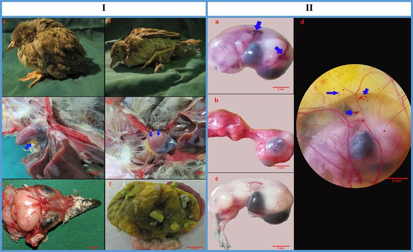
www.nature.com/scientificreports/
Recently, the chicken egg and embryo have also been considered as the laboratory models for experimental
investigations7–9. To reduce the use of mammalian or non-mammalian animals in medical experimentation,
alternative in vivo and in vitro models are increasingly necessary options.
Candida albicans (C. albicans) is the most common cause of fungal infections in humans 10,11. It generally
colonizes in regions such as the genitourinary tract, gastrointestinal track, lower respiratory and oropharynx.
Candidemia is the fourth most common cause of nosocomial bloodstream infections in the USA and in Europe
and has a significant impact on hospitalization cost and patient outcome 12,13. An increasing incidence of fungal
infections with Candida spp. has been noted in immunocompromised patients such as those staying in ICUs,
postsurgical and neutropenic patients 14,15. The systemic candidiasis has also occurred in patients with malignant
diseases in recent years 16–18. Different reports in the literature have documented that systemic candidiasis in
premature infants is frequently fatal or associated with significant morbidity in survivors 19–21. In this respect,
introduction of newer in vivo preclinical model for precise studying of the candidiasis, its pathogenesis and
treatment is an important and valuable issue nowadays.
In recent years, the partridge industry has experienced remarkable development and chukar partridge has
been widely raised industrially in many parts of the world22,23. As a research subject, chukar partridge has certain
advantages as follows:
Modest size of the bird simplifies housing in laboratory animal facilities and reduced feed costs. The bird
handling is easy and fulfilling its nutritional requirement is not a difficult task or expensive. The chukar partridge
is not very sensitive to environmental conditions and it can be easily reared in floor and cage system. Further-
more, the modest size of breeding adults facilitates breeding in the laboratory. The female partridges generally
lay 3–5 eggs per week and under favorable environments, they produce for long periods. Their eggs can be
grown in large batches in incubators, and allow the effects of particular substances on embryonic development
to be studied. The partridge embryo is also an amniote and it presents significant experimental advantages for
the study of amniotes similar to those of humans. Unlike mammillaria, partridge embryo can be easily studied
and manipulated as they monitor by removing a small section of the eggshell. This has made it possible to fol-
low their developmental stages via time-lapsed video-microscopy24–28. Therefore, we chose the chukar partridge
and embryonated partridge egg so that they could be used as laboratory models in areas that are easy to access.
On the other hand, in the past decade, some species of Phasianidae family (e.g. Japanese Quail), have become
important experimental animals for scientific researches. They are used extensively in physiology, nutrition,
genetics, embryology, toxicology, pathology and endocrinology r esearches29,30. Chukar partridge has also clas-
sified into the Phasianidae family. Considering this property, a major advantage that seems to be very promising
is the fact that it is possible to achieve a new experimental model for scientific investigations.
In this context, the objective of this study was to establish new experimental infection models for systemic
candidiasis and its pathogenesis using partridge and embryonated partridge egg. The present study was aimed
to answer the following questions:
I. Does C. albicans by human origin, cause systemic infection in the partridge and partridge embryo?
II. What are the clinical signs, gross and histopathological lesions of systemic candidiasis in partridge model?
III. Does C. albicans cause apoptotic effect in vessels?
IV. Does C. albicans alter the expression of proteins and genes which have important roles in vascular apop-
tosis?
V. How is the affinity between C. albicans virulence factors and apoptotic-regulator proteins?
Results
The results are explained in terms of A) clinical and histopathological signs and lesions following systemic
candidiasis in partridge model, B) vascular analysis, IHC and qPCR results from the EEM vasculature and C)
in silico results.
(A) Clinical and histopathological signs and lesions following systemic candidiasis in partridge model
Clinical signs. The infected partridges began to exhibit the clinical signs on the second day of infection. The
early symptoms manifested with multiple adverse health outcomes which included depression, lethargy and ruf-
fled feathers (Fig. 1I-a). Following the progression of the disease, some infected birds were unable to stand and
maintained their posture with drooping of the wings (Fig. 1I-b). Neurological disturbance such as spasmodic
tremors in the head was also seen in four sick birds, mainly on the fourth day after infection. No mortality
occurred during the experimental period in the infected group; however, two birds were culled due to severe
debilitation on days 3 and 4 following infection. No clinical signs were observed in any of the control birds.
Cloacal temperature. The normal cloacal temperature of partridge chicks, before the inoculation of C.
albicans, was 41.3 ± 0.3 °C. An increase in the cloacal temperature was a primary response to infection. The
temperature began to increase from normal value 30 h after infection. It continued to increase for another 24 h
(≥ 42 °C) and remained almost constant for 3–4 days post-infection. The mentioned temperature (≥ 42 °C) was
not observed in any of the control birds. It was also significant after statistical analysis (control bird = 41.29 ± 0.17,
infected birds = 41.95 ± 0.07, p = 0.002).
Scientific Reports | (2021) 11:2072 | https://doi.org/10.1038/s41598-021-81592-y 2
Vol:.(1234567890)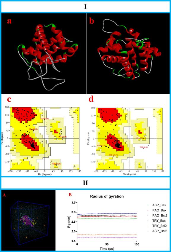
www.nature.com/scientificreports/
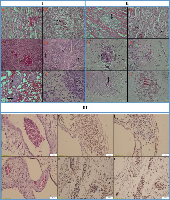
Figure 1. I (The Alectoris chukar chicks inoculated intravenously with Candida albicans. (a) The bird is
characterized by depression, lethargy and ruffled feathers. (b) Following the progression of the disease, infected
bird is unable to stand. (c) Hydro pericardium is seen in heart (blue arrow). (d) White necrotic foci are seen
in myocardium (blue arrows). (e) Congestion is seen on the brain surface. (f) The gizzard is devoid of food in
debilitated bird). II The partridge embryo model and its extra-embryonic membrane C. albicans inoculation.
(a) Vascular congestion (blue arrows) or (b) general congestion is located on the embryo skin. (c) Normal
embryo did not show any gross lesion. (d) Vascular disruption is occurred on the partridge’s EEM model witch
demonstrated by haemorrhages (blue arrows).
Body weight. Figure 2I shows the body weight of infected and control partridges at the beginning of the
experiment (day 30) and 5 days after infection (day 35). The body weights of the infected partridges were signifi-
cantly different from those of the control birds at the end of the experiment. A marked decrease was observed in
the growth rate of the inoculated birds after injection of the C. albicans (152.2 ± 0.75 g at day 30; 156.5 ± 1.3 g at
day 35, p = 0.033). The control partridges had normal weights (149.8 ± 1.03 g at day 30; 173.6 ± 0.34 g at day 35).
Gross findings. Gross lesions associated with systemic candidiasis were observed in the kidneys, liver, lungs,
heart and brain. Congestion was noticed in some internal organs including the kidneys, liver and lungs of the
affected birds. The kidneys and liver were swollen. In some affected birds, hydropericardium with white necrotic
foci occurred in myocardium (Figs. 1I-c,d). Congestion of the brain surfaces was evident in birds showing neu-
rological disturbance (Fig. 1I-e). The alimentary tract of most cases was devoid of food (Fig. 1I-f). Muscular
atrophy was observed in debilitated birds; however, the skeletal structure appeared normal.
Histopathological findings. Histological lesions were observed in the kidneys, liver, spleen, lungs, pan-
creas, heart and brain of the infected birds. Varying degrees of congestion were detected in some internal organs
including the kidneys, liver and lungs (Fig. 3I-a, c and e). In some cases, granulomas were evident in the lungs
(Fig. 3I-e), spleen (Fig. 3I-b) and brain (Fig. 3II-c to f). Histologically, granulomas were composed of both
mycelial and yeast forms of C. albicans with areas of necrosis and inflammatory cell infiltration (particularly the
macrophages). Lesions of the liver included congestion, hepatocellular degeneration, necrosis, central vein dila-
tation, bile duct hyperplasia and vascular thrombosis (Fig. 3I-c and d). The pancreas showed degeneration and
necrosis in the exocrine portion (Fig. 3I-f). Myocardial fiber necrosis with infiltration of mononuclear cells, par-
ticularly the macrophages, was noticed in myocardium (Fig. 3II-a and b). In the brain, gliosis, edema, neuronal
ischemic cell change and granuloma formation in the cerebrum, molecular/granular layer of the cerebellum and
midbrain were observed (Fig. 3II-c to f).
Susceptibility of the partridge embryo model to systemic candidiasis. To confirm the suscep-
tibility of the partridge embryo model to C. albicans by human origin, we chose the 10th embryonic day. We
observed that embryonic infection causes a decrease in survival time, and at later embryonic days (the 11–12th)
Scientific Reports | (2021) 11:2072 | https://doi.org/10.1038/s41598-021-81592-y 3
Vol.:(0123456789)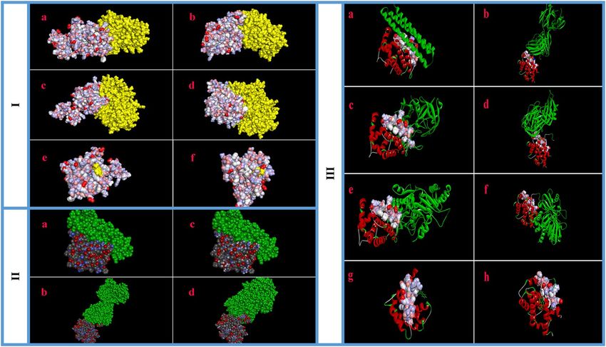
www.nature.com/scientificreports/

                                            Figure 2.  I (The body weight of the Alectoris chukar chicks at the start of experiment (day 30) and 5 days after
                                            infection (day 35) with comparison between control and Candida albicans infected birds. The infected birds
                                            showed significant weight loss compared to controls at the end of the experiment (error bars show standard
                                            error of mean; *p < 0.05).) II:(Expression levels of apoptotic-regulator genes following Candida albicans
                                            treatment. The expression level of Bax mRNA in the partridge extra-embryonic membranes is increased but the
                                            expression levels of Bcl-2, VEGF-A and VEGF-R2 are decreased in the LM-treated groups compared to controls.
                                            (Error bars show standard error of mean).III (The mean capillary area (MCA) analysis on the partridge’s extra-
                                            embryonic membrane. (a) A constant area (125 mm2) is extracted from captured image. (b) Extracted area has
                                            been converted to a binarized image. (c) Five defined areas (rectangular) without any branch vessels are selected
                                            and the percentage of the areas containing black pixels is quantified for the MCA. The black pixels of the
                                            image indicate the red color, or blood, in the original image. (d) MCA is significantly decreased in the fungus
                                            inoculated group compared to the control (error bars show standard error of mean; *p < 0.000, T test).

                                            of infection, embryos showed gross pathological lesions. The lesions were generally noticed as vascular con-
                                            gestion (Fig. 3II-a) or general congestion (Fig. 3II-b), which was located on the skin. Vascular disruption also
                                            occurred on the partridge EEM model that was expressed by haemorrhages (Fig. 3II-d).

                                            (B) Vasculature analysis, IHC and qPCR results from EEM vasculature

                                               As described previously, the EEM partridge modelwas used to evaluate the effect of C. albicans on vascular
                                            apoptosis. The results were as follows:

                                            Mean capillary area. The response to C. albicans inoculation onto the partridge EEM is presented in
                                            Fig. 2III-d. There was a significant decrease in MCA of the vasculature of the treated group (control group,
                                            29.98 ± 2.72; infected group, 9.12 ± 1.80; p = 0.034).

                                            Immunohistochemistry results. In order to prove vascular apoptosis in the partridges’ EEM vasculature,
                                            H&E and IHC staining were performed to detect apoptotic cells and their components. As illustrated in Fig. 3III,
                                            in C. albicans infected group, Bcl-2 on leukocytes, endothelial and stromal cells wereless intensely stained com-
                                            pared with Bax.

                                            Gene expression results.    The expression levels of apoptotic-regulator genes in C. albicansinfected EEM
                                            were determined by qPCR. Our results showed increased expression of Bax genes and reduced expression of
                                            Bcl-2, VEGF-A and VEGF-R2 (Fig. 2II).

          Scientific Reports |   (2021) 11:2072 |               https://doi.org/10.1038/s41598-021-81592-y                                                 4

Vol:.(1234567890)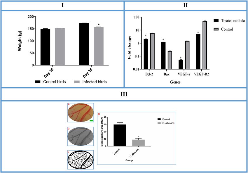
www.nature.com/scientificreports/
(C) In silico results
In silico assay, as mentioned earlier, was applied to investigate the interaction between C. albicans virulence
factors (ASP, PAO and TRY) and apoptotic-regulator proteins (Bcl-2 and Bax). The results are presented below.
Structural parameters of simulated proteins. The bioinformatics tools were applied to construct the
structures of the apoptotic-regulator proteins (Bcl-2 and Bax, Gallus gallus) (Fig. 4I-a and b). The qualities of
constructed models were checked by Ramachandran plots (Fig. 4I-c and d). Ramachandran results for Bcl-2 and
Bax were respectively as follows: 88.1% and 87.2% of the residues located in most favored regions, 9.4% and 9.9%
of the residues located in additional allowed regions and 2.5% and 2.3% of the residues located in generously
allowed regions. The Ramachandran plot is a fundamental tool in the analysis of protein structure. It locates
the amino acid residues of the simulated models R amachandran31. A good quality Ramachandran plot has over
85–90% in the most favored regions. Ramachandran plots of simulated proteins (Bcl-2 and BAX) have 88.1%
and 87.2% of residues in the most favored regions; therefore, these data exhibited that good quality models were
simulated.
The structural parameters of the simulated Bcl-2 were as follows: molecular weight = 21,422.02, total number
of atoms = 2954, total number of amino acids = 194, number of negatively charged residues (Asp + Glu) = 22 and
the number of positively charged residues (Arg + Lys) = 17.
The conformational parameters of the simulated Bax were as follows: molecular weight = 21,666.16, total num-
ber of atoms = 3085, total number of amino acids = 196, number of negatively charged residues (Asp + Glu) = 19
and number of positively charged residues (Arg + Lys) = 17.
Molecular docking. After constructing the 3D structures of Bcl-2 and Bax, active binding pockets were
determined using CASTp program. The volumes of the pockets were 6391.543 and 7094.497 for Bcl-2 and Bax,
respectively.
The dockings of ASP, PAO and TRY with Bcl-2 and Bax were performed using the HEX program. The PDB
file format of receptors and ligands were uploaded in HEX for energy minimization and structure refinement
and then, the highest scoring conformations (lowest energy) were selected. The results are described below:
The docking between ASP and Bcl-2 was successful, with a significant score (E-total score: − 564.43 kcal/
mol). The 3D docking result is demonstrated in Fig. 5I-a. When searching for binding residues on the surface
of ASP, we detected that VAL, GLU, SER, VAL, ASN, ARG, GLU, MET, ALA, GLN, MET, SER, GLY, GLN, LEU,
HIS, LEU and THR were important for the interaction with Bcl-2.
The analysis of docking data showed that ASP also interacted with Bax (E-total score: − 610.11 kcal/mol) by
the binding residues including LEU, THR, VAL, GLY, GLY, TRP, MET, ASN, SER, ILE, PRO, ALA, LEU, ALA,
CYS, PHE, SER, VAL, ASP, GLN, PHE, SER, GLY and SER (Fig. 5I-b).
As with the case of ASP, the docking between PAO and Bcl-2 was successful with a significant score (E-total
score: − 507.77 kcal/mol) (Fig. 5I-c). For PAO, various residues including THR, ALA, HIS, GLY, ARG, PHE, VAL,
ALA, VAL, VAL, GLU, GLU, LEU, PHE, ARG, ASP, GLY, ASN, TRP, ILE, GLN, ASP and ASN were predicted
to interact with Bcl-2.
After docking, the affinity between PAO and Bax was also high (E-total score: − 526.59 kcal/mol) (Fig. 5I-d)
and the protein–protein interaction successfully occurred through the binding residues including ASP, LYS, LEU,
ASP, GLN, ASP, GLN, ALA, PHE, ASN, ASP, MET, ILE, ASP, GLY, LEU and VAL.
The analysis of data showed that TRY interacted with Bcl-2 and Bax with low affinity (E-total score: − 143.62
and − 160.53 kcal/mol, respectively) (Fig. 5I-e and f).
Affinity of C. albicans virulence factors for Bcl‑2 family from different origin. As pointed out
earlier, cross-dockings were done with 5JSN and 5W5X, instead of their specific ligands, to evaluate the affinity
of ASP, PAO and TRY for Bcl-2 family proteins that originated from a different species (Human). The results are
shown in Fig. 5III and Table 1. The analysis of data revealed that after separating the original ligands from 5JSN
and 5W5X, the ASP, PAO and TRY still tended to interact with them. The 5JSN and 5W5X are crystallographic
structures of human Bcl-2 and Bax, which interacted with specific inhibitor or activator, respectively. When
searching for binding residues on the surfaces of 5JSN and 5W5X faced toward their inhibitor or activator, vari-
ous residues including ASP, PHE, SER, ARG, ARG, TYR, ARG, ARG, ASP, PHE, ALA, GLU, MET, SER, SER,
GLN, LEU as well as SER, GLU, SER, LEU, LYS, ARG, ILE, GLY, ASP, GLU, LEU, ASP, SER, ASN, MET were
predicted, respectively (Fig. 5III-a and b). When comparing these results, we found that several of these residues
are known to interact with ASP, PAO and TRY and are located near the binding sites of 5JSN and 5W5X with
ASP, PAO and TRY, suggesting that ASP, PAO and TRY may alter the activity of 5JSN and 5W5X similar to that
of their specific inhibitor or activator. Similar residues, which are located at the binding sites of 5JSN and 5W5X
faced toward their ligands, are illustrated in Fig. 5III-c to h (residues in white color).
Self‑docking. We assessed the accuracy of the results by a self-docking stage (validation stage). Among the
various conformations, the best-docked conformation was selected. Self-docking results for 5JSN and 5W5X are
listed in Table 1, and the best conformation for each docking is demonstrated in Fig. 5II. As predicted, the accu-
racy of the results was proved with an appropriate binding energy. As revealed in Fig. 5II-c and d, the specific
ligands of the 5JSN and 5W5X were docked with their receptors by correct orientations.
Scientific Reports | (2021) 11:2072 | https://doi.org/10.1038/s41598-021-81592-y 5
Vol.:(0123456789)www.nature.com/scientificreports/
Scientific Reports | (2021) 11:2072 | https://doi.org/10.1038/s41598-021-81592-y 6
Vol:.(1234567890)www.nature.com/scientificreports/
◂Figure 3. I (Histopathological lesions encountered in Alectoris chukar inoculated with Candida albicans. (a)
Congestion (arrow) is seen in kidney, H&E. (b) Mycelial and yeast forms of Candida albicans (arrows) are
seen in spleen, PAS. (c) Congestion (arrows) is seen in liver, H&E. (d) Central vein dilatation (asterix) and
hepatocellular necrosis (arrows) are seen in liver, H&E. (e) Congestion and granulomas (arrows) are seen in
lung, H&E. (f) Degeneration and necrosis (arrow) are noticed in the exocrine portion of the pancreas, H&E).
II (Histopathological lesions encountered in Alectoris chukar inoculated with Candida albicans. (a) Myocardial
necrosis with mononuclear cell infiltration (arrow) are seen in myocardium, H&E. (b) Mycelial and yeast
forms of Candida albicans (arrows) are seen in myocardium, PAS. (c) Granuloma (arrow) is seen in cerebrum,
H&E. (d and e) Granuloma (arrow) is seen in the molecular and granular layer of cerebellum, respectively,
H&E. (f) Granuloma with mycelial and yeast forms of Candida albicans are seen in cerebrum, PAS.) III:(Effects
of systemic candidiasis in partridge on vascular apoptosis and blood vessel system (× 40). (A) H&E stained
vessels in the control embryo: the embryo with normal blood vessel and adequate distribution of leukocytes.
RBC’s endothelial living cells seem normal. (B) H&E stained vessels in the infected embryo: decreasing and
compassing of embryonic vasculature compared with the control. (C) IHC staining with Bax in the control
embryo: scattered Bax positive blood cells and rarely positive endothelial and stromal cells. (D) IHC staining
with Bax in the infected embryo: compassed vessels with more positive stained leukocytes and stromal cells,
compared with the control group. (E) IHC staining with Bcl-2 in the control embryo: compassed with Bcl-2 IHC
staining more cells are stained. (F) IHC staining with Bcl-2 in the infected embryo: compassed vessels with less
positive stained leukocytes and stromal cells, compared with the control group).
Molecular dynamics results. Molecular dynamics simulation was performed to assess the conformational
stability of simulated proteins (ASP, PAO, TRY, Bcl-2 and Bax) and their interactions in the simulated body envi-
ronment. The simulation box is illustrated in Fig. 5II-A. By calculating the radius of gyration, it was revealed that
the proteins remain stable, in its compact (folded) form over the course of runs (Fig. 5II-B). The Van der Waal’s
interactions (Lennard–Jones and Coulomb interactions), solvent accessible surface areas, number of hydrogen
bonds and density of structures are presented in Table 2.
Discussion
In this work, we demonstrate that the partridge and embryonated partridge egg are new and effective preclinical
infection models for evaluating systemic candidiasis. Moreover, some mechanisms, which could be associated
with the pathogenesis of the organism, were assessed. Various disorders and lesions were noticed following
systemic candidiasis in partridge.
The clinical signs were seen over a 48–96 h period in the partridge model. It was a variety of adverse health
consequences including depression, lethargy, decreased feed and water intake and growth depression. Five days
post-infection, the analysis of body weight revealed that C. albicans caused severe growth depression. The next
indicator of the systemic candidiasis in the partridge model was neurological disturbance. Several types of neural
disturbances were observed in the Galliformes: 1) extreme opisthotonus with spasmodic tremors, 2) extreme
torticollis with cranial rotation and 3) extreme torticollis which resulted in the head being drawn in a medial-
ventral direction 32. A spasmodic tremor was seen in the head of some partridge birds on day 4 post-infection.
Birds with head tremors exhibited the microscopic appearance of C. albicans in different parts of the brain.
Various gross abnormalities and microscopic lesions in multiple tissues were also indicators of the candidiasis
in the partridge model. Based on the results of this study, it is deduced that C. albicans can affect the kidneys,
liver, spleen, lungs, pancreas, heart and brain of the partridge model. We noticed gross and microscopic lesions
in these organs, however, further data may be provided on alterations in other organs.
Our results are in agreement with the clinical presentation of bloodstream candidiasis in humans. It generally
presents as fever and worsening of clinical status, especially in a patient with risk factors who is not responding
to appropriate therapy. When C. albicans disseminates in humans, various organs are usually involved with the
kidney, liver, spleen, myocardium and eye being the most common, and less frequently the other organs (e.g.
lung, joint and bone). Candida infects both meninges and brain parenchymal tissue. The organism induces
diffuse encephalopathy with diminished consciousness in which the predominant lesion is micro abscess. The
severity of the illness and its profile (acute or chronic) may be conditioned by the size of the inoculums. In the
partridge model (our work), 5 × 10⁵cfuof C. albicans produces neurocandidiasis on the fourth day after infection.
Complex cellular and molecular processes may be related to the pathogenesis of C. albicans. In the cur-
rent research, details of the vascular apoptotic effect of C. albicans were assessed through in vivo (EEM par-
tridge model) and in silico investigations. Some highlights of our findings on the vascular alteration are herein
discussed.
Morphometrical analysis of the partridge’s EEM vasculature has revealed that C. albicans affects the vascular
plexus negatively. The method applied to assess the vascular alteration effect of C. albicans was the calculation of
the mean capillary area in the captured images. To date, this method has been widely used in vascular analysis
7,33,34
.
The next aspect to discuss is the considerable alteration in the expression of Bcl-2 and Bax proteins following
C. albicans treatment. The pathways and mechanisms by which C. albicans induced apoptotic activity in ves-
sels have not been identified explicitly. However, regarding the IHC results, which revealed that Bcl-2 was less
intensely and Bax was more intensely stained in C. albicans treated EEM, we suggest that alteration in the Bcl-2
family members could be one of the mechanisms involved in the vascular apoptotic activity of C. albicans. This
assumption was also supported by our in silico study in which C. albicans virulence factors bound to the active
site of Bcl-2 and Bax proteins.
Scientific Reports | (2021) 11:2072 | https://doi.org/10.1038/s41598-021-81592-y 7
Vol.:(0123456789)www.nature.com/scientificreports/
Figure 4. I (Structural parameters of simulated proteins (a and b), Protein models of Bcl-2 and Bax (Gallus gallus),
respectively The structures of Bcl-2 and Bax (Gallus gallus) were constructed using SWISS-MODEL (https://swiss
model.expasy.org/). (c and d) Ramachandran plots for protein model of Bcl-2 and Bax (Gallus gallus), respectively.
Ramachandran plot was created using PROCHECK server,https://servicesn.mbi.ucla.edu/PROCHECK/). II
(Molecular dynamics simulations (A) Simulated proteins solvated in SPC water (cyan) and sodium ions (orange).
(B) Radius of gyration values for simulated proteins. By calculating the radius of gyration, it is revealed that the
proteins remain stable, over the course of runs. ASP aspartic proteinase, PAO polyamine oxidase, TRYtryptophol).
Molecular dynamics simulation was performed using the GROMACS 5.4.1 package http://www.gromacs.org/.
Scientific Reports | (2021) 11:2072 | https://doi.org/10.1038/s41598-021-81592-y 8
Vol:.(1234567890)www.nature.com/scientificreports/
Figure5. I (The docking results between apoptotic-regulator proteins (yellow/right) and virulence factors of C.
albicans (left). The van der Waals surface of each atom in the protein structure is demonstrated. (a) Successful
docking between ASP and Bcl-2. (b) Protein–protein interaction \ successfully occurred between ASP and
Bax. (c) Successful docking between PAO and Bcl-2. (d) Protein–protein interaction occurred between PAO
and Bax. (e) The docking between TRY and Bcl-2 with low affinity. (f) Protein–protein interaction occurred
between TRY and Bax with low affinity. Bax; simulated Bax of Gallus gallus. Bcl-2; simulated Bcl-2 of Gallus
gallus. ASP aspartic proteinases: virulence factor of C. albicans. PAO polyamine oxidase: virulence factor of C.
albicans. TRYtryptophol: virulence factor of C. albicans. II (Validation stage through self-dockings between
apoptotic-regulator proteins (left) and their original ligands (green/right) (a) 5JSN; Crystallographic structure
of human Bcl-2 in complex with specific inhibitor. (b) 5W5X; crystallographic structure of human Bax in
complex with specific activator. (c and d) Binding orientations predicted by the self-dockings are in agreement
with the binding orientations confirmed by experiment.) III (Cross docking between C. albicans virulence
factors (left) and apoptotic-regulator proteins (green/right). Similar residues located in the binding sites of
5JSN and 5W5X facing toward their ligands are illustrated (white/blue) (a) 5JSN; Crystallographic structure
of human Bcl-2 protein in complex with specific inhibitor. (b) 5W5X; crystallographic structure of human
Bax protein in complex with activator. (c) Successful cross-docking between ASP and 5JSN. (d) Successful
cross-docking between ASP and 5W5X. (e) Successful cross-docking between PAO and 5JSN. (f) Successful
cross-docking between PAO and 5W5X. (g) Cross-docking between TRY and 5JSN with low affinity. (h) Cross-
docking between TRY and 5JSN with low affinity. ASP; aspartic proteinases: virulence factor of C. albicans. PAO
polyamine oxidase: virulence factor of C. albicans. TRYtryptophol: virulence factor of C. albicans. Figure 5.I, II
and III were created using the HEX 8.0.0 software http://hex.loria.fr/dist/index.php).
Docking Bcl-2 family Virulence factors E-total (kcal/mol) Binding affinity
Cross-docking Bcl-2 ASP − 612.57 +
Cross-docking Bcl-2 PAO − 528.99 +
Cross-docking Bcl-2 TRY − 156.31 ±
Cross-docking Bax ASP − 502.44 +
Cross-docking Bax PAO − 483.25 +
Cross-docking Bax TRY − 151.07 ±
Self-docking 5W5X Self-ligand − 391.77 +
Self-docking 5JSN Self-ligand − 1029.09 +
Table 1. Cross-dockings and self-dockings results. + ; high affinity. ± ; low affinity.
Scientific Reports | (2021) 11:2072 | https://doi.org/10.1038/s41598-021-81592-y 9
Vol.:(0123456789)www.nature.com/scientificreports/
Bcl-2 family Virulence factors LJ (kJ/mol) Coul (kJ/mol) SASA (nm2) NHB Density (kg m
−3)
Bcl-2 ASP − 589.92 − 985.23 288.66 26.66 1005.11
Bcl-2 PAO − 1265.08 − 1722.55 360.71 37.94 1005.93
Bcl-2 TRY − 57.32 − 42.71 120.68 0.99 1020.46
Bax ASP − 505.32 − 839.19 280.85 24.21 1015.24
Bax PAO − 1281.96 − 1673.63 357.00 41.90 1013.41
Bax TRY − 63.48 − 50.64 114.55 1.07 1025.48
Table 2. Molecular dynamics simulation results. LJ Lennard–Jones interaction, Coul Coulomb interaction,
SASA Solvent accessible surface areas, NHB Number of hydrogen bonds (average per timeframe).
The results of our study verified the low expression of Bcl-2, VEGF-A and VEGF-R2 and increased expression
of Bax genes in the fungus-exposed group. It seems that Akt/PKB signaling pathway is involved in this process
and consequently the VEGF-A caused a decrease in the expression of Bcl2 and increased the expressions of Bax
gene 35.
We also exploit a docking assay to reveal more details about the apoptotic activity of C. albicans. Docking assay
has recently been considered as a valuable technique to elucidate the interaction between receptor and ligand
36–38
. It is well known that the Bcl-2 family members are critical targets for apoptotic and anti-apoptotic agents
39,40
. In this regard, an important highlight in our study is the interaction of ASP, PAO and TRY with Bcl-2 and
Bax proteins. Our data suggest the modulation of Bcl-2 family members via the binding of C. albicans virulence
factors, which must be validated by further experiments.
Another highlight of the protein–protein interaction is the quantitative aspect of binding affinity between
C. albicans virulence factors and Bcl-2 family proteins. Based on docking results, which revealed the lowest
scoring energy for ASP (− 564.43 and − 610.11) compared to PAO (− 507.77 and − 526.59) and TRY (− 143.62
and − 160.53), it is predicted that the affinity of ASP for Bcl-2 and Bax is higher than that of PAO and TRY.
Therefore, ASP can be regarded as a hopeful target for designing anti-apoptotic agents to alleviate the devastat-
ing outcomes of candidiasis.
As explained in the materials and methods section, cross-docking was performed to appraise the possible
interaction of ASP, PAO and TRY with Bcl-2 family members of Homo sapiens (5JSN and 5W5X). Concerning
their successful interaction, it is worthwhile to note that the virulence factors of C. albicans are able to selectively
interact with Bax and Bcl-2 proteins originating from species other than the birds (Gallus gallus).
To the best of the authors’ knowledge, this is the first study to target different aspects of systemic candidiasis
with the help of the partridge model. Our results show that C. albicans of human origin not only causes systemic
candidiasis in the partridge model, but also exhibits adverse clinical consequences, which were similar to human
candidiasis. Therefore, partridge and embryonated partridge egg are powerful and attractive preclinical models
to study the pathogenesis of candidia spp. because of cost, ease of handling and technical feasibility. The acquired
results also indicate that C. albicans alters the normal growth of vessels and changes the expression of apoptotic-
regulator proteins and genes. The presented data on vascular alteration activity of C. albicans permit us to suggest
that this activity is one of the important pathways in the pathogenicity of organism. In this investigation, we also
employed the partridge’s EEM model for apoptotic studies. This model offers a promising approach to assess the
role of various mechanisms, which are associated with the pathogenesis of the diseases.
Methods
This section is explained in terms of A) induction of systemic candidiasis in partridge and embryonated par-
tridge egg, B) effect of C. albicans on vasculature and C) interactions between C. albicans virulence factors and
apoptotic-regulator proteins.
Materials. Partridge chicks (Alectoris chukar) as well as fertile partridge eggs, with the average egg-weight
of 20.7 ± 0.5 g, were purchased from Hasanzadeh Partridge Farm, Yazd, Iran. In the supplier farm, the par-
tridge breeders were kept under optimal condition of rearing. Paraffin was obtained from Merck, Darmstadt,
Germany. Real-time PCR materials were purchased from Qiagen, Chatsworth, CA and Takara Bio, Inc., Shiga,
Japan. Various biological databases and bioinformatics tools like National Center for Biotechnology Informa-
tion (NCBI), RCSB Protein Data Bank, CASTp (http://sts.bioe.uic.edu/castp/), SWISS-MODEL, (https://swiss
model.expasy.org/), PROCHECK (http://servicesn.mbi.ucla.edu/Verify3D/), GROMACS 5.1.4, HEX 8.0.0,
ImageJ 1.48 (National Institutes of Health, Bethesda, Maryland, USA), MATLAB (MathworksMatlabR2015a)
and Digimizer4.3.0 (MedCalc Software, Mariakerke, Belgium) were used for in silico evaluations.
(A) Induction of systemic candidiasis in partridge and embryonated partridge egg
This section was designed to clarify whether C. albicans of human origin can cause systemic disease in the
partridge model. In the next step, the clinical signs and pathological lesions of systemic candidiasis were assessed
in the partridge model.
Scientific Reports | (2021) 11:2072 | https://doi.org/10.1038/s41598-021-81592-y 10
Vol:.(1234567890)www.nature.com/scientificreports/
Feed ingredients %
Corn 49.38
Soybean meal (44% CP) 44.58
Vegetable oil 1.56
Dicalcium phosphate 1.80
Limestone 1.78
NaCl 0.3
DL-methionine 0.10
Vitamin Premix* 0.25
Mineral Premix* 0.25
Total 100
Analysis
Crude protein 24.00
Ca 1.10
P 0.51
Na 0.11
ME, Kcal/kg 2800
Table 3. Ingredients and chemical composition of diet during the experimental period (0–35 days). *Vitamin/
Mineral Premix (Talavang company, Tehran, Iran) supplied per 5 kg: vitamin A, 11,000,000 IU; cholecalciferol,
5,000,000 IU; vitamin E, 7 500 IU; K3, 3000 mg; vitamin B1, 3000 mg; vitamin B2, 8000 mg; niacin, 4000 mg;
d-pantothenic acid, 15,555 mg; vitaminB12, 16 mg; folic acid, 2000 mg; biotin, 150 mg, Mn, 120,000 mg; Fe,
40,000 mg; Zn, 100,000 mg; Cu, 16,000 mg; iodine, 125 mg; Se, 300 mg; cholin chloride, 900,000 mg.
Organism. The PTCC 5027 strain of C. albicans was kindly provided by Persian Type Culture Collection
Center (PTCC), Iran. It was obtained from a human patient and, based on antigenic properties, is categorized to
serotype A. This isolate produces D-arabinolactone oxidase, DNA topoisomerase, aspartic proteinases, aspartyl
proteinase, estrogen-binding protein, lanosterol synthase, 2, 3-oxidosqualenelanosterol cyclase, phenethyl alco-
hol, polyamine oxidase and tryptophol. The inoculum was prepared from the mentioned strain in Sabouraud’s
dextrose broth (Oxoid, Thermo Fisher Scientific, Basingstoke, UK) at 42 °C for 18 h32. The broth culture was then
centrifuged (2 min at 2500 g) and the remaining pellet was washed three times with sterile phosphate buffered
saline (pH 7.4). Finally, the cell suspension was adjusted to an optical density of 1 06 cfu/ml 41. The cell numbers
in inoculum were confirmed by plating serial dilutions on Sabouraud’s dextrose agar plates.
Birds and housing conditions. Day-old chukarpartridge chicks (Alectoris chukar) were obtained from
a commercial breeder farm (Hasanzadeh Partridge Farm, Yazd, Iran). Upon arrival at the Animal Research
Center of Shahid Bahonar University, Kerman, Iran, the birds were housed in an electrically-heated battery cage
(Belderchin Damavand Co. PLC-DQSH, Tehran, Iran) at 34 °C for 10 days and then were held in two floor pens.
The partridges were kept 30 days for acclimation. The temperature was controlled and gradually reduced from
34 °C on day 1–29 °C on day 30 and remained constant throughout the study. During the first week of brood-
ing, a photoperiod of 23 h/day was maintained and then a photoperiod of 20 h/day was used. The temperature
and photoperiod are necessary for raising and managing partridge chick especially during the first 5 weeks of
life. Poor or inadequate temperature and photoperiod is stressful for bird and causing a problem in partridge
management. Therefore, temperature and photoperiod were supplied for partridge chicks according to recom-
mended procedures28.Food and water were provided ad libitum. The food was formulated according to the
nutritional requirements of the partridge chicks from which all medications had been omitted. The ingredients
of the diet are presented in Table 3 28. The trial lasted for 35 days.
Experimental design. The experiment was performed according to the suggested European Ethical Guide-
lines for the care of animals in experimental investigations, in line with the guidelines of Kerman University of
Medical Sciences and was approved by the Animal Ethics Committee of the Research Council of Shahid Bahonar
University, Kerman, Iran (project number D.550.P.835, Ethics number IR.UK.VETMED.REC. 398.014).
At 30 days of age, twenty-four partridge chicks were selected on the basis of their overall appearance and body
weight uniformity. Blood samples were randomly collected aseptically from the brachial vein of ten birds and
serological tests (Hemagglutination-inhibition tests for H9N2 avian influenza subtype and Newcastle diseases)
as well as bacteriological cultures were done to assess the pre-challenge health status of the partridges 42. All
sampled birds showed negative results in antibody tests. Negative bacteriological cultures were also obtained.
The birds were randomly separated into two different groups of 12 birds each. The partridges of the first group
were injected intravenously (in brachial vein) with 0.5 ml of prepared suspension of C. albicans. The birds of
the second group were used as the control and were intravenously inoculated with sterile phosphate buffered
saline. Infectious dose was chosen based on our preliminary investigation and experimental study inducing
systemic candidiasis in G alliformes32. The birds were monitored twice daily during the experimental period
(5 days after inoculation). Each bird was evaluated individually and efforts were made so that just one person
Scientific Reports | (2021) 11:2072 | https://doi.org/10.1038/s41598-021-81592-y 11
Vol.:(0123456789)www.nature.com/scientificreports/
could be involved in the clinical evaluation of the birds in order to avoid subjectivity in the analysis. Cloacal
temperature and body weight were also recorded. Cloacal temperature was measured four times daily using a
digital medical thermometer (FT15/1, Beurer Company, Ulm, Germany, with a range between 35.5 and 42 °C,
measurement accuracy ± 0.1 °C). Body weight was measured by a digital scale (Sartorius TE212, Germany, with
a range up to 200 g, measurement accuracy ± 0. 01 g). On day 35 of the experiment, the birds were necropsied
and tissue samples were taken for pathological investigations. After being fixed in 10% neutral buffered formalin,
serial sections of paraffin-embedded tissues were prepared and processed routinely for hematoxylin and eosin
(H&E) as well as periodic acid-Schiff (PAS) staining. Tissues of the kidneys, liver, spleen, lungs, pancreas, heart
and brain were examined.
Induction of systemic candidiasis in partridge embryo model. Fertile partridge eggs, of the breed
Alectoris chukar, were incubated in an electrical incubator (General Cocks, Cocksmachine Company, Tehran,
Iran) at 37.5 °C and 60% relative humidity. Prior to the fungal inoculation, eggs were checked for embryonic
development by candling. On the10th day of incubation, C. albicans was inoculated into the chorioallantoic sac
and embryos were allowed to develop for a few more days. For C. albicans inoculation, embryo viability was con-
firmed prior to inoculation by candling, and infertile and nonviable embryos were removed. For all embryos, the
air cell was marked, and the egg-shell was disinfected with 70% ethanol. A 1–2-mm diameter region of the shell
just above the air cell marking was penetrated using an electric drill, and the chorioallantoic sac was inoculated
via a 1-ml syringe with a 1-inch, 22-ga needle.
The 10th day of the embryonic stage was chosen based on our preliminary experiment. On the other hand,
on the10th day of incubation, the partridge’s chorioallantoic sac is large enough to inoculate an appropriate
volume of fungus. The embryos were examined at 3 different time intervals: 24, 48 and 72 h after inoculation.
Finally, the embryos were humanely killed by cooling 43. The eggs were opened at the blunt end and embryos
were removed to study any gross abnormalities on the external body surface The experiment was repeated for a
total of two trials to ensure the repeatability of the experiment.
(B) Effect of C. albicans on vasculature
In this study, we used the EEM partridge model for evaluating the effect of C. albicans on vasculature and
apoptotic-related proteins. In recent years, the EEM of Gallus gallus has provided a valuable model for in vivo
evaluation of the vascular toxicity of the agents 44,45. It is also used as an alternative host model for fungal patho-
gens 43,46. Hence, we used the EEM partridge model to investigate the effect of C. albicans on the vasculature
and apoptotic-related proteins. This effect was assessed via morphometric analysis of vascular pattern from
the partridge EEM. Immunohistochemistry (IHC) and quantitative real-time PCR (qPCR) assays were also
performed to evaluate the expressions of apoptotic-regulator proteins and genes following fungus treatment.
The details are as follows:
Experimental design and image acquisition. Fertile partridge eggs were incubated at 37.5 °C and 60%
relative humidity. On the 10th day, C. albicans was inoculated onto the EEM. After infection, the shell was
cut with scissors and a window of 15 × 15 mm was opened to allow image acquisition. High-resolution images
(5312 × 2988 pixels) were captured from the EEM vasculature. Afterwards, an area of approximately 10 × 10 mm
of partridge’s EEM was removed and subjected to IHC staining.
Vascular pattern analysis. Image analysis was performed using computerized software such as MAT-
LAB (Mathworks Matlab R2015a) and ImageJ 1.48 (National Institutes of Health, Bethesda, Maryland, USA).
Initially, a defined area (125 mm2) of the EEM was extracted and the contrast was improved (Fig. 2III-a). Effort
has been made to select a constant area in each image. The extracted area was converted to a binarized format
(Fig. 2III-b). Then, the areas without any branch vessels were selected for analysis. Five such areas per case were
identified and the percentage of the areas containing black pixels were quantified (Fig. 2III-c). The black pixels of
the images indicate the red color, or blood, in the original images. The mean of all areas calculated in each image
is described as the mean capillary area (MCA) 33.
Immunohistochemistry assay. The samples of the partridge’s EEM were fixed in 10% buffered forma-
lin and embedded in paraffin. Tissue sections were made by the microtome (Slee Germany) and IHC staining
was performed for Bcl-2 (mouse monoclonal antibody, American, ID number: PDMO16-lotH147) and Bax
(Zytomed Germany, ID number: 502_17990) markers. The expression levels of Bcl-2 and Bax were assessed by
counting the stained cells and calculating the mean in 10 high-power fields (400 ×).
Gene expression. The effect of C. albicans on the apoptotic-regulator genes, in vessels, was evaluated by
qPCR assessment of relative expression levels of Bax, Bcl-2, VEGF-A and VEGF-R2 genes. Briefly, the total RNA
of the partridge EEMs was isolated using the RNeasy mini kit (Qiagen, USA). A nano-drop was used to evalu-
ate the quality of samples (ND-1000, Thermo Scientific Wilmington, DE, USA) and the cDNA was synthesized
using a total of 500 ng RNA by RT reagent kit (Takara, Clontech) according to the protocol. Finally, qPCR was
performed using a SYBR Green assay (SYBR Premix Ex Ta II; Japan). The specific primers and reference gene
sequences are listed in Bellow.
Scientific Reports | (2021) 11:2072 | https://doi.org/10.1038/s41598-021-81592-y 12
Vol:.(1234567890)www.nature.com/scientificreports/
1. Bax (Forward: CACAGGTGCCTACTGTCGT T)
(Reverse: CACACTGGGATTCTTCCGCT)226 bp.
2. Bcl-2 (Forward: TCGTCGCCTTCTTCGAGTTC)
(Reverse:CATCCCATCCTCCGTTGTCC)150 bp.
3. VEGF-R2 (Forward: GCCAACTCTATGGCAGAAGC).
(Reverse: CTGAACACCATGCCACTGTC)86 bp.
4. VEGF-A (Forward: CAAT TGAGACCCTGGTGGAC).
(Reverse: TCTCATCAGAGGCACACAGG)86 bp.
5. HPRT (Forward: GATGAACAAGGTTACGACCTGGA).
(Reverse: TATAGCCACCCTTGAGTACACAGAG) 103 bp.
6. GAPDH (Forward: CCTCTCTGGCAAAGTCCAAG).
(Reverse: GGTCACGCTCCTGGAAGATA)176 bp.
Expression levels were calculated in relation to the expression levels of the selected reference gene. GAPDH
and HPRT genes were demonstrated to be the most superior and stable genes in the experiments. All tests were
performed in duplicate. Using the 2–ΔCt method, the gene expression level was analyzed in fold change.
(C) Interactions between C. albicans virulence factors and apoptotic-regulator proteins
In this section, using in silico approaches, we simulated the three-dimensional structure of Gallus gallusBax
and Bcl-2 proteins and predicted their interactions with the virulence factors of C. albicans. It has been found
that some virulence factors of C. albicans such as aspartic proteinase (ASP) 47, polyamine oxidase (PAO) 48–50
and tryptophol (TRY) 51,52 can induce apoptosis in hamster, human epithelial cells and particular cell lines.
However, the roles of these factors in partridge-vessels apoptosis are not clearly defined. Therefore, we choose
these virulence factors for in silico study. The procedures are described as follows:
Model simulation and assessment. The structures of Bcl-2 and Bax (Gallus gallus) were constructed
using SWISS-MODEL (https://swissmodel.expasy.org/) based on target-template alignment. The accuracy of
the SWISS-MODEL server has been proved previously 53. At first, the domain sequence of Bcl-2 (Gallus gallus,
ID: NP_990670.2) and Bax (Gallus gallus, ID: ACR83547.1) were obtained from the NCBI server and known
homologous structures were recognized by blasting between the query sequences. In the next step, we retrieved
the closest homologous structures of proteins from the Protein Data Bank and identified the partially homolo-
gous structures to serve as a template for Bcl-2 and Bax.
The FASTA formats of the target’s sequences (Gallus gallus) are listed as follows:
> Bcl-2: NP_990670.2
MAHPGRRGYDNREIVLKYIHYKLSQRGYDWAAGEDRPPVPPAPAPAAAPAAVAAAGASSHHRPEPPG-
SAAASEVPPAEGLRPAPPGVHLALRQAGDEFSRRYQRDFAQMSGQLHLTPFTAHGRFVAVVEELFRDGVNW-
GRIVAFFEFGGVMCVESVNREMSPLVDNIATWMTEYLNRHLHNWIQDNGGWDAFVELYGNSMRPLFDF-
SWISLKTILSLVLVGACITLGAYLGHK.
> Bax: ACR83547.1
MACEASQDYQIGEALLIGVVRQELMEVMEVTEGNAAPPALPEAKPISNSQDQILVQ LNTIKVIGD-
KLDQDQAFNDMIDGLVKVADKSSFWKLVEKVFTDGQINWGRIIVLFYS GLSAKMVVARPRIVSDILSLSLDY-
FKRNLLQWILTVGGWMNSIPALACFSVDQFSGSSMRKYSPYVGVVAFTGGLLLG FIVSRFQKT.
The simulated proteins were further assessed using Ramachandran plot at PROCHECK server https://servi
cesn.mbi.ucla.edu/PROCHECK/)31.
Protein–protein interactions. On the protein surfaces, the active binding pockets were predicted based
on computational geometry theories using CASTp server (http://sts.bioe.uic.edu/castp/). Following the pocket
identification, the interactions of ASP, PAO and TRY with Bcl-2 and Bax proteins were assessed via HEX 8.0.0
software (http://hex.loria.fr/dist/index.php). This software is introduced in the Critical Assessment of Predicted
Interactions (CAPRI, http://capri.ebi.ac.uk/) as a high-quality docking program based on the fast Fourier trans-
form approach 54.
The cross-dockings were also performed to evaluate the affinity of ASP, PAO and TRY in a way that Bcl-2
and Bax proteins are linked to specific inhibitor or activator. For this purpose, the molecular structures of 5JSN
(crystal structure of Bcl-2 in complex with specific inhibitor) and 5W5X (crystal structure of Bax in complex
with specific activator) were selected from the Protein Data Bank (Fig. 5III-a and b). After the separation of the
ligands from the coordinates of the receptors, the affinity among ASP, PAO and TRY with 5JSN and 5W5X were
assessed. Eventually, a validation step was made by self-dockings among 5JSN, 5W5X and their specific ligands.
This step was considered as the key of docking accuracy.
Molecular dynamics simulation. Molecular dynamics simulation was performed using the GROMACS
5.4.1 package http://www.gromacs.org/ to assess the conformational property of simulated proteins (ASP, PAO,
TRY, Bcl-2 and Bax) and their interactions in the simulated body environment. Briefly, the docked structures of
ASP, PAO and TRY with Bcl-2 and Bax were subjected to the GROMOS 54A7 force field and the water model
SPC was used as the solvent. The solvated system was defined in a charge neutralized system by adding ions
(Na or Cl ions). The cubic solvent box was considered and solvated by explicit water using the gmx solvate
algorithm. The system was minimized for 50,000 steps using the steepest descent algorithm and then simulation
Scientific Reports | (2021) 11:2072 | https://doi.org/10.1038/s41598-021-81592-y 13
Vol.:(0123456789)www.nature.com/scientificreports/
was performed at 310 K and 1 bar under periodic boundary conditions. The v-rescale and Parrinello-Rahman
algorithms were used for temperature and pressure coupling, respectively 55,56. Electrostatic interactions were
calculated by the particle-mesh Ewald method 57. During the production phase, conformers were stored every
0.002 ps. The stability of the structure during the simulations was investigated by calculating the radius of gyra-
tion using the tool gmx gyrate of the GROMACS package. The h-bonds, Van der Waal’s interactions, solvent
accessible surface area and density of structures were also analyzed.
Statistical analysis. Statistical analysis was performed using SPSS version 20. The Fisher’s exact and
repeated measurement tests were used to determine the significant differences in lesion occurrence and the body
weight between experimental groups, respectively. The T test was applied to assess the significant differences in
the MCA values. A p value of < 0.05 was considered statistically significant.
Received: 14 March 2020; Accepted: 6 January 2021
References
1. Conti, H. R., Huppler, A. R., Whibley, N. & Gaffen, S. L. Animal models for candidiasis. Curr. Protoc. Immunol. 105, 16–19 (2014).
2. Takagi, J., Singh-Babak, S. D., Lohse, M. B., Dalal, C. K. & Johnson, A. D. Candida albicans white and opaque cells exhibit distinct
spectra of organ colonization in mouse models of infection. PLoS ONE 14, e0218037 (2019).
3. Bandi, A. & Wurster, S. Drosophila melanogaster as a Facile Model for large-scale studies of virulence mechanisms and antifungal
drug efficacy in Candida auris candidiasis. J. Infect. Dis. 193, 1014–1022 (2006).
4. Wurster, S. et al. Drosophila melanogaster as a model to study virulence and azole treatment of the emerging pathogen Candida
auris. J. Antimicrob. Chemother. 74, 1904–1910 (2019).
5. Ames, L. et al. Galleria mellonella as a host model to study Candida glabrata virulence and antifungal efficacy. Virulence 8,
1909–1917 (2017).
6. Segal, E. & Frenkel, M. Experimental in vivo models of candidiasis. J. Fungi 4, 21 (2018).
7. Khosravi, A. et al. Embryonic toxico-pathological effects of meglumine antimoniate using a chick embryo model. PLoS ONE 13,
e0196424 (2018).
8. Kunz, P., Schenker, A., Sähr, H., Lehner, B. & Fellenberg, J. Optimization of the chicken chorioallantoic membrane assay as reliable
in vivo model for the analysis of osteosarcoma. PLoS ONE 14, e0215312 (2019).
9. Tavakkoli, H. et al. Vascular alteration in relation to fosfomycine: in silico and in vivo investigations using a chick embryo model.
Biomed. Pharmacother. 118, 109240 (2019).
10. Khan, Z. et al. Changing trends in epidemiology and antifungal susceptibility patterns of six bloodstream Candida species isolates
over a 12-year period in Kuwait. PLoS ONE 14, e0216250 (2019).
11. Melo, A. P. V. et al. Virulence factors of Candida spp. obtained from blood cultures of patients with candidemia attended at tertiary
hospitals in Northeast Brazil. J. Mycol. MÚdicale 29, 132–139 (2019).
12. Herwald, S. E. & Kumamoto, C. A. Candida albicans niche specialization: features that distinguish biofilm cells from commensal
cells. Curr. Fungal Infect. Rep. 8, 179–184 (2014).
13. Amado, C., Blair, P., Keiser, J. & Siegel, M. O. The impact of infectious diseases consultation on the choice of antifungal therapy in
patients with candidemia. Infect. Dis. Clin. Pract. 25, 33–36 (2017).
14. Pfaller, M. A., Moet, G. J., Messer, S. A., Jones, R. N. & Castanheira, M. Candida bloodstream infections: comparison of species
distributions and antifungal resistance patterns in community-onset and nosocomial isolates in the SENTRY Antimicrobial
Surveillance Program, 2008–2009. Antimicrob. Agents Chemother. 55, 561–566 (2011).
15. Lindberg, E., Hammarström, H., Ataollahy, N. & Kondori, N. Species distribution and antifungal drug susceptibilities of yeasts
isolated from the blood samples of patients with candidemia. Sci. Rep. 9, 1–6 (2019).
16. Muthular, M. et al. Effects of tamoxifen on periodontal disease and Candida albicans of patients with breast cancer and other
pathologies. Future Microbiol. 14, 129–137 (2019).
17. Sun, M. et al. Increase in Candida parapsilosis candidemia in cancer patients. Mediterr. J. Hematol. Infect. Dis.11, e2019012 (2019).
18. Freifeld, A. G. & Kaul, D. R. Infection in the patient with cancer. In Abeloff ’s Clinical Oncology 544–564 (Elsevier, 2020).
19. Arsenault, A. B. et al. Dietary supplementation with medium-chain triglycerides reduces candida gastrointestinal colonization in
preterm infants. Pediatr. Infect. Dis. J. 38, 164–168 (2019).
20. Kim, J. et al. A randomized, double-blind trial investigating the efficacy of caspofungin versus amphotericin B deoxycholate in the
treatment of invasive candidiasis in neonates and infants younger than 3 months of age. J. Antimicrob. Chemother. 75, 215–220
(2020).
21. Walsh, T. J., Katragkou, A., Chen, T., Salvatore, C. M. & Roilides, E. Invasive candidiasis in infants and children: recent advances
in epidemiology, diagnosis, and treatment. J. Fungi 5, 11 (2019).
22. Tavakkoli, H., Rahmani, M., Ghanbarpoor, R. & Kheirandish, R. Induced systemic listeriosis in Alectoris chukar chicks: clinical,
histopathological and microbiological findings. Br. Poult. Sci. 56, 651–657 (2015).
23. Habibi, H., Ghahtan, N. & Brooks, D. M. Effect of sex ratio, storage time and temperature on hatching rate, fertility and embryonic
mortality in Chukar partridge (Alectoris chukar). Anim. Reprod. Sci. 203, 68–74 (2019).
24. Tavakkoli, H., Kheirandish, R., Ghanbarpoor, R. & Mohseni, Z. Systemic staphylococcosis in partridge chicks. Eurasian J. Vet. Sci.
32, 7–14 (2016).
25. Lei, J., Wu, J. & Guan, Q. The potential effects of future climate change on suitable habitat for the Taiwan partridge (Arborophila
crudigularis): an ensemble-based forecasting method. Turkish J. Zool. 41, 513–521 (2017).
26. Lennon, R. J. et al. Using long-term datasets to assess the impacts of dietary exposure to neonicotinoids on farmland bird popula-
tions in England. PLoS ONE 14, e0223093 (2019).
27. Llorente, F. et al. Influence of flavivirus co-circulation in serological diagnostics and surveillance: a model of study using West
Nile, Usutu and Bagaza viruses. Transbound. Emerg. Dis. 66, 2100–2106 (2019).
28. Woodard, A. E. Raising Chukar Partridges (Agriculture and Natural Resources University of California, California, 2002).
29. Huss, D., Poynter, G. & Lansford, R. Japanese quail (Coturnix japonica) as a laboratory animal model. Lab Anim. (NY) 37, 513–519
(2008).
30. Baer, J., Lansford, R. & Cheng, K. Japanese quail as a laboratory animal model. In Laboratory Animal Medicine 1087–1108 (Elsevier,
2015).
31. Ramachandran, G. N. T. & Sasisekharan, V. Conformation of polypeptides and proteins. In Advances in Protein Chemistry vol. 23,
283–437 (Elsevier, 1968).
Scientific Reports | (2021) 11:2072 | https://doi.org/10.1038/s41598-021-81592-y 14
Vol:.(1234567890)You can also read