Post-Traumatic Hallux Valgus: A Modified Surgical Technique
←
→
Page content transcription
If your browser does not render page correctly, please read the page content below
Technical Note

                Post-Traumatic Hallux Valgus: A Modified Surgical
                                   Technique
                  Zylyftar Gorica, M.D., Kimberly McFarland, B.S., John S. Lewis Jr., M.D.,
                          Karl M. Schweitzer Jr., M.D., and Alexander R. Vap, M.D.

Abstract: Post-traumatic hallux valgus, a turf toe variant, is a rare, yet limiting injury. According to the literature, the
deformity has been associated with acute medial collateral ligament tears, turf toe variant injuries, Lisfranc injury patterns,
and first metatarsal fractures. There have been few documented cases of post-traumatic hallux valgus secondary to medial
collateral ligament tears, and the treatment has been variable. Some authors have described direct end-to-end repair of the
ligament to address the deformity, while others have described a modified McBride bunionectomy involving a Silver
bunionectomy, lateral soft tissue release, and medial capsular and ligamentous repair. We propose a modified technique
similar to the modified McBride bunionectomy, however, with the use of an all-suture anchor in the medial capsular and
ligamentous repair. Our belief is that the all-suture anchor will allow for a stronger repair that will meet the physical
demands of everyday ambulation and athletic participation. We used this technique in an individual who had evidence of
a medial ligamentous complex injury of the hallux on MRI and failed conservative management. Postoperatively, the
patient is immobilized until they can begin working on range of motion, strengthening, and finally to achieve return to full
activity and sports.

                            Introduction                                           the MCL and address the valgus deformity is even more

P    ost-traumatic hallux valgus is a rare, yet signifi-
     cantly limiting injury in athletes and nonathletes.
While the literature is abundant regarding the etiologies
                                                                                   sparse and varied. Douglas et al.7 describe an end-to-
                                                                                   end repair of the MCL using two 0-Ethibond sutures,
                                                                                   which were subsequently removed two weeks after
of chronic hallux valgus, the literature on causes of                              surgery. Lohrer6 describes an end-to end repair of the
acute hallux valgus are limited. To date, post-traumatic                           ligament using 3-0 Vicryl reinforced by a periosteal flap.
hallux valgus has been attributed to Lisfranc injury,1                             Fabeck et al.5 and Covell et al.3 describe a modified
medial plantar neuropathy,2 turf toe variant injuries,3                            McBride bunionectomy involving a Silver bunion-
first metatarsal fracture,4 and medial collateral liga-                             ectomy, lateral soft tissue release, and medial capsular
ment (MCL) tears.5,6 Rupture of the first metatarsal                                and ligamentous repair. Our Technical Note introduces
medial collateral ligament was initially described in                              the use of an all-suture anchor in treating a MCL tear in
1997 in a professional soccer player.7 Since then, there                           order to address post-traumatic hallux valgus. Our
has been less than 10 reported documented cases of                                 belief is that the all-suture anchor will allow for a
post-traumatic hallux valgus secondary to MCL tears.                               stronger repair that will meet the physical demands of
Literature describing the surgical technique to repair                             everyday ambulation and athletic participation.

   From the Department of Orthopaedic Surgery, Virginia Commonwealth                  Received July 30, 2021; accepted August 29, 2021.
University, Richmond, Virginia, U.S.A. (A.R.V., Z.G.); Virginia Common-               Address correspondence to Zylyftar Gorica, M.D., Department of Ortho-
wealth University School of Medicine, Richmond, Virginia, U.S.A. (K.M.);           paedic Surgery, Virginia Commonwealth University, 1200 East Broad St., 9th
Louisville Orthopedic Clinic, Louisville, Kentucky, U.S.A. (J.S.L.); and           Fl., Box 980153, Richmond, VA 23298, U.S.A. E-mail: Zylyftar.Gorica@
Department of Orthopaedic Surgery, Duke University, Raleigh, North                 vcuhealth.org
Carolina, U.S.A. (K.M.S.).                                                            Ó 2021 by the Arthroscopy Association of North America. Published by
   The authors report the following potential conflicts of interest or sources of   Elsevier. This is an open access article under the CC BY-NC-ND license (http://
funding: K.M.S. reportspersonal fees from Arthrex, outside the submitted           creativecommons.org/licenses/by-nc-nd/4.0/).
work. J.S.L. reports personal fees from Stryker/Wright Medical, Medshape/             2212-6287/211110
DJO, Medline, Kinos Medical/Restor 3d, ActivOrtho, Panther Orthopaedics,              https://doi.org/10.1016/j.eats.2021.08.032
and Bioventus, outside the submitted work. Full ICMJE author disclosure
forms are available for this article online, as supplementary material.

                                             Arthroscopy Techniques, Vol 11, No 1 (January), 2022: pp e37-e42                                               e37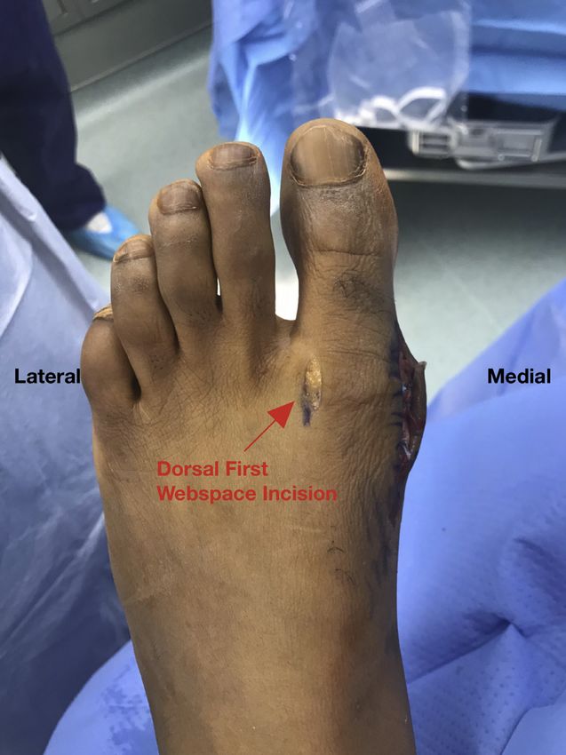
e38 Z. GORICA ET AL.
Surgical Technique (With Video Illustration)
Indications
Given the paucity of literature regarding the injury,
there are no clear indications on when to surgically
treat a MCL tear of the first MTP joint. Our indications
include hallux MTP joint instability, progressive defor-
mity, progressive symptoms, additional intra-articular
joint pathology (e.g. first metatarsal head osteochon-
dral lesion), and those who have failed conservative
therapy and continue to have pain and/or functional
limitations.
Patient Evaluation
Prior to proceeding with surgery, a thorough history
should be obtained, including mechanism and chro-
nicity of injury, attempted treatments to date, and
impact on activities. Physical examination should be
performed with attention to gait, joint alignment,
tenderness to palpation, stability of the joint, and with
comparison to the contralateral limb. Plain radiographs
(Fig 1) and MRI (Fig 2) should be obtained to evaluate
the MCL complex, as well as other anatomical consid-
erations, such as the hallux valgus angle (HVA), inter-
metatarsal angle (IMA), and distal metatarsal articular
angle (DMAA). As the decision is made to proceed with
surgery, the patient should be educated on the post-
operative rehabilitation and recovery process.
Positioning and Preparation
A regional block is performed in the preoperative Fig 1. Preoperative weight-bearing AP view of a left foot
area. Generally, regional anesthesia is avoided in demonstrating a hallux valgus angle of 19 (normal 15 ).
higher-level athletes, in favor of a general anesthetic Preoperative assessment of radiographs should be performed
(Table 1). The patient is brought into the operating in conjunction with physical exam. One should evaluate the
room and placed supine on the table. IV sedation or hallux valgus angle, intermetatarsal angle, distal metatarsal
general anesthesia is initiated. A well-padded thigh articular angle, and hallux valgus interphalangeal angle. Close
tourniquet is placed. The operative leg and foot are then consideration of these radiographic measurements is
prepped and draped in a sterile fashion. The leg is important to determine whether osteotomies are indicated.
Esmarch exsanguinated and the tourniquet is inflated.
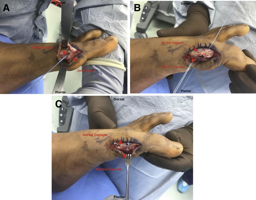
The medial metatarsal head is examined for exos-
Procedure tosis formation. A silver bunionectomy is then per-
A transverse 5-cm incision is made over the medial formed to remove the prominence on the medial
aspect of the first MTP joint (0:05). Care is taken to aspect of the metatarsal head (1:04) (Fig 4B). A 2-cm
identify and protect the dorsomedial cutaneous nerve dorsal incision is made in the first webspace at the
of the hallux. Sharp dissection is then carried out down level of the metatarsal heads (1:10) (Fig 5). Dissection
to the medial capsule. Full-thickness plantar and dorsal is carried out until the adductor hallucis tendons are
skin flaps are then developed. The capsule is then identified. These tendons are sharply released off of
examined for any disruptions, fibrosis, or adhesions. A the first metatarsal head. A 15-blade scalpel is then
transverse incision is then made in the medial capsule used to make several, short pie-crust openings
exposing the medial ligamentous complex (0:32). The through the lateral capsule of the first MTP joint. Varus
medial ligamentous complex is examined for any dis- stress is then applied to the joint to release any lateral
ruptions, attenuation, or fibrosis (Fig 3). The disrupted joint contracture and help neutralize the alignment of
MCL is released proximally off of its origin on the hallux.
the metatarsal, exposing the metatarsal head (0:52) A 1.9-mm double-loaded SUTUREFIX ULTRA anchor
(Fig 4A). (Smith & Nephew, Andover, MA) is then placed on the
POST-TRAUMATIC HALLUX VALGUS                                                       e39

Fig 2. T2 axial view of the left foot (A) demonstrating disruption of the medial collateral ligament of the first meta-
tarsophalangeal joint. The ligament originates at the medial eminence of the first metatarsal head and travels obliquely to attach
at the volar aspect of the medial base of the proximal phalanx. This can manifest with gross hallux valgus deformity (B), valgus
laxity at the joint, tenderness to palpation at the medial aspect of the joint, and pain with weight bearing or activity.

central portion of the medial aspect of the first meta-               Suture limbs are then passed through the dorsal
tarsal head (1:41) (Fig 6A). The double-loaded anchor                capsular leaflet in a mattress fashion to help imbricate
is then used to mattress sutures through the plantar                 this over the plantar capsule (2:40) (Fig 6C). 2-0 Fiber-
aspect of the capsular layer (1:53) (Fig 6B). Knots are              Wires (Arthrex, Naples, FL) are then placed in a figure-
then tied down to help re-establish the accessory liga-              eight fashion distally in a pants-over-vest fashion to
ment and the plantar capsule to the metatarsal head.

Table 1. Pearls and Pitfalls
            Pearls                             Pitfalls
Refrain from using regional        Failure to address associated
  anesthesia in athletes due to      deformities that would
  the risk of postoperative          indicate a need for
  neurologic symptoms that           osteotomies
  would effect timely
  participation in
  rehabilitation and return to
  sport.
Perform lateral release prior to   Failure to identify and protect
  medial reconstruction to           the dorsomedial cutaneous
  ensure there is appropriate        nerve of the hallux
  balancing of soft tissues.
Ensure central placement of        Failure to cast in appropriate
  the anchor in the metatarsal       position with a balance
  head to reduce the risk of         between adequate padding        Fig 3. Intraoperative view of the medial aspect of left foot
  violating the joint.               and molding                     demonstrating the developed full thickness skin flaps, incision
Obtain meticulous hemostasis       Overtensioning the medial         of the medial capsule and a disrupted medial collateral liga-
  after tourniquet let-down to       ligamentous complex             ment. This significant soft tissue dissection increases the risk of
  prevent development of a           reconstruction and/or           postoperative hematoma formation; therefore, meticulous
  hematoma.                          overaggressive release of the   hemostasis must be achieved once the tourniquet is released
                                     lateral structures              at the end of the case.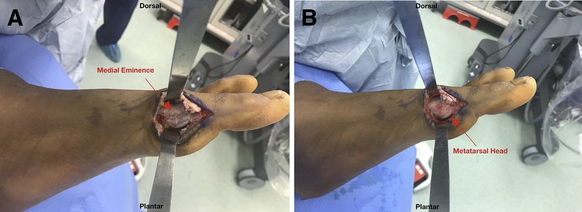
e40                                                    Z. GORICA ET AL.

Fig 4. (A) Picture shows the exposed first metatarsal head of a left foot after the medial collateral ligament has been completely
released from its proximal attachment. There is evidence of medial eminence prominence. (B) Picture shows the medial
metatarsal head after a silver bunionectomy has been performed to remove the medial prominence of the metatarsal head. This
exostectomy is performed to relieve any symptoms caused by the medial prominence, as well as for exposure for suture anchor
placement.

further complete the imbrication overlying the                    remaining proximal capsule again in a pants-over-vest
proximal phalanx (3:12). A #2 FiberWire (Arthrex,                 fashion.
Naples, FL) is used proximally to imbricate the
                                                                  Closure
                                                                    The wound is thoroughly irrigated. The tourniquet is
                                                                  released and hemostasis is obtained. The medial and
                                                                  dorsal incisions are closed with 3-0 Ethicon Nylon su-
                                                                  tures (Ethicon, Raritan, NJ) in a vertical mattress
                                                                  fashion. The operative extremity is then placed in a
                                                                  short leg splint modified with a toe spica component
                                                                  holding the great toe in adduction (Fig 7).

                                                                  Postoperative Course and Rehabilitation
                                                                    Postoperatively, the patient is kept non-weight
                                                                  bearing in a toe spica cast for 3 weeks. They are then
                                                                  transitioned into a boot with toe spacers. They are kept
                                                                  non-weight bearing for an additional week before
                                                                  initiating partial weight bearing at the 4 week post-
                                                                  operative mark, increasing by 25% of body weight
                                                                  each week. Passive range of motion of the toe will begin
                                                                  at the 4 week mark with physical therapy and active
                                                                  range of motion will begin at the 8 week mark at which
                                                                  time they will come out of the boot. At the 12 week
                                                                  mark, they will be allowed to resume running. Full
                                                                  activity including contact sports is allowed at the 4 to
                                                                  6 month mark.

                                                                                         Discussion
                                                                     In the limited literature that exists regarding post-
                                                                  traumatic hallux valgus injuries, patients often present
Fig 5. Intraoperative view of the dorsal aspect of a left foot
                                                                  with significant pain with ambulation and limited
demonstrating the first webspace incision used to perform the
adductor hallucis tendon release and lateral capsule pie-
                                                                  function with regard to athletic and nonathletic activ-
crusting. Performing this soft tissue release will help in        ities. Characterizing the mechanism and spectrum of
addressing excess hallux valgus angle. It is important to         injury, as well as the appropriate treatment options, is
perform this release prior to the medial ligamentous complex      difficult. Injuries have been described after a direct blow
reconstruction to ensure proper tensioning of soft tissues and    resulting in a valgus force to the hallux,6,8 as well as
neutral alignment of the joint.                                   after a significant valgus stress is experienced during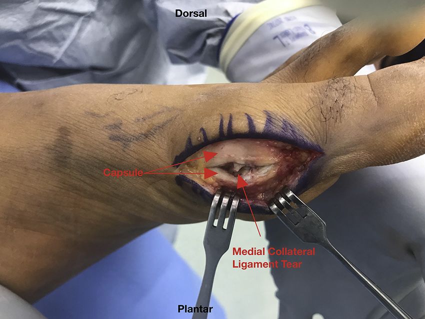
POST-TRAUMATIC HALLUX VALGUS                                                 e41

Fig 6. (A) Picture demonstrates suture anchor placement at the central aspect of the medial first metatarsal head of a left foot.
Care must be taken to avoid violating the joint with anchor placement. (B) and (C) Pictures show the suture limbs being passed
through the plantar and dorsal capsule in pants-over-vest fashion to recreate the medial ligamentous complex. This
reconstruction will allow for maintained alignment and stability to the great toe.

                                                                  push off.5 Chissell et al.8 reported a successful outcome
                                                                  with cast immobilization in an adolescent female.
                                                                  Others7 have reported successful outcomes with surgi-
                                                                  cal intervention. In athletes, especially those with hopes
                                                                  of returning to high levels of play, there is a more sig-
                                                                  nificant stress on operative repairs than there would be
                                                                  for nonathletes. In athletes who have failed nonoper-
                                                                  ative treatment and require surgical intervention, we
                                                                  believe that our surgical technique using an all-suture
                                                                  anchor will provide a repair, which once healed, will
                                                                  hold up to the stresses placed on it by an athlete’s ac-
                                                                  tivity demands (Table 2). Higher load to failure values
                                                                  in suture anchors versus sutures have been consistently
                                                                  reported in biomechanical studies.9 Specifically, the use
                                                                  of an all-suture anchor is associated with minimal peri-
                                                                  anchor cyst formation.10 The likely risks of this tech-
                                                                  nique are similar to other procedures that use a suture
                                                                  anchor; such as anchor pull-out and suture failure. The
Fig 7. A left leg in a short leg cast with great toe-spica
modification. This is applied to maintain the great toe in         technique also requires significant dissection and a long
neutral positioning and to allow the medial ligamentous           period of immobilization. This technique may not be
complex repair to heal without undue stress. It is important to   ideal for those patients with poor wound healing ability
balance adequate padding with a good mold to ensure in-           or those who are unlikely to be compliant with weight-
cisions are not threatened, and the alignment is maintained.      bearing restrictions. Furthermore, this technique has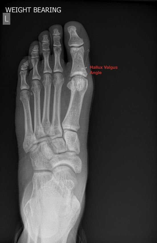
e42                                                          Z. GORICA ET AL.

Table 2. Advantages and Disadvantages                                   4. Ganel A, Israeli A, Horoszowski H. Posttraumatic
                                                                           development of hallux valgus. Orthop Rev 1987;16:667-
           Advantages                       Disadvantages
                                                                           670.
The suture anchor augmentation      Significant soft tissue              5. Fabeck LG, Zekhnini C, Farrokh D, et al. Traumatic hallux
  provides a stronger repair than     dissection
                                                                           valgus following rupture of the medial collateral ligament
  suture alone.
                                                                           of the first metatarsophalangeal joint: A case report. J Foot
The use of an all-suture anchor     Prolonged period of
  translates to a lower risk of       immobilization                       Ankle Surg 2002;41(2):125-128.
  perianchor cyst formation                                             6. Lohrer H. MP I joint giving wayeA case study. Foot Ankle
  compared to biocomposite or                                              Int 2001;22(2):153-157.
  PEEK anchors.                                                         7. Douglas DP, Davidson DM, Robinson JE, Bedi DG.
                                                                           Rupture of the medial collateral ligament of the first
                                                                           metatarsophalangeal joint in a professional soccer player.
only been used in one patient, and therefore, outcome                      J Foot Ankle Surg 1997;36:388-390.
data are limited.                                                       8. Ansari A, Tincknell LJ, Chissell H. Acute traumatic hallux
                                                                           valgus deformity in an adolescent that resolved with
                                                                           appropriate splintage: A case report. Eur J Orthop Surg
                        References                                         Traumatol 2008;18:73-75.
 1. Bohay DR, Johnson KD, Manoli A. The traumatic bunion.               9. Barber AF, Herbert MA, Coons DA, Boothby MH. Sutures
    Foot Ankle Int 1996;17:383-387.                                        and suture anchorsdUpdate 2006. Arthroscopy 2006;22(10):
 2. Johal S, Sawalha S, Pasapula C. Post-traumatic acute                   1063-1069.
    hallux valgus: A case report. Foot (Edinb) 2010;20:87-89.          10. Ergün S, Akgün U, Barber FA, Karahan M. The clinical
 3. Covell DJ, Lareau CR, Anderson RB. Operative treatment                 and biomechanical performance of all-suture anchors: A
    of traumatic hallux valgus in elite athletes. Foot Ankle Int           systematic review. Arthrosc Sports Med Rehabil 2020;2:
    2017;38:590-595.                                                       e263-e275.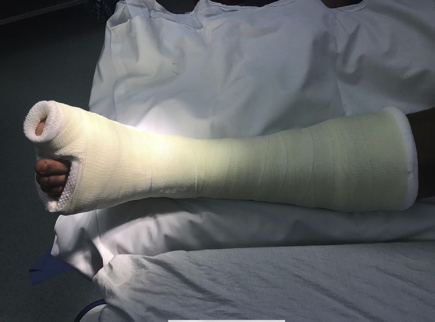
You can also read