Post-weaning Social Isolation in Male and Female Prairie Voles: Impacts on Central and Peripheral Immune System
←
→
Page content transcription
If your browser does not render page correctly, please read the page content below
ORIGINAL RESEARCH
                                                                                                                                         published: 17 January 2022
                                                                                                                                   doi: 10.3389/fnbeh.2021.802569

                                            Post-weaning Social Isolation in
                                            Male and Female Prairie Voles:
                                            Impacts on Central and Peripheral
                                            Immune System
                                            Meghan L. Donovan 1,2,3 , Eileen K. Chun 1 , Yan Liu 1 and Zuoxin Wang 1*
                                            1
                                              Program in Neuroscience, Department of Psychology, Florida State University, Tallahassee, FL, United States, 2 Rocky
                                            Mountain Mental Illness Research Education and Clinical Center, Rocky Mountain Regional VA Medical Center, Aurora, CO,
                                            United States, 3 Department of Physical Medicine and Rehabilitation, University of Colorado Anschutz Medical Campus,
                                            Aurora, CO, United States

                                            The socially monogamous prairie vole (Microtus ochrogaster) offers a unique opportunity
                                            to examine the impacts of adolescent social isolation on the brain, immune system,
                                            and behavior. In the current study, male and female prairie voles were randomly
                                            assigned to be housed alone or with a same-sex cagemate after weaning (i.e., on
                                            postnatal day 21–22) for a 6-week period. Thereafter, subjects were tested for anxiety-
                             Edited by:     like and depressive-like behaviors using the elevated plus maze (EPM) and Forced
                    Angela J. Grippo,
            Northern Illinois University,
                                            Swim Test (FST), respectively. Blood was collected to measure peripheral cytokine
                          United States     levels, and brain tissue was processed for microglial density in various brain regions,
                       Reviewed by:         including the Nucleus Accumbens (NAcc), Medial Amygdala (MeA), Central Amygdala
                          Ruiyong Wu,       (CeA), Bed Nucleus of the Stria Terminalis (BNST), and Paraventricular Nucleus of the
          Yangzhou University, China
                 Katharine Northcutt,       Hypothalamus (PVN). Sex differences were found in EPM and FST behaviors, where
      Mercer University, United States      male voles had significantly lower total arm entries in the EPM as well as lower latency
                 Monika Sadananda,
          Mangalore University, India
                                            to immobility in the FST compared to females. A sex by treatment effect was found
                  *Correspondence:
                                            in peripheral IL-1β levels, where isolated males had a lower level of IL-1β compared
                        Zuoxin Wang         to cohoused females. Post-weaning social isolation also altered microglial density in a
                  zwang@psy.fsu.edu         brain region-specific manner. Isolated voles had higher microglial density in the NAcc,
                   Specialty section:
                                            MeA, and CeA, but lower microglial density in the dorsal BNST. Cohoused male voles
         This article was submitted to      also had higher microglial density in the PVN compared to cohoused females. Taken
             Behavioral Endocrinology,
                                            together, these data suggest that post-weaning social housing environments can alter
                a section of the journal
 Frontiers in Behavioral Neuroscience       peripheral and central immune systems in prairie voles, highlighting a potential role for
        Received: 26 October 2021           the immune system in shaping isolation-induced alterations to the brain and behavior.
      Accepted: 27 December 2021
                                            Keywords: social isolation, inflammation, cytokine, microglia, anxiety
       Published: 17 January 2022
                             Citation:
    Donovan ML, Chun EK, Liu Y and
  Wang Z (2022) Post-weaning Social
                                            INTRODUCTION
  Isolation in Male and Female Prairie
             Voles: Impacts on Central
                                            Adolescence is a critical time window of development, where one’s environment can have long-
      and Peripheral Immune System.         lasting impacts on health outcomes. During this adolescent window, humans rely on a wide
  Front. Behav. Neurosci. 15:802569.        variety of social relationships to develop skills necessary for becoming successful adults. These
    doi: 10.3389/fnbeh.2021.802569          adolescent bonds, such as with peers and family, provide necessary support for optimal wellbeing

Frontiers in Behavioral Neuroscience | www.frontiersin.org                       1                                        January 2022 | Volume 15 | Article 802569
Donovan et al. Post-weaning Isolation Affects Immune System throughout life (Viner et al., 2012; Wang R.A.H. et al., 2017). suggest a critical role of both the peripheral and central immune In contrast, a lack of social support, or social isolation, during systems in shaping outcomes from isolation stressors. Although adolescence can be associated with profoundly negative effects these studies lay out a strong foundation for uncovering on mental health, and can correlate with increased rates of mechanisms of isolation, most studies to date have been done in both anxiety and depression in adulthood (Butler et al., 2016; species that do not exhibit some of the social behaviors humans Cruz et al., 2016; Matthews et al., 2016; Tzanoulinou and show (i.e., forming social preferences, biparental care, etc.). To Sandi, 2016; Walker et al., 2019). Further, the perception of fully understand how isolation during developmental windows social rejection and social isolation is often correlated with can alter central and peripheral immune function and behaviors, dysregulation of immune and stress response systems in the body a highly social animal model that displays these translational (Slavich et al., 2010; Hawkley et al., 2012; Murphy et al., 2013). behaviors must be used. Studies in humans have found that increased social isolation The prairie vole (Microtus ochrogaster) is an established rodent and perceived loneliness are also associated with elevated model that displays a myriad of social behaviors. As a socially inflammation (Eisenberger et al., 2017; Leschak and Eisenberger, monogamous species, the prairie vole has been used to investigate 2019). These long-term changes to physiological systems can have the neurobiological mechanisms underlying the regulation of severe impacts on health and mortality. However, the specific social monogamy, and several neurochemical systems, including underlying mechanisms of how isolation during adolescence can DA and OT, have been implicated in regulating these social lead to detrimental behaviors and immune dysregulation remains behaviors (Young et al., 2011; Lieberwirth and Wang, 2014; largely unknown. Tabbaa et al., 2017). Similar to humans, social isolation in Traditional laboratory rodents have been used to better prairie voles can be a powerful stressor. Data from previous understand how adolescent social isolation alters behavior, studies have shown that isolation in adult prairie voles alters the brain, and the immune system. In both mice and rats, the gut-immune-brain axis (Donovan et al., 2020), increases social isolation immediately after weaning (a time window depressive-like behaviors (Grippo et al., 2008), and alters both representative of adolescence, prior to adulthood) can result plasma OT and central OT expression in the PVN (Grippo et al., in increased anxiety-like and depressive-like behaviors (Lukkes 2007a). Interestingly, adult prairie voles housed in isolation show et al., 2009a; Amiri et al., 2015; Huang et al., 2017; Walker alterations in both immune function in the peripheral nervous et al., 2019). Social isolation not only increases maladaptive system (Scotti et al., 2015) and autonomic function (McNeal et al., behaviors, but also changes the underlying neurocircuitry of 2014), supporting the notion that post-weaning isolation may the brain. For example, social isolation stress alters several have system-wide effects in the periphery as well. However, it neurochemical systems, including oxytocin (OT), and dopamine is still unknown how social isolation during development can (DA), in brain regions involved in stress responses, such as affect central and/or peripheral immune function and how these the paraventricular nucleus of the hypothalamus (PVN) and alterations may differ across sexes. dentate gyrus of the hippocampus (DG) (Tanaka et al., 2010; As prairie voles often live in the nest through adulthood in Oliveira et al., 2019). Furthermore, traditional animal models the wild (Getz et al., 1993; Getz and Carter, 1996), the lack of have been useful for understanding specific central and peripheral social environment during adolescence (i.e., post-weaning, but immune alterations that can result from developmental social prior to reaching adulthood) can be detrimental. For example, isolation. For example, microglia, the primary immune cells of a previous study showed that male prairie voles exposed to the brain, play a vital role in homeostatic regulation of the 6 weeks of isolation after weaning display increased anxiety- brain and are critical in synaptic pruning in early life for proper like behaviors and altered mRNA levels for OT, vasopressin development of social play in male rats (Bilbo and Schwarz, (AVP), corticotrophin releasing hormone (CRH), and tyrosine 2009). Early life social stressors can also increase both microglial hydroxylase (TH) in the PVN (Pan et al., 2009), suggesting that activation and cytokine release in the hippocampus in both mice post-weaning isolation can alter a wide variety of neurochemical and rats (Delpech et al., 2016; Dunphy-Doherty et al., 2018). systems in prairie voles. This 6-week period after weaning Further, these microglial alterations have been shown to play a can be representative of a juvenile, developmental window for causal role in the resulting alterations in neuronal signaling and prairie voles, as they reach adulthood/reproductive maturation ultimate increases in depressive-like behaviors from isolation in around postnatal day 60–80. Moreover, the manipulation of rats (Wang H. T. et al., 2017). social factors (i.e., presence of siblings, population density, etc.) Other social isolation studies have shown long-term has been shown to alter juvenile vole behaviors (Getz and dysregulation in both central and peripheral immune function Carter, 1980, 1996; McGuire et al., 1993), emphasizing the as well (Chida et al., 2005; Ko and Liu, 2015). Interestingly, importance of social cues during this time frame. Therefore, manipulation of the human immune system via peripheral in the present study, we examined how social isolation via inflammatory challenge increases perceived social disconnection single housing during a developmental period that begins (Eisenberger et al., 2010). Likewise, individual housing in adult at PND 21–22 (i.e., post-weaning) and lasts until prairie rats elevated the levels of peripheral cytokines and increased voles reach adulthood (after PND 60) may differentially alter display of “despair” behavior (Krügel et al., 2014). Peripheral microgliosis in the brain and circulating levels of cytokines cytokines have also been found to be essential for driving social in both male and female prairie voles. We hypothesized behavior in traditional rodent models (Hoogenraad and Riol- that post-weaning social isolation has enduring effects on Blanco, 2020; Reed et al., 2020). Taken together, these studies behaviors as well as on peripheral and central immune marker Frontiers in Behavioral Neuroscience | www.frontiersin.org 2 January 2022 | Volume 15 | Article 802569
Donovan et al.                                                                                         Post-weaning Isolation Affects Immune System

expression, and such effects may differ between male and                   Behaviors quantified included the duration and frequency of
female prairie voles.                                                      swimming and immobility. Total activity bouts (locomotor
                                                                           measure) and the latency to immobility were also calculated.

MATERIALS AND METHODS                                                      Blood Sample Preparation and Plasma
Subjects and Social Isolation Procedure                                    Cytokine Analysis
                                                                           After the FST, subjects were deeply anesthetized via euthasol
Subjects were male and female prairie voles (Microtus
                                                                           and cardiac blood samples were collected immediately prior to
ochrogaster) captive-bred at Florida State University and were
                                                                           perfusion. Blood was collected in microcentrifuge vials with 20
kept at 20◦ C under a 14:10 h light:dark cycle (lights on at 0700).
                                                                           µl EDTA and were placed immediately on ice. Samples were
At weaning (PND 21-22), subjects were placed into a clean,
                                                                           then centrifuged at 6,000 rpm for 15 min at 4◦ C, and plasma
Plexiglas cage (20 × 25 × 45 cm) with a standard metal cage lid
                                                                           was then transferred to new tubes. Plasma was re-centrifuged at
and were randomly assigned to either be housed alone (social
                                                                           6,000 rpm for 10 min at 4◦ C. Plasma samples were then analyzed
isolation) or with a same-sex cagemate (a standard housing
                                                                           for cytokines on a 96-well plate in duplicates using the Milliplex
condition for prairie voles laboratory settings) (n = 10 per sex per
                                                                           MAP Cytokine magnetic bead panel (#MCYTOMAG-70k; EDM
treatment; n = 40 total), using our previously established methods
                                                                           Millipore, Billerica, MA, United States) as described in previous
(Pan et al., 2009). All cages contained cedar chip bedding with
                                                                           studies (Lu et al., 2017; Corsi-Zuelli et al., 2019). Previous
food (Purina 5326 pelleted rabbit food, large pellet form) and
                                                                           cytokine studies have shown that prairie vole immune responses
water provided ad libitum by Laboratory Animal Resources staff.
                                                                           are similar to traditional rodent species (Klein and Nelson, 1999;
Isolated subjects were in the same room as cohoused subjects but
                                                                           Curtis et al., 2011). Moreover, these studies demonstrate that
were placed on separate rows of housing racks from cohoused
                                                                           mouse cytokine antibodies can cross react with prairie vole
animals. Although isolated animals were singly housed to prevent
                                                                           cytokines and can be measured via ELISA, which is what we used
social contact throughout the treatment period, they could still
                                                                           in the current study. The assay was performed at the University of
possibly receive olfactory/auditory/visual cues from the other
                                                                           Maryland School of Medicine’s Center for Innovative Biomedical
animals in the housing room. The isolation/cohousing procedure
                                                                           Resources Cytokine Core Laboratory according to manufacturer’s
lasted 6 weeks, after which all subjects underwent behavioral
                                                                           instructions. Cytokines analyzed included Interleukin (IL)-17,
testing. All procedures were approved by the Institutional
                                                                           IL-4, IL-6, IL-1β, Interferon (IFN)γ, and Vascular Endothelial
Animal Care and Use Committee at Florida State University and
                                                                           Growth Factor (VEGF).
were in accordance with the guidelines set forth by the National
Institute of Health.
                                                                           Brain Tissue Preparation
Behavioral Testing                                                         Immediately following cardiac blood sample collection, subjects
All subjects underwent the elevated plus maze (EPM) on day one             were transcardially perfused via 0.9% saline followed by
of behavioral testing. The EPM has been established and validated          4% paraformaldehyde solution in 0.1M phosphate PB buffer
in our previous vole studies (Pan et al., 2009; Smith and Wang,            (PB). Their brains were then collected and fixed in 4%
2014). Briefly, the apparatus is elevated 45 cm off the ground and         paraformaldehyde for 2 h until placed in 30% sucrose in PB at
consists of two open (35 × 6.5 cm) and two closed arms [35 ×               4◦ C for storage. The brains were cut into 40 µm coronal sections
5 × 15 (H) cm] that cross in the middle. All subjects underwent            via sliding microtome.
EPM testing in the morning (between the hours of 0800 and 1000)
on day one of behavioral testing. Subjects were placed onto the            Immunocytochemistry
center of the maze facing an open arm, and their behaviors on the          Ionized calcium binding adaptor molecule 1 (Iba-1) is
EPM were recorded for 5 min using Active Webcam software and               macrophage/microglial-specific molecule involved in the
were subsequently quantified by an observer blind to treatment             phagocytotic process (Ohsawa et al., 2000). Iba-1 is constitutively
via J-Watcher. Behaviors quantified included the duration and              expressed in all microglia and is upregulated in active microglia in
frequency in the open arms, closed arms, and the center of the             the brain. The Iba-1 antibody (Wako Chemicals Inc., Richmond,
maze. Locomotor activity (as indicated by total arm entries) was           VA) used in this study has been used previously in prairie vole
also calculated. Immediately after the EPM test, subjects were             studies (Rebuli et al., 2016; Pohl et al., 2021). Iba-1 staining was
returned to their respective housing cages/conditions.                     performed on sets of coronal sections with 200 µm intervals
    All subjects underwent the forced swim test (FST) in the               using previously established methods from our lab (Liu et al.,
morning (between the hours of 0800 and 1000) on day two of                 2019). Briefly, sections were rinsed in 0.1M phosphate-buffered
behavioral testing. The FST has been established and validated             saline (PBS) 3 times for a total 15 min (each rinsing was kept
in our previous studies (Lieberwirth et al., 2012, 2013). Briefly, a       at the rate of 3 times for a total of 15 min below) and then
large, Plexiglas tub [25 × 45 × 20 (H) cm] is filled with lukewarm         incubated with 1% NaBH4 in 0.1 PBS for 10 min. Sections were
water (32 ± 1◦ C). Subjects were placed into the tub and allowed           rinsed and then treated with 0.3% H2 O2 in 0.1M PBS for 20
to swim freely for 5 min. Subjects’ behaviors were recorded for the        min. After rinsing in 0.1M PBS, sections were incubated in 10%
entire test using Active Webcam software and were subsequently             normal goat serum (NGS, Sigma-Aldrich, St. Luis, MO) in 0.3%
quantified by an observer blind to treatment via J-Watcher.                Triton X-100 in 0.1M PB (TPBS) for 1 h at room temperature.

Frontiers in Behavioral Neuroscience | www.frontiersin.org             3                                   January 2022 | Volume 15 | Article 802569
Donovan et al. Post-weaning Isolation Affects Immune System
Subsequently, sections were incubated in rabbit anti-Iba-1 total arm entries in comparison to female voles [F (1 , 31) = 4.64,
(1:10,000, Wako Chemicals) in 0.3% TPB with 2% NGS at 4◦ C p < 0.05]. No other main effects of sex, treatment, nor sex-by-
overnight. Sections were then placed at room temperature for treatment interactions were found in any of the EPM measures.
1 h, rinsed in 0.3% TPBS, and incubated in biotinylated goat Depressive-like behavior was examined in the FST test
antirabbit IgG (1:300, Vector Laboratories, Burlingame, CA) for (Figure 1B). No treatment effects were found in the duration
2 h at room temperature. Thereafter, sections were rinsed in 0.3% [F (1 , 34) = 1.64, p = 0.21) or frequency [F (1 , 34) = 0.98, p = 0.33] of
TPBS followed by 0.1M PBS and then incubated in the ABC immobility. However, isolated voles showed a higher frequency
Elite HRP Kit (Vector Laboratories) in 0.1M PBS for 90 min of total bouts in comparison to cohoused voles [F (1 , 34) = 9.42,
at room temperature, and Iba-1 immunostaining was revealed p < 0.01]. We also found a main effect of sex, where males had a
using 30 -diaminobenzidine (DAB, Sigma-Aldrich). lower latency to immobility in comparison to female voles [F (1 ,
34) = 9.54, p < 0.01]. No other significant effects were found in
Data Quantification and Analysis any of the FST measures.
All behaviors were scored via JWatcher software program (V1.0,
Macquarie University and UCLA)1 and analyzed via two-way Post-weaning Isolation Alters
ANOVA (SEX x TREATMENT) via GraphPad PRISM. Plasma Interleukin-1β Plasma Cytokine Levels
cytokine levels (pg/ml) were calculated via Luminex Exponent The plasma samples were assayed for a variety of cytokine
Software. Cytokine data failed tests of normality and were thus markers previously found to be associated with social isolation
analyzed via non-parametric analysis. Means of cytokine levels (Figure 2). No main effects were found for any cytokines.
per animal were analyzed via Kruskal-Wallis tests. Iba-1 labeled A significant sex-by-treatment interaction was found for IL-
cells were quantified in selected brain regions defined by the 1β (H3 = 9.45, p < 0.05; Figure 2A), where isolated males
Paxinos and Watson, 7th edition (Paxinos and Watson, 2014), had a significantly lower level compared to cohoused females.
including the PVN (Plate 23-27), Nucleus Accumbens (NAcc: the Further, there was trending interaction significance for IL-17
core and shell subnuclei; Plate 10-13), Bed Nucleus of the Stria (H3 = 6.72, p = 0.08; Figure 2B) and IFNγ (H3 = 6.25, p = 0.10;
Terminalis (BNST: anterior dorsal and anterior ventral subnuclei; Figure 2C). No other sex-by-treatment interactions were found
Plate 18-20), Amygdala (AMYG: the cortical (CoA), medial for any measured cytokines (Figure 2).
(MeA), and central (CeA) subnuclei; Plate 28-32), Prefrontal
Cortex (PFC; Plate 8-11), Dorsal Raphe (DR; Plate 49-53),
Post-weaning Isolation Impacts
Lateral Septum (LS; Plate 13-17), and Dentate Gyrus (DG) of
the hippocampus: the granular (GrDG), molecular (MoDG), and Microglial Density in a Brain
polymorphic (PoDG) layers; Plate 30-34). These brain regions Region-Specific Manner
were chosen due to their demonstrated roles in social behaviors, Iba-1 cell density was quantified from the selected brain regions.
stress responses, and learning/memory (Sandi and Haller, 2015; Significant treatment effects were found in multiple brain regions
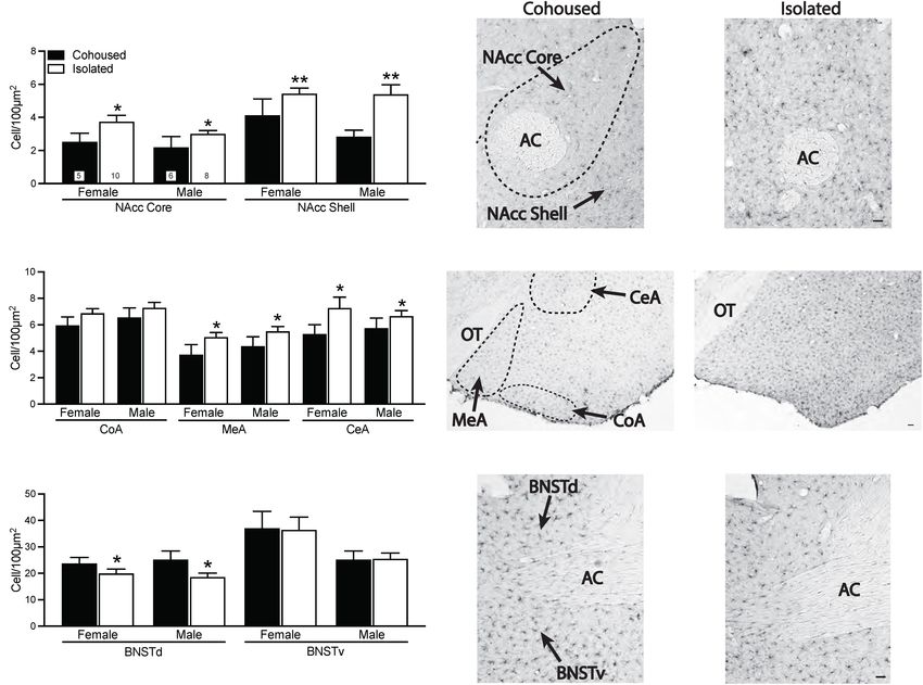
Tabbaa et al., 2017; Yavas et al., 2019). The boundary of each (Figure 3). In both the NAcc core [F (1 , 25) = 5.16, p < 0.05]
nucleus was traced and area was measured by using Stereo and NAcc shell [F (1 , 25) = 11.76, p < 0.01], isolated voles
Investigator software (MBF Bioscience). Iba-1 positive cells were had significantly higher microglia cell densities compared to
counted bilaterally from two brain sections per area per subject cohoused voles (Figures 3A–C). No sex or sex-by-treatment
by using Zeiss Axioskop 2, microscope (Carl Zeiss Microscopy interactions were found in the NAcc core or shell. Isolated voles
Ltd., Germany), and subsequently converted to cell density (i.e., also had significantly higher density of microglia compared to
number of cells per 100 µm2 for each brain region) as described cohoused voles in both the MeA [F (1 , 29) = 4.92, p < 0.05] and
previously (Gobrogge et al., 2007; Lieberwirth et al., 2013). The CeA [F (1 , 29) = 4.34, p < 0.05] of the amygdala, but this was not
means of Iba-1 density per brain region per animal were analyzed seen in the CoA [F (1 , 29) = 2.33, p = 0.14; Figures 3D–F]. No main
by two-way ANOVA via GraphPad PRISM. Significant sex-by- effects of sex or sex-by-treatment interactions were found in any
treatment interactions were further analyzed by the Student- measured subnuclei of the amygdala. A main effect of treatment
Newman-Keuls post hoc tests. was found in the BNST, where isolated voles showed a significant
lower density of microglia in the dorsal BNST [F (1 , 31) = 5.49,
p < 0.05], but not the ventral BNST [F (1 , 31) = 0.42, p = 0.52]
RESULTS in comparison to cohoused voles (Figures 3G–I). There were no
main effects of sex or sex-by-treatment interactions in the dorsal
Post-weaning Isolation Did Not Affect or ventral BNST.
Anxiety—and Depressive-Like Behavior Iba-1 cells were also quantified in the PVN. No significant
Anxiety-like behavior was assessed in the EPM test (Figure 1A). main effects were present, but a significant sex-by-treatment
No treatment effects were found in the duration of time spent in interaction was found [F (1 , 28) = 4.61, p < 0.01], where cohoused
the open arms of the maze [F (1 , 31) = 0.10, p = 0.76] or in the male and female voles differed significantly from each other in
frequency of open arm entries [F (1 , 31) = 1.98, p = 0.17]. A main the microglial density, but not from isolated males or females
effect of sex was found, where males showed a lower number of (Figure 4). No significant effects of treatment, sex, or sex-by-
treatment interactions were found in the PFC (Figure 4) or other
1
http://www.jwatcher.ucla.edu/ quantified brain regions, including the DR, LS, and DG (Table 1).
Frontiers in Behavioral Neuroscience | www.frontiersin.org 4 January 2022 | Volume 15 | Article 802569Donovan et al. Post-weaning Isolation Affects Immune System
FIGURE 1 | Post-weaning isolation effects on elevated plus maze (EPM) (A) and forced swim test (FST) (B) behaviors. Six weeks of post-weaning social isolation did
not affect the duration on the open arms nor the frequency of open arm entries on the EPM in male and female prairie voles. Females displayed higher levels of
overall locomotor activity during the EPM test in comparison to male prairie voles. In the FST, post-weaning social isolation did not affect the duration of immobility in
male and female prairie voles. Male prairie voles had significantly lower latency to immobility compared to females, and socially isolated animals had higher overall
levels of activity in comparison to cohoused animals. Bars indicate mean ± SEM. The number within each bar on (A) indicates the number of animals for each
group. * represents p < 0.05.
FIGURE 2 | Post-weaning alterations to IL-1β plasma cytokine levels. Isolated males had significantly lower levels of IL-1β (A) compared to cohoused females.
Although not significant, similar patterns were seen for levels of IL-17 (B), IFNγ (C), IL-4 (D), IL-6 (E), and VEGF (F). IL-17, Interleukin 17, IFNγ, interferon gamma,
VEGF, vascular endothelial growth factor, IL-1β, Interleukin 1 beta, IL-4, interleukin 4, IL-6, interleukin 6. Bars indicate mean ± SEM. The number within each bar on
Panel A indicates the number of animals for each group. ∗ represents p < 0.05.
DISCUSSION physiological stress responses—especially in social animals (Fone
and Porkess, 2008; Matsumoto et al., 2019; Walker et al., 2019).
Post-weaning social isolation, a chronic stressor during In the present study, post-weaning isolation did not significantly
development, has been shown to induce behavioral and alter anxiety-like or depressive-like behaviors compared to
Frontiers in Behavioral Neuroscience | www.frontiersin.org 5 January 2022 | Volume 15 | Article 802569Donovan et al. Post-weaning Isolation Affects Immune System FIGURE 3 | Post-weaning isolation alters ionized calcium binding adaptor molecule 1 (Iba-1) density in a brain region-specific manner. Post-weaning isolation increased Iba-1 cell density in the NAcc Core and Shell (A–C) and in the MeA and CeA (D–F) of the amygdala. Social isolation also resulted in decreased Iba-1 cell density in the BNSTd, but not BNSTv (G–I). Photomicrographs show representative images of Iba-1 staining in the NAcc (B,C), amygdala (E,F), and BNST (H,I). AC, anterior commissure; BNSTd, dorsal region of the bed nucleus stria terminalis; BNSTv, ventral region of the bed nucleus stria terminalis; CeA, central amygdala; CoA, cortical amygdala; MeA, medial amygdala; NAcc, Nucleus Accumbens; OT, optic tract. Bars indicate mean ± SEM. The number within each bar on Panel A indicates the number of animals for each group. * represents p < 0.05, ** represents p < 0.01. Scale bar = 50 µm. cohoused male and female prairie voles. However, isolated male experimental designs and procedures. The type, number, timing, voles showed a lower level of IL-1β in compared to cohoused and order of behavioral tests conducted in previous studies females. Isolated voles also showed higher microglial densities were different from our current study, which may contribute in the NAcc, MeA, CeA, but a lower microglial density in the to these differences. For example, in a previous study (Pan dorsal BNST compared to cohoused voles. These results suggest et al., 2009), male prairie voles underwent the social affiliation, that chronic social isolation during a juvenile window may open field, and EPM tests (over three consecutive days) after have distinctive effects on both peripheral and central immune 6 weeks of post-weaning isolation. Moreover, the majority of function between male and female prairie voles. social isolation studies in prairie voles have been done in adult Previous findings have shown that isolation increases anxiety- voles, which may contribute to the difference in results. In like and depressive-like behaviors in prairie voles (Grippo et al., addition, our vole breeding colony has recently been outbred 2008; Pan et al., 2009) as well as in traditional rodent models with wild-captured prairie voles. This outbreeding process is (Amiri et al., 2015; Huang et al., 2017). Therefore, we were essential for maintaining genetic variation across prairie vole surprised to see no significant changes in anxiety-like and laboratories. Whether such outbreeding has generated colony- depressive-like behaviors by post-weaning social isolation in specific behavioral responses to social isolation needs to be the present study. We don’t have ready explanations for such further examined. Nevertheless, post-weaning isolation increased discrepancies and can only speculate that these differences total bouts in the FST in both male and female voles in the in results may have been due to subtle differences in the present study. Although much of the social isolation literature Frontiers in Behavioral Neuroscience | www.frontiersin.org 6 January 2022 | Volume 15 | Article 802569
Donovan et al.                                                                                                        Post-weaning Isolation Affects Immune System

  FIGURE 4 | Sex differences in ionized calcium binding adaptor molecule 1 (Iba-1) cell density in the paraventricular nucleus of the hypothalamus (PVN). Cohoused
  males had higher Iba-1 cell density in the PVN compared to cohoused females, but no differences were found across isolated male or female prairie voles. No group
  differences in Iba-1 cell density were found in the prefrontal cortex (PFC). Photomicrographs show representative images of Iba-1 labeling in the PVN. 3V, third
  ventricle. Bars indicate mean ± SEM. The number within each bar on (A) indicates the number of animals for each group. ∗ represents p < 0.05. Scale bar = 50 µm.

show that social isolation increases immobility in the FST                           post-weaning isolation (Fone and Porkess, 2008; Ashby et al.,
(Lukkes et al., 2009b; Kokare et al., 2010; Sargin et al., 2016),                    2010; Hickey et al., 2012). It should be noted that we chose to
there are numerous studies that demonstrate hyperactivity after                      administer the FST second, as it is more stress-inducing than

Frontiers in Behavioral Neuroscience | www.frontiersin.org                       7                                         January 2022 | Volume 15 | Article 802569
Donovan et al.                                                                                                       Post-weaning Isolation Affects Immune System

TABLE 1 | Iba-1 cell density (cell/100 µm2 ) in various brain regions.

Group                              Cohoused                                              Isolated                                            p

Sex                       M                           F                       M                          F               Group              Sex             G×S

DR                   17.1 ± 4.6*                 10.7 ± 0.7               10.2 ± 1.3                 9.8 ± 1.1             ns                ns               ns
LS                    6.4 ± 0.8                  6.6 ± 1.0                7.3 ± 1.0                  8.2 ± 0.6             ns                ns               ns
PoDG                184.9 ± 81.3               193.1 ± 59.2              234.5 ± 57.3               202.6 ± 47.7           ns                ns               ns
MoDG                130.8 ± 28.9               193.2 ± 56.8              235.8 ± 51.9               173.6 ± 32.4           ns                ns               ns
GrDG                 81.8 ± 25.2               121.9 ± 36.0              131.7 ± 40.8               95.4 ± 16.6            ns                ns               ns

Iba-1, ionized calcium-binding adapter molecule 1; DR, dorsal raphe; LS, lateral septum; PoDG, polymorphic layer of the dentate gyrus; MoDG, molecular layer of the
dentate gyrus; GrDG, granular layer of the dentate gyrus. *Mean ± SEM.

the EPM test. However, we cannot exclude the possibly that                              production of IL-17 (Kuwabara et al., 2017; Pyrillou et al., 2020).
the EPM test might still have residual effects 24 h later, or that                      Likewise, IL-17 can also induce production of IL-1β (Xu and Cao,
post-weaning isolation also may have resulted in dysregulated                           2010). Therefore, this bidirectional feedback between cytokines
behaviors not illustrated by the EPM and FST tests.                                     in their release and production may be an important factor when
   To our knowledge, the present study is the first study to                            considering the functional importance of peripheral cytokines in
examine peripheral cytokines post-isolation in male and female                          shaping central function and resulting behaviors.
prairie voles. In mice, cytokine expression peaks 6–12 h after                              Although only trending toward significance, our additional
one acute stressor, and 1 h after two acute stressors spaced 24 h                       data show a similar pattern, where female voles isolated after
apart (similar to our current study paradigm) (Cheng et al., 2015).                     weaning tend to have lower plasma levels of IFNγ and IL-
As plasma cytokines were collected immediately after the 5-min                          17 compared to cohoused females. The lack of significance
FST, our data should be better explained by the effects of social                       in these results may be due to either small sample sizes or
housing conditions rather than from the acute FST stressor. Our                         large individual variation, which is common in prairie voles
data indicate a sex by treatment interaction, where isolated male                       (Hammock et al., 2005; Barrett et al., 2013; Donovan et al.,
voles had lower levels of IL-1β compared to cohoused females.                           2020). Although high levels of plasma cytokines may suggest
This is surprising, given that IL-1β is a key cytokine in the                           a proinflammatory state, metadata analysis of transcriptomes
induction of the sickness behavioral responses, which includes                          found that group-housed rats and mice have upregulated IFNγ
HPA axis activation, fever, and social withdrawal (Dantzer, 2001,                       gene signature, and social isolation greatly reduces IFNγ (Krügel
2006). However, a previous post-weaning isolation experiment                            et al., 2014). Moreover, IFNγ- and IFNγ receptor-deficient mice
in rats found that contrary to the expected proinflammatory                             display social deficits, and IFNγ signaling controls neural circuits
phenotype seen in socially isolated adults, the measured cytokines                      shaping social behavior (Filiano et al., 2016). Further, IL-17 has
(e.g., IL-10, IL-6, TNF-α) either stayed the same or decreased                          been dubbed as the “social cytokine” (Hoogenraad and Riol-
(Corsi-Zuelli et al., 2019), supporting our data that post-weaning                      Blanco, 2020) due to its role in rescuing social deficits in mouse
isolation can result in lower levels of peripheral cytokines.                           models of neurodevelopmental disorders (Reed et al., 2020).
Perhaps this reduction in IL-1β also plays a role in the lack                           Given the critical role the immune system plays in shaping
of significant differences in behavior through compensatory                             neuronal circuits during development (Morimoto and Nakajima,
mechanisms resulting from the stressful isolation experience. IL-                       2019), high levels of IFNγ and IL-17 in cohoused females may be
1β’s relationship with the stress response, and more specifically                       shaping typical behaviors, whereas a reduction in these peripheral
corticosterone (CORT), is also worth noting. Previous prairie                           immune markers may point to the potentially damaging effect
vole studies using the 6-week isolation paradigm have found                             of social isolation on neuronal health and development during
elevated CORT levels from the isolation experience in adult, male                       a critical period.
prairie voles (Donovan et al., 2020), but not in adult females                              As social isolation has been shown to alter immune function
(Lieberwirth et al., 2012; Donovan et al., 2020). Intriguingly, it                      in adult prairie voles (Scotti et al., 2015) and is also associated
has been shown that pretreating animals with varying levels of                          with sex- and brain region-specific alterations in microgliosis
CORT changes the levels of IL-1β released in response to an                             (Donovan et al., 2020), we were interested in seeing how post-
immune challenge—emphasizing their dynamic relationship (Liu                            weaning isolation altered microglial density in key brain regions
et al., 2018). The high level of IL-1β seen in cohoused females                         known to be affected by isolation stress. We found significant
in the present study may be due to the fact that prairie voles,                         differences in microglial density in a brain region-specific pattern,
and particularly female voles, naturally have much higher levels                        where isolated voles had significantly higher levels of microglial
of circulating CORT than other traditional laboratory rodents                           densities in the NAcc, MeA, and CeA, but lower microglial
(Taymans et al., 1997). Further, IL-1β is expressed at high levels                      density in the dorsal BNST compared to cohoused voles. These
in the brain during development (Bilbo and Schwarz, 2009)                               data add to the growing literature that isolation during adolescent
and is essential for normal cognitive function (Giulian et al.,                         timeframes can alter central immune function in various rodent
1988; Bilbo and Schwarz, 2012). IL-1β is responsible for many                           species (Wang H. T. et al., 2017; Dunphy-Doherty et al., 2018;
downstream immune targets and effects, one of which is the                              Gong et al., 2018).

Frontiers in Behavioral Neuroscience | www.frontiersin.org                        8                                      January 2022 | Volume 15 | Article 802569
Donovan et al.                                                                                         Post-weaning Isolation Affects Immune System

   The NAcc has been linked to microglial-specific alterations             (Donovan et al., 2020). A recent study demonstrated that
after various stressors, with both chronic restraint stress and            parvocellular PVN microglial priming occurs in male, but not
social isolation resulting in increased levels of microglial               female, prairie voles after partner loss (Pohl et al., 2021). In
activation in this brain region (Tynan et al., 2010; Donovan               addition, neurochemical and gene expression in the PVN is
et al., 2020). Moreover, it has been shown that neurochemical              altered by social isolation during adolescence in male voles (Pan
systems in the NAcc, such as CRF and OT, are critical for                  et al., 2009). Together, these data support the notion that the PVN
partner loss in prairie voles (Bosch et al., 2016), and microglia          may be particularly important for driving sexually dimorphic,
can also directly interact with reward systems critical for pair           immune-related mechanisms in the brain.
bonding (Loth and Donaldson, 2021). As the NAcc has been                       In summary, our data, for the first time, demonstrate that 6
shown to be an important node for post-weaning-induced                     weeks of social isolation immediately after weaning is associated
alterations (Bendersky et al., 2021), we speculate that changes            with central and peripheral immune alterations in prairie voles
in microglia in the NAcc play a role in alterations to neuronal            in a sex-, brain region-, and cytokine-specific manner. These
circuits and behaviors. Further, it has been reported that post-           data add to the growing literature suggesting that social isolation
weaning isolation reduces the size of neurons and volume of the            can have long-lasting impacts on central and peripheral immune
MeA (Cooke et al., 2000), and microglia play a causal role in              function, and that these, along with other system-wide effects
modulating underlying neurochemical alterations and behavioral             (Grippo et al., 2007b; McNeal et al., 2014), may be ultimately
outcomes from isolation stress (Wang H. T. et al., 2017; Gong              leading to negative health outcomes. As previous research
et al., 2018). Given that our data show an isolation-induced               demonstrate a casual role of microglia shaping sex-specific social
increase in microglial density in the MeA, it would be interesting         behaviors during development (Kopec et al., 2018), more research
to assess whether the increased presence of microglia plays a              must be done to explore how microglia and/or peripheral
causal role in amygdala neuronal alterations and their underlying          cytokines may modulate sex differences in male and female
behaviors. Finally, although there are little data focusing on             prairie vole social behavior as well.
microglial alterations after isolation in the BNST, one study found
that aged rats have significantly higher density of microglia in
the BNST, indicating that BNST microglial alterations may play             DATA AVAILABILITY STATEMENT
a role in shaping age-related circuits in the brain (Perkins et al.,
2018). A study in mice also showed that acute social isolation             The raw data supporting the conclusions of this article will be
can blunt long-term potentiation in the dorsal-lateral BNST                made available by the authors, without undue reservation.
(Conrad et al., 2011). Thus, our data showing lower microglial
density in the dorsal BNST after isolation during development              ETHICS STATEMENT
support the idea that the BNST may serve as a critical node
in the underlying circuitry driving age- and isolation-induced             The animal study was reviewed and approved by the Institutional
changes to behavior.                                                       Animal Care and Use Committee at Florida State University.
   Another interesting finding in our study is the notable sex
differences in behavior as well as in both peripheral and central
immune measures. We saw sex differences in both behavioral                 AUTHOR CONTRIBUTIONS
assays, where females displayed higher levels of locomotor
activity on the EPM and a longer latency to immobility in the              MD and ZW designed the study. MD, EC, and YL conducted
FST. Sex differences in activity levels after stress have also been        the experiments. MD and ZW analyzed the data and wrote the
found in previous studies, suggesting that males and females               manuscript. All authors read and approved the manuscript.
may have differential outcomes with hyperactivity (West and
Michael, 1988; Yamaura et al., 2013). We have also found a
sex by treatment interaction in IL-1β cytokine levels. These
                                                                           FUNDING
differences seen in levels of IL-1β were particularly fascinating          This work was supported by grants from National Institutes
given previously established sex differences in inflammation               of Health (NIMH R01-108527, NIMH R01-109450, and NIMH
and immunity (Ahnstedt and McCullough, 2019). These sex                    R21-111998) to ZW. EC was supported by the NIH program
differences in our data could reflect differences in gonadal               training grant (T32 MH093311, P.K. Keel and L.A. Eckel) and
steroids between males and females, given that gonadal steroids            the National Science Foundation Graduate Research Fellowship
can promote the expression of immune genes and can thus                    under (grant no. 1449440). MD was supported by the VA Office of
mediate the immune response (Klein and Flanagan, 2016;                     Academic Affiliations, Advanced Fellowship Program in Mental
Ahnstedt and McCullough, 2019). Interestingly, a previous study            Illness Research and Treatment, Department of Veterans Affairs.
found that developmental exposure to synthetic estrogen can
alter sex-specific colonization by microglia in a brain region-
specific manner in prairie voles (Rebuli et al., 2016). Further,           ACKNOWLEDGMENTS
in the present study, we found that control males have higher
microglial density in the PVN compared to control females,                 We would like to thank MacKenzie Ellis, Gabriela Petasne, and
which is consistent with our finding in adult prairie voles                Amanda Zint for assistance with data collection and analyses.

Frontiers in Behavioral Neuroscience | www.frontiersin.org             9                                   January 2022 | Volume 15 | Article 802569
Donovan et al. Post-weaning Isolation Affects Immune System
REFERENCES Delpech, J. C., Wei, L., Hao, J., Yu, X., Madore, C., Butovsky, O., et al. (2016). Early
life stress perturbs the maturation of microglia in the developing hippocampus.
Ahnstedt, H., and McCullough, L. D. (2019). The impact of sex and age on T Brain. Behav. Immun. 57, 79–93. doi: 10.1016/j.bbi.2016.06.006
cell immunity and ischemic stroke outcomes. Cell. Immunol. 345:103960. doi: Donovan, M., Mackey, C. S., Platt, G. N., Rounds, J., Brown, A. N., Trickey,
10.1016/j.cellimm.2019.103960 D. J., et al. (2020). Social isolation alters behavior, the gut-immune-brain axis,
Amiri, S., Haj-Mirzaian, A., Rahimi-balaei, M., Razmi, A., Kordjazy, N., Shirzadian, and neurochemical circuits in male and female prairie voles. Neurobiol. Stress
A., et al. (2015). Co-occurrence of anxiety and depressive-like behaviors 13:100278. doi: 10.1016/j.ynstr.2020.100278
following adolescent social isolation in male mice; possible role of nitrergic Dunphy-Doherty, F., O’Mahony, S. M., Peterson, V. L., O’Sullivan, O., Crispie, F.,
system. Physiol. Behav. 145, 38–44. doi: 10.1016/j.physbeh.2015.03.032 Cotter, P. D., et al. (2018). Post-weaning social isolation of rats leads to long-
Ashby, D. M., Habib, D., Dringenberg, H. C., Reynolds, J. N., and Beninger, term disruption of the gut microbiota-immune-brain axis. Brain Behav. Immun.
R. J. (2010). Subchronic MK-801 treatment and post-weaning social isolation 68, 261–273. doi: 10.1016/j.bbi.2017.10.024
in rats: Differential effects on locomotor activity and hippocampal long-term Eisenberger, N. I., Inagaki, T. K., Mashal, N. M., and Irwin, M. R. (2010).
potentiation. Behav. Brain Res. 212, 64–70. doi: 10.1016/j.bbr.2010.03.041 Inflammation and social experience: an inflammatory challenge induces
Barrett, C. E., Keebaugh, A. C., Ahern, T. H., Bass, C. E., Terwilliger, E. F., and feelings of social disconnection in addition to depressed mood. Brain Behav.
Young, L. J. (2013). Variation in vasopressin receptor (Avpr1a) expression Immun. 24, 558–563. doi: 10.1016/j.bbi.2009.12.009
creates diversity in behaviors related to monogamy in prairie voles. Horm. Eisenberger, N. I., Moieni, M., Inagaki, T. K., Muscatell, K. A., and Irwin, M. R.
Behav. 63:5. doi: 10.1016/j.yhbeh.2013.01.005 (2017). In Sickness and in health: the co-regulation of inflammation and social
Bendersky, C. J., Milian, A. A., Andrus, M. D., De La Torre, U., and Walker, behavior. Neuropsychopharmacology 42, 242–253. doi: 10.1038/npp.2016.141
D. M. (2021). Long-Term impacts of post-weaning social isolation on nucleus Filiano, A. J., Xu, Y., Tustison, N. J., Marsh, R. L., Baker, W., Smirnov, I., et al.
accumbens function. Front. Psychiatry 12:745406. doi: 10.3389/fpsyt.2021. (2016). Unexpected role of interferon-γ 3 in regulating neuronal connectivity
745406 and social behaviour. Nature 535, 425–429. doi: 10.1038/nature18626
Bilbo, S. D., and Schwarz, J. M. (2009). Early-life programming of later-life brain Fone, K. C. F., and Porkess, M. V. (2008). Behavioural and neurochemical
and behavior: a critical role for the immune system. Front. Behav. Neurosci. 3:14. effects of post-weaning social isolation in rodents-Relevance to developmental
doi: 10.3389/neuro.08.014.2009 neuropsychiatric disorders. Neurosci. Biobehav. Rev. 32, 1087–1102. doi: 10.
Bilbo, S. D., and Schwarz, J. M. (2012). The immune system and developmental 1016/j.neubiorev.2008.03.003
programming of brain and behavior. Front. Neuroendocrinol. 33:267–286. doi: Getz, L. L., and Carter, C. S. (1980). Social organization in Microtus ochrogaster
10.1016/j.yfrne.2012.08.006 populations. Biol 62, 56–59.
Bosch, O. J., Dabrowska, J., Modi, M. E., Johnson, Z. V., Keebaugh, A. C., Barrett, Getz, L. L., and Carter, C. S. (1996). Prairie-vole partnerships. Am. Sci. 84:56–62.
C. E., et al. (2016). Oxytocin in the nucleus accumbens shell reverses CRFR2- Getz, L. L., McGuire, B., Pizzuto, T., Hofmann, J. E., and Frase, B. (1993). Social
evoked passive stress-coping after partner loss in monogamous male prairie organization of the prairie vole (Microtus ochrogaster). J. Mammal. 74, 44–58.
voles. Psychoneuroendocrinology 64, 66–78. doi: 10.1016/j.psyneuen.2015.1 doi: 10.2307/1381904
1.011 Giulian, D., Young, D. G., Woodward, J., Brown, D. C., and Lachman, L. B. (1988).
Butler, T. R., Karkhanis, A. N., Jones, S. R., and Weiner, J. L. (2016). Adolescent Interleukin-1 is an astroglial growth factor in the developing brain. J. Neurosci.
social isolation as a model of heightened vulnerability to comorbid alcoholism 8, 709–714. doi: 10.1523/jneurosci.08-02-00709.1988
and anxiety disorders. Alcohol. Clin. Exp. Res. 40, 1202–1214. doi: 10.1111/acer. Gobrogge, K. L., Liu, Y., Jia, X., and Wang, Z. (2007). Anterior hypothalamic neural
13075 activation and neurochemical associations with aggression in pair-bonded male
Cheng, Y., Jope, R. S., and Beurel, E. (2015). A pre-conditioning stress accelerates prairie voles. J. Comp. Neurol. 502:21364. doi: 10.1002/cne.21364
increases in mouse plasma inflammatory cytokines induced by stress. BMC Gong, Y., Tong, L., Yang, R., Hu, W., Xu, X., Wang, W., et al. (2018). Dynamic
Neurosci. 16:31. doi: 10.1186/s12868-015-0169-z changes in hippocampal microglia contribute to depressive-like behavior
Chida, Y., Sudo, N., and Kubo, C. (2005). Social isolation stress exacerbates induced by early social isolation. Neuropharmacology 135, 223–233. doi: 10.
autoimmune disease in MRL/lpr mice. J. Neuroimmunol. 158, 138–144. doi: 1016/j.neuropharm.2018.03.023
10.1016/j.jneuroim.2004.09.002 Grippo, A. J., Gerena, D., Huang, J., Kumar, N., Shah, M., Ughreja, R., et al. (2007a).
Conrad, K. L., Louderback, K. M., Gessner, C. P., and Winder, D. G. (2011). Social isolation induces behavioral and neuroendocrine disturbances relevant
Stress-induced alterations in anxiety-like behavior and adaptations in plasticity to depression in female and male prairie voles. Psychoneuroendocrinology 32,
in the bed nucleus of the stria terminalis. Physiol. Behav. 104, 248–256. doi: 966–980. doi: 10.1016/j.psyneuen.2007.07.004
10.1016/j.physbeh.2011.03.001 Grippo, A. J., Lamb, D. G., Carter, C. S., and Porges, S. W. (2007b). Social isolation
Cooke, B. M., Chowanadisai, W., and Breedlove, S. M. (2000). Post-weaning social disrupts autonomic regulation of the heart and influences negative affective
isolation of male rats reduces the volume of the medial amygdala and leads to behaviors. Biol. Psychiatry 62, 1162–1170. doi: 10.1016/j.biopsych.2007.04.011
deficits in adult sexual behavior. Behav. Brain Res. 117, 107–113. doi: 10.1016/ Grippo, A. J., Wu, K. D., Hassan, I., and Carter, C. S. (2008). Social isolation
S0166-4328(00)00301-6 in prairie voles induces behaviors relevant to negative affect: toward the
Corsi-Zuelli, F., Fachim, H. A., Loureiro, C. M., Shuhama, R., Bertozi, G., Joca, development of a rodent model focused on co-occurring depression and
S. R. L., et al. (2019). Prolonged periods of social isolation from weaning reduce anxiety. Depress. Anx. 25, E17–E26. doi: 10.1002/da.20375
the anti-inflammatory cytokine IL-10 in blood and brain. Front. Neurosci. Hammock, E. A. D., Lim, M. M., Nair, H. P., and Young, L. J. (2005). Association of
13:1011. doi: 10.3389/fnins.2018.01011 vasopressin 1a receptor levels with a regulatory microsatellite and behavior. in
Cruz, F. C., Duarte, J. O., Leão, R. M., Hummel, L. F. V., Planeta, C. S., and Genes, Brain and Behavior. Genes Brain Behav. 4:289–301. doi: 10.1111/j.1601-
Crestani, C. C. (2016). Adolescent vulnerability to cardiovascular consequences 183X.2005.00119.x
of chronic social stress: Immediate and long-term effects of social isolation Hawkley, L. C., Cole, S. W., Capitanio, J. P., Norman, G. J., and Cacioppo,
during adolescence. Dev. Neurobiol. 76, 34–46. doi: 10.1002/dneu.22297 J. T. (2012). Effects of social isolation on glucocorticoid regulation in social
Curtis, J. T., Chen, Y., Buck, D. J., and Davis, R. L. (2011). Chronic inorganic mammals. Horm. Behav. 62, 314–323. doi: 10.1016/j.yhbeh.2012.05.011
mercury exposure induces sex-specific changes in central TNFα expression: Hickey, A. J., Reynolds, J. N., and Beninger, R. J. (2012). Post-weaning social
Importance in autism? Neurosci. Lett. 504, 40–44. doi: 10.1016/j.neulet.2011. isolation and subchronic NMDA glutamate receptor blockade: Effects on
08.053 locomotor activity and GABA signaling in the rat suggest independent
Dantzer, R. (2001). Cytokine-induced sickness behavior: Where do we stand? mechanisms. Pharmacol. Biochem. Behav. 101, 231–238. doi: 10.1016/j.pbb.
Brain. Behav. Immun. 15, 7–24. doi: 10.1006/brbi.2000.0613 2012.01.015
Dantzer, R. (2006). Cytokine, sickness behavior, and depression. Neurol. Clin. 24, Hoogenraad, C. C., and Riol-Blanco, L. (2020). Interleukin-17: a social cytokine.
441–460. doi: 10.1016/j.ncl.2006.03.003 Cell 181, 517–519. doi: 10.1016/j.cell.2020.03.060
Frontiers in Behavioral Neuroscience | www.frontiersin.org 10 January 2022 | Volume 15 | Article 802569Donovan et al. Post-weaning Isolation Affects Immune System Huang, Q., Zhou, Y., and Liu, L. Y. (2017). Effect of post-weaning isolation on population density, season, and natal social environment. Behav. Ecol. Sociobiol. anxiety- and depressive-like behaviors of C57BL/6J mice. Exp. Brain Res. 235, 32:183784. doi: 10.1007/BF00183784 2893–2899. doi: 10.1007/s00221-017-5021-5 McNeal, N., Scotti, M. A. L., Wardwell, J., Chandler, D. L., Bates, S. L., LaRocca, M., Klein, S. L., and Flanagan, K. L. (2016). Sex differences in immune responses. Nat. et al. (2014). Disruption of social bonds induces behavioral and physiological Rev. Immunol. 16, 626–638. doi: 10.1038/nri.2016.90 dysregulation in male and female prairie voles. Auton. Neurosci. Basic Clin. 180, Klein, S. L., and Nelson, R. J. (1999). Activation of the immune-endocrine system 9–16. doi: 10.1016/j.autneu.2013.10.001 with lipopolysaccharide reduces affiliative behaviors in voles. Behav. Neurosci. Morimoto, K., and Nakajima, K. (2019). Role of the immune system in the 113, 1042–1048. doi: 10.1037//0735-7044.113.5.1042 development of the central nervous system. Front. Neurosci. 13:916. doi: 10. Ko, C. Y., and Liu, Y. P. (2015). Isolation rearing impaired sensorimotor gating 3389/fnins.2019.00916 but increased pro-inflammatory cytokines and disrupted metabolic parameters Murphy, M. L. M. M., Slavich, G. M., Rohleder, N., and Miller, G. E. (2013). in both sexes of rats. Psychoneuroendocrinology 55, 173–183. doi: 10.1016/j. Targeted rejection triggers differential pro- and anti-inflammatory gene psyneuen.2015.02.007 expression in adolescents as a function of social status. Clin. Psychol. Sci. 1, Kokare, D. M., Dandekar, M. P., Singru, P. S., Gupta, G. L., and Subhedar, N. K. 30–40. doi: 10.1177/2167702612455743 (2010). Involvement of α-MSH in the social isolation induced anxiety- and Ohsawa, K., Imai, Y., Kanazawa, H., Sasaki, Y., and Kohsaka, S. (2000). depression-like behaviors in rat. Neuropharmacology 58, 1009–1018. doi: 10. Involvement of Iba1 in membrane ruffling and phagocytosis of 1016/j.neuropharm.2010.01.006 macrophages/microglia. J. Cell Sci. 113, 3073. doi: 10.1242/jcs.113.17.3073 Kopec, A. M., Smith, C. J., Ayre, N. R., Sweat, S. C., and Bilbo, S. D. Oliveira, V. E., de, M., Neumann, I. D., and de Jong, T. R. (2019). Post-weaning (2018). Microglial dopamine receptor elimination defines sex-specific nucleus social isolation exacerbates aggression in both sexes and affects the vasopressin accumbens development and social behavior in adolescent rats. Nat. Commun. and oxytocin system in a sex-specific manner. Neuropharmacology 156:107504. 9:3769. doi: 10.1038/s41467-018-06118-z doi: 10.1016/j.neuropharm.2019.01.019 Krügel, U., Fischer, J., Bauer, K., Sack, U., and Himmerich, H. (2014). The impact Pan, Y., Liu, Y., Young, K. A., Zhang, Z., and Wang, Z. (2009). Post-weaning social of social isolation on immunological parameters in rats. Arch. Toxicol. 88, isolation alters anxiety-related behavior and neurochemical gene expression in 853–855. doi: 10.1007/s00204-014-1203-0 the brain of male prairie voles. Neurosci. Lett. 454, 67–71. doi: 10.1016/j.neulet. Kuwabara, T., Ishikawa, F., Kondo, M., and Kakiuchi, T. (2017). The Role of IL- 2009.02.064 17 and related cytokines in inflammatory autoimmune diseases. Med. Inflamm. Paxinos, G., and Watson, C. (2014). The Rat Brain in Stereotaxic Coordinates 2017:8061. doi: 10.1155/2017/3908061 Seventh Edition. Amsterdam: Elsevier Acad. Press. Leschak, C. J., and Eisenberger, N. I. (2019). Two distinct immune pathways Perkins, A. E., Piazza, M. K., and Deak, T. (2018). Stereological analysis of microglia linking social relationships with health: inflammatory and antiviral processes. in aged male and female fischer 344 rats in socially relevant brain regions. Psychosom. Med. 81, 711–719. doi: 10.1097/PSY.0000000000000685 Neuroscience 377, 40–52. doi: 10.1016/j.neuroscience.2018.02.028 Lieberwirth, C., and Wang, Z. (2014). Social bonding: regulation by neuropeptides. Pohl, T. T., Jung, O., Di Benedetto, B., Young, L. J., and Bosch, O. J. (2021). Front. Neurosci. 8:171. doi: 10.3389/fnins.2014.00171 Microglia react to partner loss in a sex- and brain site-specific manner in prairie Lieberwirth, C., Liu, Y., Jia, X., and Wang, Z. (2012). Social isolation impairs adult voles. Brain. Behav. Immun. 96, 168–186. doi: 10.1016/j.bbi.2021.05.026 neurogenesis in the limbic system and alters behaviors in female prairie voles. Pyrillou, K., Burzynski, L. C., and Clarke, M. C. H. (2020). Alternative Pathways of Horm. Behav. 62, 357–366. doi: 10.1016/j.yhbeh.2012.03.005 IL-1 activation, and its role in health and disease. Front. Immunol. 11:613170. Lieberwirth, C., Wang, Y., Jia, X., Liu, Y., and Wang, Z. (2013). Fatherhood reduces doi: 10.3389/fimmu.2020.613170 the survival of adult-generated cells and affects various types of behavior in Rebuli, M. E., Gibson, P., Rhodes, C. L., Cushing, B. S., and Patisaul, H. B. (2016). the prairie vole (Microtus ochrogaster). Eur. J. Neurosci. 38, 3345–3355. doi: Sex differences in microglial colonization and vulnerabilities to endocrine 10.1111/ejn.12323 disruption in the social brain. Gen. Comp. Endocrinol. 238, 39–46. doi: 10.1016/ Liu, J. J., Mustafa, S., Barratt, D. T., and Hutchinson, M. R. (2018). Corticosterone j.ygcen.2016.04.018 preexposure increases NF-κB translocation and sensitizes IL-1β responses in Reed, M. D., Yim, Y. S., Wimmer, R. D., Kim, H., Ryu, C., Welch, G. M., et al. BV2 microglia-like cells. Front. Immunol. 9:3. doi: 10.3389/fimmu.2018.00003 (2020). IL-17a promotes sociability in mouse models of neurodevelopmental Liu, Y., Donovan, M., Jia, X., and Wang, Z. (2019). The ventromedial hypothalamic disorders. Nature 577, 249–253. doi: 10.1038/s41586-019-1843-6 circuitry and male alloparental behaviour in a socially monogamous rodent Sandi, C., and Haller, J. (2015). Stress and the social brain: Behavioural effects and species. Eur. J. Neurosci. 50:14550. doi: 10.1111/ejn.14550 neurobiological mechanisms. Nat. Rev. Neurosci. 16:3918. doi: 10.1038/nrn3918 Loth, M. K., and Donaldson, Z. R. (2021). Oxytocin, dopamine, and opioid Sargin, D., Oliver, D. K., and Lambe, E. K. (2016). Chronic social isolation reduces interactions underlying pair bonding: highlighting a potential role for 5-HT neuronal activity via upregulated SK3 calcium-activated potassium microglia. Endocrinology 162:bqaa223. doi: 10.1210/endocr/bqaa223 channels. Elife 5:21416. doi: 10.7554/eLife.21416 Lu, Y., Ho, C. S., Liu, X., Chua, A. N., Wang, W., McIntyre, R. S., et al. (2017). Scotti, M.-A. L., Carlton, E. D., Demas, G. E., and Grippo, A. J. (2015). Social Chronic administration of fluoxetine and pro-inflammatory cytokine change isolation disrupts innate immune reponses in both male and female prairie voles in a rat model of depression. PLoS One 12:1867. doi: 10.1371/journal.pone. and enhances agonistic behavior in female prairie voles (Microtus ochrogaster). 0186700 Horm. Behav. 70, 7–13. doi: 10.1016/j.yhbeh.2015.01.004 Lukkes, J. L., Vuong, S., Scholl, J., Oliver, H., and Forster, G. (2009b). Slavich, G. M., Way, B. M., Eisenberger, N. I., and Taylor, S. E. (2010). Neural Corticotropin-releasing factor receptor antagonism within the dorsal raphe sensitivity to social rejection is associated with inflammatory responses to nucleus reduces social anxiety-like behavior after early-life social isolation. social stress. Proc. Natl. Acad. Sci. USA 107, 14817–14822. doi: 10.1073/pnas. J. Neurosci. 29, 9955–9960. doi: 10.1523/JNEUROSCI.0854-09.2009 1009164107 Lukkes, J. L., Watt, M. J., Lowry, C. A., and Forster, G. L. (2009a). Consequences Smith, A. S., and Wang, Z. (2014). Hypothalamic oxytocin mediates social of post-weaning social isolation on anxiety behavior and related neural buffering of the stress response. Biol. Psychiatry 76, 281–288. doi: 10.1016/j. circuits in rodents. Front. Behav. Neurosci. 3:18. doi: 10.3389/neuro.08.018. biopsych.2013.09.017 2009 Tabbaa, M., Paedae, B., Liu, Y., and Wang, Z. (2017). Neuropeptide regulation Matsumoto, K., Fujiwara, H., Araki, R., and Yabe, T. (2019). Post-weaning of social attachment: the prairie vole model. Compr. Physiol. 7, 81–104. doi: social isolation of mice: A putative animal model of developmental disorders. 10.1002/cphy.c150055 J. Pharmacol. Sci. 141:2. doi: 10.1016/j.jphs.2019.10.002 Tanaka, K., Osako, Y., and Yuri, K. (2010). Juvenile social experience regulates Matthews, T., Danese, A., Wertz, J., Odgers, C. L., Ambler, A., Moffitt, T. E., central neuropeptides relevant to emotional and social behaviors. Neuroscience et al. (2016). Social isolation, loneliness and depression in young adulthood: a 2010:29. doi: 10.1016/j.neuroscience.2010.01.029 behavioural genetic analysis. Soc. Psychiatry Psychiatr. Epidemiol. 51, 339–348. Taymans, S. E., Devries, A. C., Devries, M. B., Nelson, R. J., Friedman, T. C., Castro, doi: 10.1007/s00127-016-1178-7 M., et al. (1997). The hypothalamic-pituitary-adrenal axis of prairie voles McGuire, B., Getz, L. L., Hofmann, J. E., Pizzuto, T., and Frase, B. (1993). Natal (Microtus ochrogaster): Evidence for target tissue glucocorticoid resistance. dispersal and philopatry in prairie voles (Microtus ochrogaster) in relation to Gen. Comp. Endocrinol. 106:6849. doi: 10.1006/gcen.1996.6849 Frontiers in Behavioral Neuroscience | www.frontiersin.org 11 January 2022 | Volume 15 | Article 802569
You can also read