Prevalence of Alpha(α)-Thalassemia in Southeast Asia (2010-2020): A Meta-Analysis Involving 83,674 Subjects - MDPI
←
→
Page content transcription
If your browser does not render page correctly, please read the page content below
International Journal of
           Environmental Research
           and Public Health

Article
Prevalence of Alpha(α)-Thalassemia in Southeast
Asia (2010–2020): A Meta-Analysis Involving
83,674 Subjects
Lucky Poh Wah Goh , Eric Tzyy Jiann Chong and Ping-Chin Lee *
 Biotechnology Programme, Faculty of Science and Natural Resources, Universiti Malaysia Sabah,
 Kota Kinabalu 88400, Sabah, Malaysia; luckygoh@hotmail.com (L.P.W.G.); eric_ctj@live.com (E.T.J.C.)
 * Correspondence: leepc@ums.edu.my

 Received: 7 August 2020; Accepted: 24 September 2020; Published: 9 October 2020

 Abstract: Alpha(α)-thalassemia is a blood disorder caused by many types of inheritable α-globin
 gene mutations which causes no-to-severe clinical symptoms, such as Hb Bart’s hydrops fetalis that
 leads to early foetal death. Therefore, the aim of this meta-analysis was to provide an update from
 year 2010 to 2020 on the prevalence of α-thalassemia in Southeast Asia. A systematic literature search
 was performed using PubMed and SCOPUS databases for related studies published from 2010 to
 2020, based on specified inclusion and exclusion criteria. Heterogeneity of included studies was
 examined with the I2 index and Q-test. Funnel plots and Egger’s tests were performed in order
 to determine publication bias in this meta-analysis. Twenty-nine studies with 83,674 subjects were
 included and pooled prevalence rates in this meta-analysis were calculated using random effect
 models based on high observed heterogeneity (I2 > 99.5, p-value < 0.1). Overall, the prevalence
 of α-thalassemia is 22.6%. The highest α-thalassemia prevalence was observed in Vietnam (51.5%)
 followed by Cambodia (39.5%), Laos (26.8%), Thailand (20.1%), and Malaysia (17.3%). No publication
 bias was detected. Conclusions: This meta-analysis suggested that a high prevalence of α-thalassemia
 occurred in selected Southeast Asia countries. This meta-analysis data are useful for designing
 thalassemia screening programs and improve the disease management.

 Keywords: prevalence; α-thalassemia; Southeast Asia; meta-analysis; haematological disorder

1. Introduction
      Thalassemia is the most common hereditary red blood cell disorder which causes anemias due
to defective genes that code for globin proteins synthesis [1]. The inheritance of the thalassemia
genotype could result in the individual being either a carrier or a patient. There are two major types of
thalassemia: (1) alpha (α) and (2) beta (β), in which the former is the most common form of thalassemia
worldwide especially in Southeast Asia populations [2,3]. Both α- and β-thalassemia arise from genetic
defects in α and/or β-globin genes, which regulate the number of globin chains in red blood cells.
Genetic defects in either α and/or β-globin genes can cause imbalance in numbers of α and β chains in
red blood cells. This leads to the manifestation of clinical conditions known as α- or β-thalassemia.
The most common genetic defect in α-thalassemia is a deletion in the α-globin gene involving one or
both globin genes such as -α3.7 , -α4.2 , –SEA , –THAI , -αCD59 , -α20.5 , -αIVS I-1 and others (Figure 1) [4–6].
A highly severe form of deletional α-thalassemia, known as Haemoglobin (Hb) Bart’s hydrops fetalis,
is a homozygous α0 -thalassemia deletion with a complete loss of functional α-globin that leads to
foetal death or death shortly after birth. Currently, there is no effective treatment for this disease [7,8].

Int. J. Environ. Res. Public Health 2020, 17, 7354; doi:10.3390/ijerph17207354       www.mdpi.com/journal/ijerph
Int. J. Environ. Res. Public Health 2020, 17, 7354                                                                               2 of 11
  Int. J. Environ. Res. Public Health 2020, 17, x                                                                             2 of 13

       Figure1.1.Some
      Figure      Somecommon
                         commonchanges
                                    changes in
                                             in α-thalassemia
                                                α-thalassemia in
                                                              in Southeast
                                                                 Southeast Asia.
                                                                             Asia.The
                                                                                   Thegenes
                                                                                       genesare
                                                                                             areshown
                                                                                                 shownininboxes
                                                                                                             boxes
       with  a scale in kilobases  (kb). The  most  common   deletions of α-thalassemia mutations
      with a scale in kilobases (kb). The most common deletions of α-thalassemia mutations are     areindicated
                                                                                                        indicatedby
       by grey
      grey bars bars  indicating
                 indicating      the length
                             the length      of deletion.
                                         of deletion.     (Adapted
                                                      (Adapted  fromfrom  Farashi
                                                                       Farashi    & Harteveld,
                                                                               & Harteveld,    2018
                                                                                            2018    [6])
                                                                                                  [6]).

         Thereare
       There      arealso
                        alsocases
                              casesofofnon-deletional
                                         non-deletional mutations
                                                           mutations in   in the
                                                                             the α-globin
                                                                                  α-globingenegenesuch
                                                                                                     suchas asHbHbConstant
                                                                                                                     ConstantSpring
                                                                                                                                 Spring
(CS), which is caused by a nucleotide substitution in the termination codon TAA→CAA and alsoHb
  (CS),   which     is caused     by  a nucleotide   substitution     in  the  termination     codon    TAA→CAA         and   also   Hb
  Pakse(-α
Pakse      (-α 4PS), which is caused by the termination codon (UAA→CAA). This results in the elongation
             4PS  ), which is caused by the termination codon (UAA→CAA). This results in the elongation
ofofthe
      theα-globin
           α-globinchain chainprotein
                                 proteinandandcauses
                                                causes severe
                                                         severe anemia
                                                                   anemia withwith serious
                                                                                    seriouscomplications
                                                                                               complicationsthat   thatinclude
                                                                                                                         includeliver
                                                                                                                                   liver
  impairment,        cardiac     disease   and  endocrine     disorder     [9,10]. Some    cases
impairment, cardiac disease and endocrine disorder [9,10]. Some cases of Hb CS require red blood    of Hb   CS   require   red  blood
  cell transfusions when Hb drop to dangerously low levels [11]. Most importantly, an individual can
cell  transfusions when Hb drop to dangerously low levels [11]. Most importantly, an individual can
  carry a single or multiple type form of the mutation (deletional/non-deletional), which give rise to
carry a single or multiple type form of the mutation (deletional/non-deletional), which give rise to
  different clinical manifestations and complicates diagnosis as well as treatments for α-thalassemia
different clinical manifestations and complicates diagnosis as well as treatments for α-thalassemia [12].
  [12]. Both deletional and non-deletional α-thalassemia prevalence rates are highly important in
Both deletional and non-deletional α-thalassemia prevalence rates are highly important in determining
  determining the overall severity of clinical symptoms in a region or a specific population.
the overall severity of clinical symptoms in a region or a specific population.
         Despite the detrimental effects of thalassemia, evidence shows that thalassemia confers a
       Despite the detrimental effects of thalassemia, evidence shows that thalassemia confers a protective
  protective effect against hyperparasitemia due to malaria infection [13]. Plasmodium vivax parasitemia
effect against hyperparasitemia due to malaria infection [13]. Plasmodium vivax parasitemia were
  were two to three times lower in thalassemia patients as compared to malaria cases in people without
two to three times lower in thalassemia patients as compared to malaria cases in people without
  thalassemia. However, the protective effect of thalassemia against parasitemia was not observed in a
thalassemia.
  study conducted  However,        the protective
                            in Papua      New Guinea effectinofchildren
                                                                thalassemia agedagainst    parasitemia
                                                                                   3–21 months               was not observed
                                                                                                      [14]. Therefore,     there isina a
study   conducted
  natural    selectioninforcePapua     Newleads
                                    which    Guinea   in children
                                                   to the  prevalence aged  of3–21  months [14].
                                                                               thalassemia     casesTherefore,      there is a natural
                                                                                                       in malaria-endemic        areas
selection
  [15].      force    which     leads   to the prevalence      of thalassemia      cases  in  malaria-endemic         areas [15].
       Most
         Mostofofthe       α-thalassemiacases
                     theα-thalassemia        casesreveal
                                                     revealsome
                                                              some abnormalities
                                                                     abnormalities    inin
                                                                                         their
                                                                                            theirred  cell
                                                                                                    red    index.
                                                                                                         cell index.According
                                                                                                                       According  to to
                                                                                                                                      the
British   Committee
  the British    Committee for Standards     in Haematology,
                                   for Standards    in Haematology, a value    of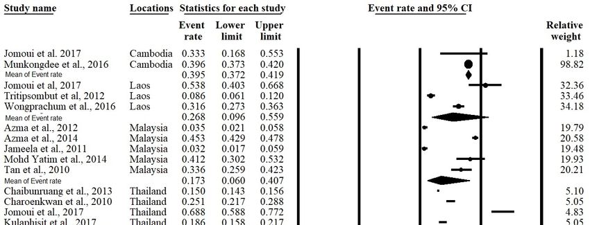
Int. J. Environ. Res. Public Health 2020, 17, x                                                                   3 of 13
Int. J. Environ. Res. Public Health 2020, 17, 7354                                                                 3 of 11

meta-analyzed the prevalence and epidemiology of α-thalassemia—where the results of these similar
studies are
country      quantitatively
         to country          combined—for
                     with different           this [4,5,17,18].
                                      ethnicities  region. Therefore,   the aim
                                                                 However,       of this meta-analysis
                                                                            no studies                  was to
                                                                                         have systematically
provide an update
meta-analyzed          (from 2010
                the prevalence  and until   2020) using
                                      epidemiology         data concerning α-thalassemia
                                                      of α-thalassemia—where      the results ofprevalence   in
                                                                                                 these similar
Southeast   Asia, focusing  on Cambodia,    Laos,  Malaysia,    Thailand, and  Vietnam.   The study
studies are quantitatively combined—for this region. Therefore, the aim of this meta-analysis was to  outcome
on the perspective
provide              of α-thalassemia
         an update (from                prevalence
                           2010 until 2020) using datain Southeast
                                                         concerningAsia   could aid prevalence
                                                                      α-thalassemia  in designing in healthcare
                                                                                                     Southeast
policies
Asia,     for α-thalassemia
      focusing  on Cambodia,  screening   in large Thailand,
                                 Laos, Malaysia,     populations   and
                                                                 and     provideThe
                                                                     Vietnam.    better   genetic
                                                                                      study        counselling
                                                                                             outcome    on the
programs.
perspective of α-thalassemia prevalence in Southeast Asia could aid in designing healthcare policies
for α-thalassemia screening in large populations and provide better genetic counselling programs.
 2. Materials and Methods
2. Materials and Methods
 2.1. Study Guidelines and Literature Search
2.1. Study Guidelines and Literature Search
       PRISMA guidelines were followed for conducting and reporting the results of this meta-analysis
      PRISMA
 (Table          guidelines
          S1) [19].  PubMedwere  (Tablefollowed
                                          S2) andfor  conducting
                                                    SCOPUS         andS3)
                                                               (Table    reporting
                                                                            databases the were
                                                                                          resultssearched
                                                                                                   of this meta-analysis
                                                                                                              up to March
(Table  S1) [19].  PubMed      (Table   S2) and  SCOPUS    (Table  S3)  databases    were  searched
 2020 with the lower limit set to January 2010 using related terms, including alpha, thalassemia,      up   to March 2020
with   the lower   limit
 southeast, and Asian.   set  to  January    2010 using  related  terms,  including     alpha, thalassemia,     southeast,
and Asian.
 2.2. Selection of Studies and Data Extraction
2.2. Selection of Studies and Data Extraction
       All search results were screened by two investigators, and all potential studies were
      All search results were screened by two investigators, and all potential studies were independently
 independently reviewed to be included in this meta-analysis. The main inclusion criteria were: (1)
reviewed    to be included
 studies published             in thisinmeta-analysis.
                       in English                        The mainofinclusion
                                          which the prevalence                   criteria(including
                                                                       α-thalassemia       were: (1) studies    published
                                                                                                       all deletional  and
in  English   in which     the  prevalence     of α-thalassemia     (including    all  deletional
 non-deletional mutations) in Southeast Asian countries were reported; and (2) the study was a peer-and    non-deletional
mutations)     in Southeast
 reviewed publication.           Asianthat
                             Studies      countries   were reported;
                                             did not report              and (2)
                                                             cross-sectional,       the study was
                                                                                observational,          a peer-reviewed
                                                                                                  cohort,    or prevalence
publication.     Studies    that   did   not  report  cross-sectional,   observational,     cohort,
 of α-thalassemia were excluded. We identified additional eligible studies based on references         or   prevalencethat
                                                                                                                         of
α-thalassemia     were   excluded.     We   identified additional   eligible studies    based  on  references
 were cited in the relevant articles. When publications overlapped, only the study with the largest or           that were
cited  in the recent
 the most     relevantdata
                        articles.
                              was When      publications
                                     included    in this overlapped,
                                                          meta-analysis. onlyData,
                                                                               the study   with the
                                                                                      including       largest
                                                                                                   first       or thename,
                                                                                                          author’s    most
recent
 publication year, country, sample size, and prevalence of α-thalassemia of the included studies, year,
         data  was  included     in this  meta-analysis.   Data, including    first  author’s  name,    publication   were
country,   sample    size,  and   prevalence    of  α-thalassemia    of the included     studies,
 extracted and documented by the reviewing investigators. A total number of 278 articles were       were    extracted  and
documented
 included in this meta-analysis (Tables S4 and S5). The study selection and review processthis
                 by  the  reviewing      investigators.   A  total number    of  278   articles were     included   in  are
meta-analysis     (Tables2.S4 and S5). The study selection and review process are illustrated in Figure 2.
 illustrated in Figure

                         Figure 2.
                         Figure 2. Flow
                                   Flow diagram
                                        diagram of
                                                of the
                                                   the systemic
                                                       systemic literature
                                                                literature search
                                                                           search in
                                                                                   in this
                                                                                       this study.
                                                                                            study.
Int. J. Environ. Res. Public Health 2020, 17, 7354 4 of 11
2.3. Statistical Analyses
The prevalence of α-thalassemia was calculated for each study with the number of reported
α-thalassemia cases as the numerator and the total sample size as the denominator. Homogeneity
across studies was investigated with the I2 index (represented as percentage) and Q-test (represented
as a p-value) that indicated heterogeneity between studies. It was reported that an I2 value > 75% and
Q-test with a p-value < 0.1 was regarded as high heterogeneity [20,21]. A random effects model was
used to combine individual effect sizes to create pooled α-thalassemia prevalence if a significantly high
heterogeneity was observed. If other results were obtained, a fixed effects model was utilized. A forest
plot was generated to illustrate the prevalence of each study with a 95% confidence interval (95% CI)
that contributed to the analysis along with the combined prevalence rate. A subsequent meta-analysis
was also performed based on each respective country. Funnel plots and Egger’s tests of asymmetry
were performed to identify any bias within the results [22,23]. All analyses were performed with
Comprehensive Meta-Analysis version 2 software (Biostat, Inc., New Jersey, USA) [24].
3. Results
3.1. Study Characteristics
Twenty-nine studies with a total number of 83,674 subjects were included in this meta-analysis after
a detailed assessment of records obtained from the database and additional searching. These studies
were published between January 2010 and October 2019. Among all included studies, two were carried
out in Cambodia, three in Laos, five in Malaysia, 20 in Thailand, and two in Vietnam (Table 1). The main
characteristics of the studies included in the meta-analysis were recorded and are shown in Table 1.
Table 1. Characteristics of studies included in the meta-analysis.
α-Thalassemia Genotypes Found in
Author [Reference] Country Specific Ethnic 1 Events 2 Total 3
Genotyping Method the Study
Munkongdee et al., Polymerase chain
-α3.7 , -α4.2 , –SEA , αCS , αPs Cambodia N/A 646 1631
2016 [25] reaction (PCR)
Jomoui et al.,
PCR –SEA Cambodia N/A 7 21
2017 [26]
Wongprachum et al., -α3.7 , -α4.2 , –SEA , –THAI ,
PCR Laos N/A 130 411
2012 [27] αCS , αPs , αQ-Thailand
Jomoui et al.,
PCR –SEA Laos N/A 28 52
2017 [26]
Tritipsombut et al.,
PCR -α3.7 , -α4.2 , –SEA , αCS , αPs Laos N/A 30 349
2012 [28]
Azma et al.,
PCR Malaysia N/A 14 400
2012 [29]
-α3.7 , -α4.2 , –SEA , αCS , Malay, Chinese,
Azma et al., 2014 [4] PCR Malaysia 736 1623
αCD59 , αIVS I-1 Indian, Other
Jameela et al., Malay, Chinese, Indian,
PCR -α3.7 , -α4.2 , –SEA , –FIL , α125 Malaysia 10 310
2011 [30] Sikh, Iban
Mohd Yatim et al.,
PCR -α3.7 , –SEA , αCS , αCD59 , Malaysia Malay 28 68
2014 [31]
-α3.7 , -α4.2 , –SEA , –THAI ,
Tan et al., 2010 [3] PCR Malaysia Kadazandusun 42 125
–FIL , αCS , α125 ,
Charoenkwan et al., -α3.7 , -α4.2 , –SEA ,
PCR Thailand N/A 142 566
2010 [32] -αQ-Thailand , αCS
Yong, Yuan, Lue, Khuen,
Lithanatudom et al., -α3.7 , -α4.2 , –SEA , –THAI ,
PCR Thailand Blang, Mon, 33 141
2016 [17] αCS , αPs
Paluang, Lawa
Nillakupt et al.,
PCR -α3.7 , –SEA , αCS , αPs Thailand N/A 47 266
2012 [33]
Pongjantharasatien
PCR –SEA , –THAI , –FIL , -αthal-1 Thailand N/A 4555 31,632
et al., 2016 [34]
Pichanun et al.,
PCR -α3.7 , αCS , αPs Thailand N/A 36 587
2010 [35]
Pharephan et al.,
PCR -α3.7 , -α4.2 , –SEA , αCS Thailand N/A 229 638
2016 [36]Int. J. Environ. Res. Public Health 2020, 17, 7354 5 of 11
Table 1. Cont.
α-Thalassemia Genotypes Found in
Author [Reference] Country Specific Ethnic 1 Events 2 Total 3
Genotyping Method the Study
Panyasai et al.,
PCR -α3.7 , -αQT , –SEA , Thailand N/A 51 23,914
2016 [37]
Panomai et al., -α3.7 , -α4.2 , –SEA , –THAI ,
PCR Thailand N/A 40 190
2010 [38] αCS , αPs
Prayalaw et al., -α3.7 , -α4.2 , –SEA ,
PCR Thailand N/A 75 300
2014 [39] αCS , -αQ-Thailand
Seeratanachot et al.,
Realtime-PCR -α3.7 , -α4.2 , –SEA Thailand N/A 62 250
2015 [40]
Wisedpanichkij et al.,
PCR -α3.7 , -α4.2 , –SEA , αCS Thailand N/A 409 578
2015 [41]
Uaprasert et al.,
PCR -α3.7 , -α4.2 , αCS Thailand N/A 67 241
2013 [42]
Srivorakun et al.,
PCR -α3.7 , –SEA , αCS Thailand N/A 44 226
2011 [43]
Tritipsombut et al.,
PCR -α3.7 , -α4.2 , –SEA , αCS , αPs Thailand N/A 85 1460
2012 [28]
Chaibunruang et al.,
PCR –SEA , –THAI Thailand N/A 1874 12,525
2013 [44]
Yong, Lue, Yuan, Shan,
Khuen, Htin, Paluang,
Kulaphisit et al., -α3.7 , -α4.2 , –SEA , –THAI ,
PCR Thailand Blang, Lawa, Mon, Skaw 124 668
2017 [5] αCS αPs
Karen, Pwo Karen,
Padong Karen
Thanyaornwanya et
PCR -α3.7 , -α4.2 , αCS , αPs Thailand N/A 676 1192
al., 2019 [45]
Jomoui et al.,
PCR –SEA Thailand N/A 66 96
2017 [26]
Mankhenthong et al., -α3.7 , -α4.2 , –SEA , –THAI ,
PCR Thailand N/A 118 1290
2019 [46] αCS
-α3.7 , -α4.2 , –SEA , –THAI ,
Pata et al., 2019 [47] PCR Thailand N/A 82 195
αCS
Kinh, Dao, Tay, Nung,
O’Riordan et al., -α3.7 , -α4.2 , –SEA , –THAI ,
PCR Vietnam S’Tieng, M’Nong, Rac Iay, 996 1431
2010 [18] –FIL , αCS
E De
Hoa Nguyen et al., -α3.7 , -α4.2 , –SEA , –THAI ,
PCR Vietnam Có-Tu 98 298
2014 [48] –SEA , αCS , αPs
Total 11,580 83,674
1 N/A: Not available due to ethnicities were not reported by the study. 2 Events: Number of subjects carrying
alpha-thalassemia. 3 Total: Total number of subjects.
3.2. Meta-Analysis Outcomes
The meta-analysis was conducted using a random effects model found significant heterogeneity
showed an I2 > 99.5% and p-value < 0.001 in overall and all subgroups except Cambodia (Table 2).
The forest plot showed that the overall prevalence rate of α-thalassemia occurrence in this meta-analysis
was 0.226 (95% CI = 0.168–0.296; I2 = 99.5%; p-value < 0.1) (Figure 3). In the subgroup analysis based
on country, Vietnam had the highest prevalence rate (51.5%) of α-thalassemia followed by Cambodia
(39.5%) Laos (26.8%), Thailand (20.1%), and Malaysia (17.3%) (Figure 4).
Table 2. Prevalence rate and heterogeneity of α-thalassemia in overall and subgroups of the study.
Heterogeneity
Prevalence Rate (95% CI) Sample Size (N) No. of Studies (N) Subgroups
I2 (%) p-Value
99.5399.53
Int. J. Environ. Res. Public Health 2020, 17, 7354 7 of 11
Int. J. Environ. Res. Public Health 2020, 17, x 8 of 13
3.3. Publication Bias
3.3. Publication Bias
Funnel plots and Egger’s tests were performed to estimate the publication bias of the included
Funnel plots and Egger’s tests were performed to estimate the publication bias of the included
literature. The shape of the funnel plot revealed obvious evidence of symmetry (Figure 5). The value
literature. The shape of the funnel plot revealed obvious evidence of symmetry (Figure 5). The value
for Egger’s test was t-value = 1.24 with a p-value = 0.112.
for Egger’s test was t-value = 1.24 with a p-value = 0.112.
Figure
Figure 5. Funnel
5. Funnel plotofofthe
plot theoverall
overall prevalence
prevalence ofofα-thalassemia
α-thalassemiain this study.
in this study.
4. Discussion
4. Discussion
ThisThis
studystudy is the
is the firstfirst
to to reportthe
report theprevalence
prevalence of α-thalassemia
α-thalassemiainin the Southeast
the SoutheastAsia region
Asia overover
region
the past
the past 10 years
10 years (2010–2020).
(2010–2020). WeWediddidnotnotobtain
obtain any
any α-thalassemia-related
α-thalassemia-related studies thatthat
studies fulfilled our our
fulfilled
inclusion and exclusion criteria from other Southeast Asian countries, including
inclusion and exclusion criteria from other Southeast Asian countries, including Brunei, Indonesia, Brunei, Indonesia,
Myanmar,
Myanmar, Philippines,
Philippines, Singapore,and
Singapore, and Timor-Leste.
Timor-Leste. UsingUsinga arandom
randomeffects model,
effects model,the the
overall
overall
prevalence rate of α-thalassemia in the included countries was 22.6%, which indicated a significant
prevalence rate of α-thalassemia in the included countries was 22.6%, which indicated a significant
reduction of ~50% of the prevalence in the Southeast Asia region since 2008. The World Health
reduction of ~50% of the prevalence in the Southeast Asia region since 2008. The World Health
Organization had reported the α-thalassemia prevalence as 44.6% in 2008 [49]. India and Brazil, were
Organization
reported athadaboutreported
12% andthe respectively prevalence
α-thalassemia
19.2%, as 44.6% in
[50,51]. The prevalence 2008
rates [49].
were highIndia and Brazil,
in countries
weresuch
reported at about
as UAE, Oman12% andandSaudi 19.2%,
Arabia respectively
at 50% [52].[50,51]. The prevalence rates were high in countries
such as UAE, Oman
The high and Saudi
prevalence Arabia at 50%in[52].
of α-thalassemia the past was due to the lack of knowledge regarding
The high prevalence of α-thalassemia
the seriousness of this disease among the populations in the past was duecountries,
in these to the lack of knowledge
especially regarding
those living in
rural areas with
the seriousness limited
of this diseaseaccess to education
among and those in
the populations who could
these not afford
countries, to obtain those
especially the proper
living in
ruraleducation
areas with similar
limitedto that
accessfound in urban areas.
to education α-thalassemia
and those who could is annotinherited
afford todisease
obtainand the the
proper
mutations may pass from parent to child, affecting the haemoglobin
education similar to that found in urban areas. α-thalassemia is an inherited disease and the mutationsproduction. Hence, the
may educational
pass from andparentscreening
to child,campaigns
affecting regarding this diseaseproduction.
the haemoglobin conducted byHence,the representative bodies and
the educational
(either government or non-government organization) have successfully reduced the prevalence of α-
screening campaigns regarding this disease conducted by the representative bodies (either government
thalassemia in the Southeast Asia region.
or non-government organization) have successfully reduced the prevalence of α-thalassemia in the
Random effects models were used, which are based on the assumption that the true effect could
Southeast Asia region.
vary between studies [51]. The existence of publication bias in this meta-analysis was determined
Random effects
using a funnel models
plot were used,
and Egger’s which
tests. The areofbased
shape on theplot
the funnel assumption that the true
in this meta-analysis effectan
showed could
varyobvious
between studies [51].
symmetry, The existence
indicating the risk ofof publication
publication biasbias in this meta-analysis
is significantly was determined
low. This hypothesis was
usingalso
a funnel
supportedplotbyand Egger’s
statistical tests. The
evidence fromshape
Egger’sof test
the (t-value
funnel plot inp-value
= 1.24; this meta-analysis showed
= 0.112) in which the an
publication bias did not significantly exist in this meta-analysis. Therefore,
obvious symmetry, indicating the risk of publication bias is significantly low. This hypothesis was we concluded that there
was no publication
also supported bias detected
by statistical evidence in this
from Egger’s test (t-value = 1.24; p-value = 0.112) in which the
meta-analysis.
publication bias did not significantly exist in this meta-analysis. Therefore, we concluded that there
was no publication bias detected in this meta-analysis.Int. J. Environ. Res. Public Health 2020, 17, 7354 8 of 11
When stratified according to country, Vietnam has the leading α-thalassemia prevalence rate of
51.5%. The high prevalence rate is probably due to one of the observational studies conducted in
Vietnam focusing on the country’s minority ethnic groups and thus, this likely skewed the actual
prevalence rate [51]. The prevalence of α-thalassemia in Cambodia was the second highest (39.5%)
when compared to other countries included in this meta-analysis. However, since there was only
two studies that reported the prevalence of α-thalassemia Cambodia and Vietnam, more data are
required to estimate the actual prevalence rate of this disease in both of these countries. The prevalence
of α-thalassemia in Laos, Malaysia, and Thailand was quite similar, ranging from 17.3% to 26.8%.
Alpha thalassemia is an inheritable disease where the presence of multiple deletional and non-deletional
mutations can cause severe clinical complications. The presence of α-thalassemia major causes hydrops
fetalis and prenatal deaths [4,5]. Therefore, the low prevalence of α-thalassemia in these countries
and this region indicates that genetic screening for α-thalassemia mutations in the parents could be
done in a population focused approach. Allele frequency and genetic diversity amongst the different
populations provide information that can be used effectively in designing thalassemia prevention
programs [53].
Thalassemia patients suffers from anemia caused by the imbalance of globin chains and impairment
of haemoglobin solubility of erythrocytes. The reduced globin chains were shown to impair the
cytoadherence of Plasmodium [54,55]. These abnormalities of erythrocytes have been shown to confer a
protective effect against malaria infection [12,56]. Hence, there is a natural selection pressure which
causes thalassemia becoming prevalent in countries with high incidence of malaria. Our meta-analysis
was not exhaustive, however it shows that Vietnam had the highest prevalence rate (51.5%) and the
lowest malaria cases (5794 cases) among the countries included in our study which supported the
protective factor of thalassemia [57].
There are several limitations that should be addressed in this meta-analysis. Firstly, only data
from certain Southeast Asian countries were available to be included in this meta-analysis; therefore,
the calculated α-thalassemia-related prevalence rate in this study was specific to selected Southeast
Asian regions. Besides, only studies from 2010 to 2020 were included in this meta-analysis, and it
is possible for studies published before the year 2010 that meet the inclusion criteria but were not
included in this meta-analysis, as this study focused on the prevalence rates from recent past 10 years
only. We also did not include ethnic stratification because the majority of the studies included in
this analysis did not report the ethnic group in the subject population. Alpha thalassemia genotype
stratification was not be performed due to inconsistencies in the reporting of genotypes in the studies
included in this analysis.
5. Conclusions
This is the first meta-analysis that investigated α-thalassemia prevalence in Southeast Asian
countries, and the findings suggest high prevalence of α-thalassemia events in certain countries which
warrants attention as α-thalassemia major could cause severe health complications and impose a
substantial burden to the health authority and families. The data in this meta-analysis may be beneficial
to the representative bodies in designing educational and screening campaigns regarding this disease
in order to further reduce α-thalassemia rates in these countries.
Supplementary Materials: The following are available online at http://www.mdpi.com/1660-4601/17/20/7354/s1,
Table S1: PRISMA checklist of items to include when reporting a systematic review or meta-analysis, Table S2:
Initial literature search from Pubmed, Table S3: Initial literature search from SCOPUS, Table S4: The SCOPUS
literature (N = 152) included in the analysis, Table S5: The Pubmed literature (N = 126) included in the analysis.
Author Contributions: Conceptualization, L.P.W.G. and P.-C.L.; methodology, L.P.W.G. and E.T.J.C.; validation,
E.T.J.C.; investigation, L.P.W.G. and E.T.J.C.; writing—original draft, L.P.W.G.; writing—reviewing and editing,
L.P.W.G. and P.-C.L.; resources, P.-C.L.; supervision, P.-C.L. All authors have read and agreed to the published
version of the manuscript.
Funding: This research received no external funding.Int. J. Environ. Res. Public Health 2020, 17, 7354 9 of 11
Conflicts of Interest: The authors declare no conflict of interest.
References
1. Higgs, D.R.; Engel, J.D.; Stamatoyannopoulos, G. Thalassemia. Lancet 2012, 379, 73–383. [CrossRef]
2. Rosnah, B.; Rosline, H.; Zaidah, A.W.; Noor Haslina, M.N.; Marini, R.; Shafini, M.Y.; Nurul Ain, F.A. Detection
of common deletional alpha-thalassemia spectrum by molecular technique in Kelantan, Northeastern
Malaysia. ISRN Hematol. 2010, 2012, 462969. [CrossRef] [PubMed]
3. Tan, J.I.M.A.; Lee, P.C.; Wee, Y.C.; Tan, K.L.; Mahali, N.F.; George, E.; Chua, K.H. High prevalence of alpha-
and beta-thalassemia in the Kadazadusuns in East Malaysia: Challenges in providing effective health care
for an indigenous group. J. Biomed. Biotechnol. 2010, 2010, 706872. [CrossRef] [PubMed]
4. Azma, R.Z.; Ainoon, O.; Hafiza, A.; Azlin, I.; Noor Farisah, A.R.; Nor Hidayati, S.; Noor Hamidah, H.
Molecular characteristic of alpha thalassemia among patients diagnosed in UKM medical centre.
Malays. J. Pathol. 2014, 36, 27–32.
5. Kulaphisit, M.; Kampuansai, J.; Leecharoenkiat, K.; Wathikthinnakon, M.; Kangwanpong, D.; Munkongdee, T.;
Svasti, S.; Fucharoen, S.; Smith, D.R.; Lithanatudom, P. A comprehensive ethnic-based analysis of alpha
thalassemia allele frequency in northern Thailand. Sci. Rep. 2017, 7, 4690. [CrossRef]
6. Farashi, S.; Harteveld, C.L. Molecular basis of α-thalassemia. Blood Cells Mol. Dis. 2018, 70, 43–53. [CrossRef]
7. Chui, D.H.K. Alpha-Thalassemia: Hb H disease and Hb Bart’s hydrops fetalis. Ann. N. Y. Acad. Sci. 2005,
1054, 25–32. [CrossRef]
8. Weatherall, D.J.; Clegg, J.B. The Thalassemia Syndrome, 4th ed.; Blackwell Scientific Publication:
Oxford, UK, 2011.
9. Casale, M.; Meloni, A.; Filosa, A.; Cuccia, L.; Caruso, V.; Palazzi, G.; Rita Gamberini, M.; Pitrolo, L.;
Caterina Putti, M.; Giuseppe D’Ascola, D.; et al. Multiparametric Cardiac Magnetic Resonance Survey in
Children with Thalassemia Major. Circ. Cardiovasc. Imaging 2015, 8, e003230. [CrossRef]
10. Kurtoglu, A.U.; Kurtoglu, E.; Temizkan, A.K. Effect of iron overload on endocrinopathies in patients with
beta-thalassaemia major and intermedia. Endokrynol. Pol. 2012, 63, 260–263.
11. Suthat, F.; Pranee, W. Haemoglobinopathies in Southeast Asian. Indian J. Med. Res. 2011, 134, 498–506.
12. Galanello, R.; Cao, A. Alpha-thalassemia. Gen. Med. 2011, 13, 83–88. [CrossRef]
13. Kuesap, J.; Chaijaroenkul, W.; Rungsihirunrat, K.; Pongjantharasatien, K.; Na-Bangchang, K. Coexistance
of Malaria and Thalassemia in malaria endemic areas of Thailand. Korean J. Parasitol. 2015, 53, 265–270.
[CrossRef] [PubMed]
14. Rosanas-Uegell, A.; Senn, N.; Raru, P.; Aponte, J.J.; Reeder, J.C.; Siba, P.M.; Michon, P.; Mueller, I. Lack of
associations of α(+)-thalassemia with the risk of Plasmodium falciparum and Plasmodium vivax infection and
disease in a cohort of children aged 3-21 months from Papua New Guinea. Int. J. Parasitol. 2012, 42, e1000097.
[CrossRef]
15. Vento, S.; Cainelli, F.; Cesario, F. Infections and thalassemia. Lancet Infect. Dis. 2006, 6, 226–233. [CrossRef]
[PubMed]
16. Ryan, K.; Bain, B.J.; Worthington, D.; James, J.; Plews, D.; Mason, A.; Roper, D.; Rees, D.C.; de la Salle, B.;
Streetly, A.; et al. Significant haemoglobinopathies: Guidelines for screening and diagnosis. Br. J. Haematol.
2010, 149, 35–49. [CrossRef]
17. Lithanatudom, P.; Khampan, P.; Smith, D.R.; Svasti, S.; Fucharoen, S.; Kangwanpong, D.; Kampuansai, J.
The prevalence of alpha-thalssemia amongst Tai and Mon-Khmer ethnic groups residing in northern Thailand:
A population-based study. Hematology 2016, 21, 480–485. [CrossRef]
18. O’Riordan, S.; Hien, T.T.; Miles, K.; Allen, A.; Quyen, N.N.; Hung, N.Q.; Anh, D.Q.; Tuyen, L.N.; Khia, D.B.;
Thai, C.Q.; et al. Large scale screening for haemoglobin disorders in southern Vietnam: Implications for
avoidance and management. Br. J. Haematol. 2010, 150, 359–364. [CrossRef]
19. Moher, D.; Loberati, A.; Tetzlaff, J.; Altman, D.G. The PRISMA Group. Preferred reporting items for
systematic reviews and meta-analyses: The PRISMA statement. PLoS Med. 2009, 6, e1000097. [CrossRef]
20. Dersimonian, R.; Laird, N. Meta-analysis in clinical trials. Control. Clin. Trials. 1986, 7, 177–188. [CrossRef]
21. Higgins, J.P.; Thompson, S.G.; Deeks, J.J.; Altman, D.G. Measuring inconsistency in meta analyses. BMJ 2003,
327, 557–560. [CrossRef]Int. J. Environ. Res. Public Health 2020, 17, 7354 10 of 11
22. Egger, M.; Smith, G.D.; Schneider, M.; Minder, C. Bias in meta-analysis detected by a simple, graphical test.
BMJ 1997, 315, 629–634. [CrossRef] [PubMed]
23. Light, R.J.; Pillemer, D.B. Summing up: The Science of Reviewing Research; Harvard University Press: Cambridge,
MA, USA, 1984.
24. Borenstein, M.; Hedges, L.; Higgins, J.; Rothstein, H.R. Comprehensive meta-analysis version 2. Englewood
2005, 104, 188–191.
25. Munkongdee, T.; Tanakulmas, J.; Butthep, P.; Winichagoon, P.; Main, B.; Yiannakis, M.; George, J.; Devenish, R.;
Fucharoen, S.; Svasti, S. Molecular epidemiology of hemoglobinpathies in Cambodia. Hemoglobin 2016, 40,
163–167. [CrossRef] [PubMed]
26. Jomoui, W.; Fucharoen, G.; Sanchaisuriya, K.; Charoenwijitkul, P.; Maneesarn, J.; Xu, X.; Fucharoen, S. Genetic
origin of α0 -thalassemia (SEA deletion) in Southeast Asian populations and application to accurate prenatal
diagnosis of Hb Bart’s hydrops fetalis syndrome. J. Hum. Gen. 2017, 62, 747–754. [CrossRef] [PubMed]
27. Wongprachum, K.; Sanchaisuriya, K.; Dethvongphanh, M.; Norcharoen, B.; Vidamaly, V.; Sanchaisuriya, P.;
Fucharoen, S.; Fucharoen, G.; Schelp, F.P. Molecular heterogeneity of thalassemia among pregnant Laotian
women. Acta Hematol. 2016, 135, 65–69. [CrossRef] [PubMed]
28. Tritipsombut, J.; Sanchaisuriya, K.; Phollarp, P.; Bouakhasith, D.; Sanchaisuriya, P.; Fucharoen, G.; Fucharoen, S.;
Schelp, F.P. Micromapping of thalassemia and hemoglobinopathies in different regions of northeast Thailand
and Vientaine, Laos people’s democratic republic. Hemoglobin 2012, 36, 47–56. [CrossRef] [PubMed]
29. Azma, R.Z.; Ainoon, O.; Azlin, I.; Hamenuddin, H.; Hadi, N.A.; Tatt, W.K.; Syazana, I.N.; Asmaliza, A.M.;
Das, S.; Hamidah, N.H. Prevalence of iron deficiency anaemia and thalassemia trait among undergraduate
medical students. Clin. Ter. 2012, 163, 287–291.
30. Jameela, S.; Sharifah Sabirah, S.O.; Babam, J.; Phan, C.L.; Visalachy, P.; Chang, K.M.; Salwana, M.A.;
Zuridah, A.; Subramanian, Y.; Rahimah, A. Thalassemia screening among students in a secondary school in
Ampang, Malaysia. Med. J. Malays. 2011, 66, 522–524.
31. Mohd Yatim, N.F.; Abd Rahim, M.; Menon, K.; Al-Hassan, F.M.; Ahmad, R.; Manocha, A.B.; Saleem, M.;
Yahaya, B.H. Molecular characterization of α and β-thalassaemia among malay patients. Int. J. Mol. Sci.
2014, 15, 8835–8845. [CrossRef]
32. Charoenkwan, P.; Taweephol, R.; Sirichotiyakul, S.; Tantiprabha, W.; Sae-Tung, R.; Suanta, S.;
Sakdasirisathaporn, P.; Sanguansermsri, T. Cord blood screening for α-thalassemia and hemoglobin variants
by isoelectric focusing in northern Thai neonates: Correlation with genotypes and hematologic parameters.
Blood Cells Mol. Dis. 2010, 45, 53–57. [CrossRef]
33. Nillakupt, K.; Nathalang, O.; Arnutti, P.; Jindadamrongwech, S.; Boonsiri, T.; Panichkul, S.; Areekul, W.
Prevalence and hematological parameters of thalassemia in the Kradarn subdistrict Chachoengsao province,
Thailand. J. Med. Assoc. Thai. 2011, 95, S124–S132.
34. Pongjantharasatien, K.; Banyatsuppasin, W.; Pounsawat, S.; Jindadamrongwech, S. Occurrence of the –SEA ,
–THAI , and –FIL α-thalassemia-1 carriers from a 7-year study at Ramathibodi hospital, Bangkok, Thailand.
Hemoglobin 2016, 40, 283–284. [CrossRef] [PubMed]
35. Pichanun, D.; Munkongdee, T.; Klamchuen, S.; Butthep, P.; Winichagoon, P.; Fucharoen, S.; Svasti, S.
Molecular screening of the Hbs constant spring (codon 142, TAA>CAA, α2) and paksé (codon 142, TAA>TAT,
α2) mutations in Thailand. Hemoglobin 2010, 34, 582–586. [CrossRef] [PubMed]
36. Pharephan, S.; Sirivatanapa, P.; Makonkawkeyoon, S.; Tuntiwechapikul, W.; Makonkawkeyoon, L. Prevalence
of α-thalassemia genotypes in pregnant women in northern Thailand. Indian J. Med. Res. 2016, 143, 315–322.
[CrossRef]
37. Panyasai, S.; Fucharoen, G.; Fucharoen, S. Hemoglobin variants in Northern Thailand: Prevalence,
heterogeneity and molecular characteristics. Genet. Test Mol. Biomark. 2016, 20, 37–43. [CrossRef]
38. Panomai, N.; Sanchaisuriya, K.; Yamsri, S.; Sanchaisuriya, P.; Fucharoen, S.; Schelp, F.P. Thalassemia and
iron deficiency in a group of northeast Thai school children: Relationship to the occurrence of anaemia.
Eur. J. Pediatr. 2010, 169, 1317–1322. [CrossRef]
39. Prayalaw, P.; Fuchafoen, G.; Fucharoen, S. Routine screening for α-thalassemia using an
immunochromatographic strip assay for haemoglobin Bart’s. J. Med. Screen. 2014, 21, 120–125. [CrossRef]
40. Seeratanachot, T.; Shimbhu, D.; Charoenkwan, P.; Sanguansermsri, T. Detection of deletion α+ -thalassemia
mutation [-α (3.7), -α (4.2)] by quantitative PCR assay. Southeast Asian J. Trop. Med. Public Health 2015, 46,
110–115.Int. J. Environ. Res. Public Health 2020, 17, 7354 11 of 11
41. Wisedpanichkij, R.; Jindadamrongwech, S.; Butthep, P. Identification of Hb constant spring (HBA2: c.427T>C)
by an automated high performance liquid chromatography method. Hemoglobin 2015, 39, 190–195. [CrossRef]
42. Uaprasert, N.; Settapiboon, R.; Amomsiriwat, S.; Sarnthammakul, P.; Thanapat, T.; Rojnuckarin, P.;
Sutcharitchan, P. Diagnostic utility of isoelectric focusing and high performance liquid chromatography in
neonatal cord blood screening for thalassemia and non-sickling hemoglobinopathies. Clin. Chim. Acta. 2013,
427, 23–26. [CrossRef]
43. Srivorakun, H.; Fucharoen, G.; Changtrakul, Y.; Komwilaisak, P.; Fucharoen, S. Thalassemia and
hemoglobinopathies in South East Asia newborns: Diagnostic assessment using capillary electrophoresis
system. Clin. Biochem. 2011, 44, 406–411. [CrossRef] [PubMed]
44. Chaibunruang, A.; Prommetta, S.; Yamsri, S.; Fucharoen, G.; Sae-Ung, N.; Sanchaisuriya, K.; Fucharoen, S.
Molecular and hematological studies in a large cohort of α0 -thalassemia in northeast Thailand: Data from a
single referral centre. Blood Cells Mol. Dis. 2013, 51, 89–93. [CrossRef] [PubMed]
45. Thanyaornwanya, C.; Singha, K.; Fucharoen, G.; Sanchaisuriya, K.; Thepphitak, P.; Wintachai, P.; Karnpean, R.;
Fucharoen, S. Molecular characteristics of α+ -thalassemia (3.7 kb deletion) in Southeast Asia: Molecular
subtypes, haplotypic heterogeneity, multiple founder effects and laboratory diagnosis. Clin. Biochem. 2019,
71, 31–37. [CrossRef]
46. Mankhenthong, K.; Phusua, A.; Suantan, S.; Srisittipoj, P.; Charoenkwan, P.; Sanguansermsri, T. Molecular
characteristics of thalassemia and haemoglobin variants in prenatal diagnosis program in northern Thailand.
Int. J. Hematol. 2019, 110, 474–481. [CrossRef]
47. Pata, S.; Laopajon, W.; Pongpaiboon, M.; Thongkum, W.; Polpong, N.; Munkongdee, T.; Paiboonsukwong, K.;
Fucharoen, S.; Tayapiwatana, C.; Kasinrerk, W. Impact of the detection of ζ-globin chains and haemoglobin
Bart’s using immunochromatographic strip test for α0 -thalassemia (–SEA ) differential diagnosis. PLoS ONE
2019, 14, e0223996. [CrossRef]
48. Hoa Nguyen, V.; Sanchaisuriya, K.; Wongprachum, K.; Nguyen, M.D.; Phan, T.T.; Vo, V.T.; Sanchaisuriya, P.;
Fucharoen, S.; Schelp, F.P. Hemoglobin constant spring is markedly high in women of an ethnic minority
group in Vietnam: A community-based survey and hematologic features. Blood Cell Mol. Dis. 2014, 52, 161–165.
[CrossRef]
49. Modell, B.; Darlison, M. Global epidemiology in haemoglobin disorders and derived service indicators.
Bull. World Health Organ. 2008, 86, 480–487. [CrossRef]
50. Nadkarni, A.; Phanasgaonkar, S.; Colah, R.; Mohanty, D.; Ghosh, K. Prevalence and Molecular
Characterization of α-Thalassemia Syndromes among Indians. Genet. Test. 2008, 12, 177–180. [CrossRef]
51. Souza, A.E.S.; Cardoso, G.L.; Takanashi, S.Y.L.; Guerreiro, J.F. α-Thalassemia (3.7 kb deletion) in a population
from the Brazilian Amazon region: Santarém, Pará State. Genet. Mol. Res. 2009, 8, 477–481. [CrossRef]
52. AL-Awamy, B.H. Thalassemia syndromes in Saudi Arabia. Meta-analysis of local studies. Saudi Med. J. 2000,
21, 8–17.
53. Kee, B.P.; Lian, L.H.; Lee, P.C.; Lai, T.X.; Chua, K.H. Genetic data for 15 STR loci in a Kadazan-Dusun
population from East Malaysia. Genet. Mol. Res. 2011, 10, 739–743. [CrossRef] [PubMed]
54. Forget, B.G.; Bunn, H.F. Classification of the disorders of haemoglobin. Cold Spring Harb. Perspect. Med. 2013,
3, a011684. [CrossRef]
55. Krause, M.A.; Diakite, S.A.S.; Lopera-Mesa, T.M.; Amaratunga, C.; Arie, T.; Traore, K.; Doumbia, S.; Konate, D.;
Keefer, J.R.; Diakite, M.; et al. α-thalassemia impairs the cytoadherence of Plasmodium falciparum-infected
erythrocytes. PLoS ONE 2012, e37214. [CrossRef] [PubMed]
56. Gundula, M.-O.; Gros, P. Erthrocyte variants and the nature of their malaria protective effect. Cell. Microbiol.
2005, 7, 753–763. [CrossRef]
57. World Health Organization. World Malaria Report 2019; World Health Organization: Geneva,
Switzerland, 2018.
© 2020 by the authors. Licensee MDPI, Basel, Switzerland. This article is an open access
article distributed under the terms and conditions of the Creative Commons Attribution
(CC BY) license (http://creativecommons.org/licenses/by/4.0/).You can also read