Protective Effects of N-acetyl-L-cysteine on the Reduction of Dopamine Transporters in the Striatum of Monkeys Treated with Methamphetamine
←
→
Page content transcription
If your browser does not render page correctly, please read the page content below

Neuropsychopharmacology (2004) 29, 2018–2023
   & 2004 Nature Publishing Group All rights reserved 0893-133X/04 $30.00
                                www.neuropsychopharmacology.org

Protective Effects of N-acetyl-L-cysteine on the Reduction
of Dopamine Transporters in the Striatum of Monkeys
Treated with Methamphetamine

     Kenji Hashimoto*,1, Hideo Tsukada2, Shingo Nishiyama2, Dai Fukumoto2, Takeharu Kakiuchi2, Eiji Shimizu1
     and Masaomi Iyo1
     1
      Department of Psychiatry, Chiba University Graduate School of Medicine, Chiba, Japan; 2Central Research Laboratory, Hamamatsu Photonics
     K.K., Hamakita, Shizuoka, Japan

     Several lines of evidence suggest that oxidative stress might contribute to neurotoxicity in the dopaminergic nerve terminals after
     administration of methamphetamine (MAP). We undertook the present study to determine whether intravenous administration of
     N-acetyl-L-cysteine (NAC), a potent antioxidant drug, could attenuate the reduction of dopamine transporter (DAT) in the striatum of
     monkey brain after administration of MAP. Positron emission tomography studies demonstrated that repeated administration of MAP
     (2 mg/kg as a salt, four times at 2-h intervals) significantly decreased the accumulation of radioactivity in the striatum after intravenous
     administration of [11C]b-CFT. In contrast, the binding of [11C]SCH 23390 to dopamine D1 receptors in the monkey striatum was not
     altered after the administration of MAP. A bolus injection of NAC (150 mg/kg, i.v.) 30 min before MAP administration and a subsequent
     continuous infusion of NAC (12 mg/kg/h, i.v.) over 8.5 h significantly attenuated the reduction of DAT in the monkey striatum 3 weeks
     after the administration of MAP. These results suggest that NAC could attenuate the reduction of DAT in the monkey striatum after
     repeated administration of MAP. Therefore, it is likely that NAC would be a suitable drug for treatment of neurotoxicity in dopaminergic
     nerve terminals related to chronic use of MAP in humans.
     Neuropsychopharmacology (2004) 29, 2018–2023, advance online publication, 16 June 2004; doi:10.1038/sj.npp.1300512
     Keywords: methamphetamine; neurotoxicity; oxidative stress; N-acetyl-L-cysteine; dopamine transporter; monkey; positron emission
     tomography (PET)

INTRODUCTION                                                                 2003; Volkow et al, 2001a, b), suggesting the neurotoxic
                                                                             effects of MAP in the human brain. These findings are
The abuse of methamphetamine (MAP), a potent psycho-
                                                                             supported by a report demonstrating that the densities of
stimulant, is an extremely serious and growing problem in
                                                                             DAT are significantly decreased in the postmortem brain
the world. The action of MAP is thought to involve rapid
                                                                             striatum of chronic MAP users (Wilson et al, 1996).
entry into the brain, followed by influx into monoaminergic                  However, the precise mechanisms underlying MAP-induced
terminals, interaction with vesicular monoaminergic trans-
                                                                             neurotoxicity in dopaminergic nerve terminals of the brain
porter, entry into monoaminergic vesicles and displacement
                                                                             are currently not known (Davidson et al, 2001; Cadet et al,
of monoamines into the cytoplasm of the terminals, and                       2003).
subsequent release of the monoamines into the synaptic
                                                                                Oxidative stress generated by an imbalance between
cleft (Cadet et al, 2003). Recent studies using positron
                                                                             reactive oxygen species (ROS; hydrogen peroxide, super-
emission tomography (PET) suggest that chronic use of
                                                                             oxide radical, and hydroxyl radical) and antioxidants might
MAP causes the reduction of dopamine transporter (DAT)
                                                                             contribute to the neurotoxicity of MAP in the brain (Imam
in the human brain (McCann et al, 1998; Sekine et al, 2001,
                                                                             et al, 2001; Davidson et al, 2001; Cadet et al, 2003). N-acetyl-
                                                                             L-cysteine (NAC), the acetylated variant of the amino acid
*Correspondence: Dr K Hashimoto, Department of Psychiatry, Chiba             L-cysteine, is an excellent source of sulfhydryl (SH) groups
University Graduate School of Medicine, 1-8-1 Inohana, Chiba                 and is converted in the body into metabolites capable of
260-8670, Japan, Tel: þ 81 43 226 2147, Fax: þ 81 43 226 2150,
E-mail: hashimoto@faculty.chiba-u.jp                                         stimulating glutathione synthesis, promoting detoxification,
Received 18 November 2003; revised 1 April 2004; accepted 12 May             and acting directly as free radical scavengers (Kelly, 1998).
2004                                                                         In addition to its mucolytic action, NAC has been studied
Online publication: 19 May 2004 at http://www.acnp.org/citations/            and utilized to treat conditions characterized by decreased
Npp05190403530/default.pdf                                                   glutathione or oxidative stress such as HIV infection,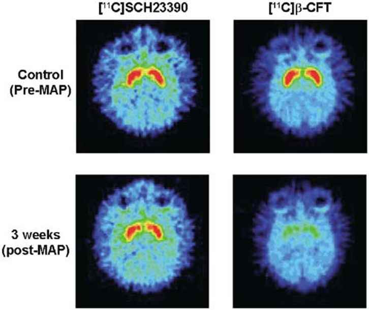
Protective effects of NAC on reduction of dopamine transporters
                                                                 K Hashimoto et al
                                                                                                                                 2019
cancer, and heart disease (Kelly, 1998). Owing to its known
characteristics, NAC has been used as a tool for investigat-
ing the role of ROS in numerous biological and pathological
processes (Kelly, 1998; Zafarullah et al, 2003). Recently, we
reported that NAC significantly attenuated 6-hydroxydopa-
mine-induced apoptotic neuronal cell death in human
neuroblastoma SK-N-SH cells, suggesting that NAC could
work as a beneficial dopaminergic neuron-survival factor
(Shimizu et al, 2002). In addition, NAC directly modifies the    Figure 1 Schedule of treatment of MAP and/or NAC in monkeys.
activity of several proteins by its reducing activity
(Zafarullah et al, 2003). Based on the role of oxidative         (20–40 mg every 2–3 h) (Konuma, 1994). For administration
stress in the neurotoxicity of MAP in the brain (Imam et al,     of NAC, subjects received a bolus of NAC (Wako Pure
2001; Davidson et al, 2001; Cadet et al, 2003), it is            Chemicals Ltd, Tokyo, Japan, 150 mg/kg, i.v.) 30 min before
interesting to study the effects of the antioxidant NAC on       administration of MAP and a subsequent continuous
MAP-induced neurotoxicity in the brain.                          infusion of NAC (12 mg/kg/h, i.v.) over 8.5 h, with a slight
  We reported recently that pretreatment with NAC                modification of the method reported previously (Molnar
significantly attenuates hyperlocomotion, development of         et al, 1999; Rank et al, 2000) (Figure 1).
sensitization, and neurotoxicity after the administration of
MAP in rats, suggesting that NAC might be a suitable drug
for treatment of MAP abuse (Fukami et al, 2004). The             Synthesis of [11C][-]Labeled Compounds
purpose of the present study was to discover therapeutic
                                                                 Carbon-11 (11C) was produced by 14N (p,a)11C nuclear
drugs to prevent or protect against neurotoxicity in
                                                                 reaction using the cyclotron (HM-18, Sumitomo Heavy
dopaminergic terminals in chronic MAP abusers. We
                                                                 Industry, Tokyo, Japan) at the Hamamatsu Photonics PET
performed the present PET study to determine whether
                                                                 Center and obtained as [11C]CO2, which was converted to
intravenous administration of NAC could attenuate the
                                                                 [11C]methyl iodide. [11C]2b-carbomethoxy-3b-(4-fluoro-
reduction of DAT in the striatum of monkey brain after
                                                                 phenyl)tropane (b-CFT) (for DAT) and [11C]SCH 23390
administration of MAP. To minimize the effects of
                                                                 (for DA D1 receptors) were synthesized as previously
anesthetics on the behavior of a labeled compound in vivo,
                                                                 reported (Tsukada et al, 2001; Harada et al, 2002). The
we performed PET scans of monkeys in the conscious state
                                                                 radiochemical and chemical purities of labeled compounds
(Onoe et al, 1994; Tsukada et al, 1999, 2000, 2001, 2002).
                                                                 were greater than 98 and 99%, respectively. After analysis
                                                                 for identification, the solution was passed through a
MATERIALS AND METHODS                                            0.22-mm pore size filter before intravenous administration
                                                                 to the monkey.
Subjects
Six young-adult male rhesus monkeys (Macaca mulatta)
                                                                 PET Scans
weighing from 4 to 6 kg were used for PET measurements.
Monkeys were maintained and handled in accordance with           PET data were collected before and 3 weeks after admin-
the recommendations of the US National Institutes of             istration of MAP or MAP/NAC. Data were collected on a
Health and also the guidelines of the Central Research           high-resolution PET scanner (SHR-7700, Hamamatsu
Laboratory, Hamamatsu Photonics (Hamakita, Shizuoka,             Photonics KK, Hamamatsu, Japan) with a transaxial
Japan). Over the course of 3 months, the monkeys were            resolution of 2.6 mm full-width at half-maximum (FWHM)
trained to sit on a chair twice a week. The magnetic             and a center-to-center distance of 3.6 mm (Watanabe et al,
resonance images (MRIs) of all monkeys were obtained with        1997). The PET camera allowed 31 slices for imaging to be
a Toshiba MRT-50A/II (0.5 T) under anesthesia with               recorded simultaneously. After an overnight fast, animals
pentobarbital. The stereotactic coordinates of PET and           were fixed to the monkey chair with stereotactic coordinates
MRI were adjusted based on the orbitomeatal (OM) line            aligned parallel to the OM line. A cannula was implanted in
with monkeys secured in a specially designed head holder         the posterior tibial vein of the monkey for administration of
(Takechi et al, 1994). At least 1 month before the PET study,    [11C]-labeled compounds. [11C]b-CFT or [11C]SCH 23390
an acrylic plate, with which the monkey was fixed to the         was injected through the posterior tibial vein cannula. For
monkey chair, was attached to the head under pentobarbital       [11C]SCH 23390, a PET scan was performed for 64 min with
anesthesia as described previously (Onoe et al, 1994).           six time frames at 10-s intervals, six time frames at 30-s
                                                                 intervals, 12 time frames at 1-min intervals, followed by 16
                                                                 time frames at 3-min intervals. For [11C]b-CFT, additional
Drug Administration
                                                                 scans of nine time frames at 3-min intervals were carried
MAP hydrochloride (Dainippon Pharmaceuticals Ltd, Osa-           out to collect data for 91 min total. After completion of the
ka, Japan, 2 mg/kg as a salt, four times at 2-h intervals) was   first scan with [11C]b-CFT, scans with [11C]SCH 23390 were
administered intramuscularly into each monkey (Figure 1).        continuously performed at 3-h intervals. Due to the very
We used that dose because it is reported to produce long-        short half-life of 11C (20.4 min), a time lag of at least 3 h
term neurotoxic effects on the brain of baboons (Ville-          between scans provided a sufficient decay time of radio-
magne et al, 1998). Also, such a dose regimen closely            activity in monkeys (approximately 1/400 of the injected
approximates the binge use of MAP by some humans                 dose). Therefore, the level of radioactivity associated with

                                                                                                       Neuropsychopharmacology
Protective effects of NAC on reduction of dopamine transporters
                                                   K Hashimoto et al
2020
       the previous injection of labeled compound would not                          Table 1 Effects of NAC on Reduction of DAT and DA D1
       interfere with the next scan.                                                 Receptors in the Monkey Striatum after Administration of MAP

                                                                                                                 Control                    MAP
       Data Analysis and Statistical Analysis
                                                                                     Monkey-1
       For quantitative analysis, time–activity curves of radio-                       DAT                     3.563 (100%)             1.022 (28.7%)
       activity in the cerebellum, used as an input function because
                                                                                       DA D1                   1.824 (100%)             1.678 (92.0%)
       of the much lower density of dopamine receptors (Creese
       et al, 1975), and each region of interest (ROI) were fitted to                Monkey-2
       a two-compartment model using the least-squares fitting                         DAT                     3.255 (100%)             1.025 (31.5%)
       method to estimate the kinetic parameters, and the binding                      DA D1                   1.836 (100%)             1.927 (105%)
       potential in each ROI was calculated as described previously                  Monkey-3
       (Lammertsma and Hume, 1996). The differences between                            DAT                     4.827 (100%)             1.700 (35.2%)
       the control (pre-MAP) monkeys and the MAP or NAC plus                           DA D1                   2.272 (100%)             2.171 (95.6%)
       MAP (post MAP) monkeys were determined using a paired
       two-tailed t-test. The difference between monkeys in the
                                                                                     DAT (mean7SEM)        3.88270.481 (100%) 1.24970.226 (31.871.88%)*
       MAP-treated group and those in the NAC plus MAP-treated
       group was determined using an unpaired two-tailed t-test.                     DA D1 (mean7SEM) 1.97770.147 (100%) 1.92570.142 (97.573.88%)
       Significance was set at po0.05.
                                                                                                                 Control                 NAC+MAP

       RESULTS                                                                       Monkey-4
                                                                                       DAT                     4.269 (100%)             2.648 (62.0%)
       PET studies using [11C]b-CFT (for DAT) or [11C]SCH 23390
                                                                                       DA D1                   2.780 (100%)             2.587 (93.1%)
       (for DA D1 receptor) were performed before and 3 weeks
                                                                                     Monkey-5
       after repeated administration of MAP. High accumulation
       of radioactivity in the striatum after intravenous adminis-                     DAT                     2.649 (100%)             1.696 (64.0%)
       tration of [11C]b-CFT or [11C]SCH 23390 was detected in the                     DA D1                   1.680 (100%)             1.788 (106%)
       control monkeys, whereas levels of radioactivity in the                       Monkey-6
       cerebellum were much lower compared to those in the                             DAT                     2.524 (100%)             1.743 (69.1%)
       striatum (Figure 2). At 3 weeks after administration of MAP                     DA D1                   1.742 (100%)             1.667 (95.7%)
       (2 mg/kg  4, 2-h intervals), the binding of [11C]b-CFT in
       the striatum was significantly (t ¼ 10.01, p ¼ 0.010) de-
                                                                                     DAT (mean7SEM)        3.14770.562 (100%) 2.02970.310 (65.072.11%)*,#
       creased, whereas the binding of [11C]SCH 23390 to DA D1
                                                                                     DA D1 (mean7SEM) 2.06770.357 (100%) 2.01470.289 (98.373.94%)

                                                                                     Values of binding potential are shown.
                                                                                     *Po0.05 as compared with the control group (a paired t-test).
                                                                                     #
                                                                                      Po0.001 as compared with the MAP-treated group (a unpaired t-test).

                                                                                     receptor in the striatum was not altered (t ¼ 0.716,
                                                                                     p ¼ 0.549) (Figure 2 and Table 1). Time–activity curves of
                                                                                     radioactivity after intravenous administration of [11C]b-
                                                                                     CFT indicated a high accumulation of radioactivity in the
                                                                                     striatum, with a low level of radioactivity in the cerebellum,
                                                                                     of the control monkeys (Figure 3). At 3 weeks after
                                                                                     administration of MAP (2 mg/kg  4, 2-h intervals), time–
                                                                                     activity curves of radioactivity in the striatum after
                                                                                     intravenous administration of [11C]b-CFT were markedly
                                                                                     decreased, whereas those of radioactivity in the cerebellum
                                                                                     were not altered (Figure 3).
                                                                                       As shown in Figure 4, the accumulation of radioactivity in
                                                                                     the striatum of monkeys treated with NAC plus MAP after
                                                                                     administration of [11C]b-CFT was higher than that of
       Figure 2 PET images of [11C]SCH 23390 and [11C]b-CFT in the brains            radioactivity in the striatum of monkeys treated with MAP
       of a rhesus monkey (Macaca mulatta). PET data were collected on an            alone (Figure 4). The binding potential in the striatum of
       animal PET scanner (Hamamatsu SHR-7700) with a transaxial resolution of
                                                                                     monkeys treated with NAC plus MAP after administration
       2.6 mm (FWHM). The PET image of [11C]SCH23390 was generated by the
       summation of data from 37 to 64 min after injection. PET images for           of [11C]b-CFT was significantly (t ¼ 11.74, po0.001) higher
       [11C]b-CFT were generated by the summation of data from 61 to 91 min          than that of the striatum of monkeys treated with MAP
       after injection. The stereotactic coordinates of PET were adjusted based on   alone, although the binding potential in the striatum of
       the OM line. These PET images were from the same monkey.                      monkeys treated with NAC plus MAP was significantly

       Neuropsychopharmacology
Protective effects of NAC on reduction of dopamine transporters
K Hashimoto et al
2021
(t ¼ 4.367, p ¼ 0.049) lower than that of the striatum of neurotoxic effects on dopaminergic terminals in the brain
control monkeys (Table 1). after administration of MAP (Cadet et al, 2003; LaVoie and
Hastings, 1999; Stokes et al, 1999). Two metabolic routes
seem possible: (1) formation of 3,4-dihydroxyphenyl acetic
acid (DOPAC) and hydrogen peroxide (H2O2) by mono-
DISCUSSION
amine oxidase (MAO), or (2) formation of reactive
The major finding of the present study is that infusion of DA-quinones and free radicals by auto-oxidation (Asanuma
the antioxidant NAC could attenuate the reduction of DAT et al, 2003). As DA-induced modifications of protein
in the monkey striatum after administration of MAP. structure and function may result in cellular toxicity, it is
Repeated administration of MAP caused a marked reduc- likely that DA quinones produced by auto-oxidation
tion of DAT in the monkey striatum, which is consistent contribute to MAP-induced neurotoxicity to dopaminergic
with previous reports (Melega et al, 1998, 2000; Villemagne nerve terminals, supporting the evidence of oxidative stress
et al, 1998). In contrast, we found that the binding of in this model (LaVoie and Hastings, 1999; Stokes et al,
[11C]SCH 23390 to DA D1 receptors in the monkey striatum 1999). In addition, high ROS, including hydrogen peroxide,
was not altered by administration of MAP, suggesting that superoxide radical, and hydroxyl radical, are generated not
administration of MAP could damage the dopaminergic only during the oxidation of DA but also during the decay
nerve terminals but not the postsynaptic neurons. In of redox-active DA quinones, suggesting that superoxide
addition, it seems that the drug schedule (2 mg/kg 4, 2-h radicals, hydrogen peroxide, and nitric oxide might be
intervals) used in this study is neurotoxic to DAT in the involved in the neurotoxicity of MAP (Davidson et al, 2001;
monkey brain. As described in Introduction, the marked Imam et al, 2001; Cadet et al, 2003).
release of DA produced by MAP could be implicated in the It is well known that NAC can act as a precursor for
glutathione synthesis, as well as a stimulator of the cytosolic
enzymes involved in glutathione regeneration. Furthermore,
NAC can act by direct reaction between its reducing thiol
group and ROS. It has been shown that NAC can prevent
programmed cell death in cultured neuronal cells and that
NAC increases mitochondrial complex I and IV specific
activities both in vitro and in vivo in synaptic mitochondrial
preparations from aged mice (Banaclocha, 2001). Therefore,
it should be noted that a potent antioxidant NAC could
attenuate the reduction of DAT in the monkey striatum
after the repeated administration of MAP.
DAT knockout mice are resistant to MAP-induced
neurotoxicity of dopaminergic nerve terminals, suggesting
the role of DAT in the MAP-induced neurotoxicity in these
nerve terminals (Fumagalli et al, 1998). Nevertheless, it is
Figure 3 Time–activity curves of radioactivity in the striatum and
cerebellum of the monkey before and 3 weeks after administration of MAP. possible that MAP-induced DA released within the cyto-
PET data were collected for 91 min. The radioactivity in the striatum and plasm of dopaminergic terminals might be a more critical
cerebellum was plotted against time after intravenous administration of trigger of MAP-induced neurotoxicity in the dopaminergic
[11C]b-CFT into a rhesus monkey. terminals, since vesicular monoamine transporter 2
Figure 4 PET images of [11C]b-CFT in the brains of a rhesus monkey 3 weeks after administration of MAP or NAC plus MAP. Each PET image was
generated by the summation of data from 61 to 91 min after injection of [11C]b-CFT.
NeuropsychopharmacologyProtective effects of NAC on reduction of dopamine transporters
K Hashimoto et al
2022
(VMAT2) knockout mice are more susceptible to the toxic neurotoxicity: evidence from mice lacking the transporter.
manifestations of MAP (Fumagalli et al, 1999). These J Neurosci 18: 4861–4869.
findings are supported by the report that MAP-induced Fumagalli F, Gainetdinov RR, Wang YM, Valenzano KJ, Miller GW,
neurotoxicity might be related to the formation of DA Caron MG (1999). Increased methamphetamine neurotoxicity in
quinones, a process dependent on increased DA levels heterozygous vesicular monoamine transporter 2 knock-out
mice. J Neurosci 19: 2424–2431.
within dopaminergic nerve terminals (LaVoie and Hastings,
Harada N, Nishiyama S, Satoh K, Fukumoto D, Kakiuchi T,
1999). It is likely that NAC binds to reactive DA quinones Tsukada H (2002). Age-related changes in the striatal dopami-
and ROS by auto-oxidation, resulting in the protection of nergic system in the living brain: a multiparametric PET study in
the neurotoxicity by DA quinones. conscious monkeys. Synapse 45: 38–45.
The frequency of emergency room visits due to acute Imam SZ, El-Yazal J, Newport GD, Itzhak Y, Cadet JL, Slikker W
MAP intoxication has increased dramatically in the past few et al (2001). Methamphetamine-induced dopaminergic neuro-
years (Lan et al, 1998; Cadet et al, 2003). In toxic doses, toxicity: role of peroxynitrite and neuroprotective role of
MAP can cause agitation, anxiety, hallucinations, delirium, antioxidants and peroxynitrite decomposition catalysts. Ann
psychosis, cognitive and psychomotor impairment, sei- NY Acad Sci 939: 366–380.
zures, and death (Lan et al, 1998; Cadet et al, 2003). NAC is Kelly GS (1998). Clinical applications of N-acetylcysteine. Altern
Med Rev 3: 114–127.
currently the ‘gold-standard’ treatment approach for Konuma K (1994). Use and abuse of amphetamines in Japan In:
management of acetaminophen-induced hepatotoxicity. Cho AK, Segal DS (eds). Amphetamine and Its Analogs.
Furthermore, NAC appears to have some clinical usefulness Academic Press: San Diego. pp 415–435.
as a chelating agent in the treatment of acute heavy-metal Lammertsma A, Hume S (1996). Simplified reference tissue model
poisoning, both as an agent capable of protecting the liver for PET receptor studies. Neuroimage 4: 153–158.
and kidney from damage and as an intervention to enhance Lan KC, Lin YF, Yu FC, Lin CS, Chu P (1998). Clinical
elimination of the metals (Kelly, 1998). Given these two manifestations and prognostic features of acute methampheta-
advantages, it is likely that NAC is a useful drug for mine intoxication. J Formos Med Assoc 97: 528–533.
treatment of neurotoxicity in dopaminergic nerve terminals LaVoie MJ, Hastings TG (1999). Dopamine quinone formation and
in the human brain caused by chronic use of MAP. protein modification associated with the striatal neurotoxicity of
methamphetamine: evidence against a role for extracellular
In conclusion, our findings demonstrate that the potent dopamine. J Neurosci 19: 1484–1491.
antioxidant NAC could attenuate the reduction of DAT in McCann UD, Wong DF, Yokoi F, Villemagne V, Dannals RF,
the monkey striatum after repeated administration of MAP. Ricaurte GA (1998). Reduced striatal dopamine transporter
Therefore, it is possible that NAC would be a suitable drug density in abstinent methamphetamine and methcathinone
for treatment of MAP abusers, since NAC has been widely users: evidence from positron emission tomography studies
used as a therapeutic drug or a nutritional supplement. with [11C]WIN-35,428. J Neurosci 18: 8417–8422.
Melega WP, Lacan G, Harvey DC, Huang SC, Phelps ME (1998).
Dizocilpine and reduced body temperature do not prevent
methamphetamine-induced neurotoxicity in the vervet monkey:
[11C]WIN 35,428Fpositron emission tomography studies.
ACKNOWLEDGEMENTS Neurosci Lett 258: 17–20.
This work was supported in part by Grant-in-Aid for Melega WP, Lacan G, Desalles AA, Phelps ME (2000). Long-term
methamphetamine-induced decreases of [11C]WIN 35,428 bind-
Creative Scientific Research of Japan Society for the
ing in striatum are reduced by GDNF: PET studies in the vervet
Promotion of Science. monkey. Synapse 35: 243–249.
Molnar Z, Shearer E, Lowe D (1999). N-acetylcysteine treatment to
prevent the progression of multisystem organ failure: a
REFERENCES prospective, randomized, placebo-controlled study. Crit Care
Asanuma M, Miyazaki I, Ogawa N (2003). Dopamine- or L-DOPA- Med 27: 1100–1104.
induced neurotoxicity: the role of dopamine quinone formation Onoe H, Inoue O, Suzuki K, Tsukada H, Ito T, Magata N et al
and tyrosinase in a model of Parkinson’s disease. Neurotox Res (1994). Ketamine increases the striatal N-11C-methylspiperone
5: 165–176. binding in vivo: positron emission tomography study using
Banaclocha MM (2001). Therapeutic potential of N-acetylcysteine conscious rhesus monkey. Brain Res 663: 191–198.
in age-related mitochondrial neurodegenerative diseases. Med Rank N, Michel C, Haertel C, Lenhart A, Welte M, Meier-Hellmann
Hypotheses 56: 472–477. A et al (2000). N-acetylcysteine increases liver blood flow and
Cadet JL, Jayanthi S, Deng X (2003). Speed kills: cellular and improves liver function in septic shock patients: results of a
molecular bases of methamphetamine-induced nerve terminal prospective, randomized, double-blind study. Crit Care Med 28:
degeneration and neuronal apoptosis. FASEB J 17: 1775–1788. 3799–3807.
Creese I, Burt DR, Snyder SH (1975). Dopamine receptor binding: Sekine Y, Iyo M, Ouchi Y, Matsunaga T, Tsukada H, Okada H et al
differentiation of agonist and antagonist states with [3H]dopa- (2001). Methamphetamine-related psychiatric symptoms and
mine and [3H]haloperidol. Life Sci 17: 993–1002. reduced brain dopamine transporters studied with PET. Am J
Davidson C, Gow AJ, Lee TH, Ellinwood EH (2001). Methamphe- Psychiatry 158: 1206–1214.
tamine neurotoxicity: necrotic and apoptotic mechanisms and Sekine Y, Minabe Y, Ouchi Y, Takei N, Iyo M, Nakamura K et al
relevance to human abuse and treatment. Brain Res Rev 36: 1–22. (2003). Association of dopamine transporter loss in the orbito-
Fukami G, Hashimoto K, Koike K, Okamura N, Shimizu E, Iyo M frontal and dorsolateral prefrontal cortices with methamphetamine-
(2004). Effect of antioxidant N-acetyl-L-cysteine on behavioral related psychiatric symptoms. Am J Psychiatry 160: 1699–1701.
changes and neurotoxicity in rats after administration of Shimizu E, Hashimoto K, Komatsu N, Iyo M (2002). Roles of
methamphetamine. Brain Res. in press. endogenous glutathione levels on 6-hydroxydopamine-induced
Fumagalli F, Gainetdinov RR, Valenzano KJ, Caron MG (1998). apoptotic neuronal cell death in human neuroblastoma SK-N-SH
Role of dopamine transporter in methamphetamine-induced cells. Neuropharmacology 43: 434–443.
NeuropsychopharmacologyProtective effects of NAC on reduction of dopamine transporters
K Hashimoto et al
2023
Stokes AH, Hastings TG, Vrana KE (1999). Cytotoxic and Villemagne V, Yuan J, Wong DF, Dannals RF, Hatzidimitriou G,
genotoxic potential of dopamine. J Neurosci Res 55: 565–659. Mathews WB et al (1998). Brain dopamine neurotoxicity in
Takechi H, Onoe H, Imamura K, Onoe K, Kakiuchi T, Nishiyama S baboons treated with doses of methamphetamine comparable to
et al (1994). Brain activation study by use of positron emission those recreationally abused by humans: evidence from
tomography in unanesthetized monkey. Neurosci Lett 182: [11C]WIN-35,428 positron emission tomography studies and
279–282. direct in vitro determinations. J Neurosci 18: 419–427.
Tsukada H, Harada N, Nishiyama S, Ohba H, Sato K, Fukumoto D Volkow ND, Chang L, Wang GJ, Fowler JS, Franceschi D, Sedler M
et al (2000). Ketamine decreased striatal [11C]raclopride binding et al (2001a). Loss of dopamine transporters in methampheta-
with no alterations in static dopamine concentrations in the mine abusers recovers with protracted abstinence. J Neurosci 21:
striatal extracellular fluid in the monkey brain: multi-parametric 9414–9418.
PET studies combined with microdialysis analysis. Synapse 37: Volkow ND, Chang L, Wang GJ, Fowler JS, Leonido-Yee M,
95–103. Franceschi D et al (2001b). Association of dopamine transporter
Tsukada H, Miyasato K, Kakiuchi T, Nishiyama S, Harada N, reduction with psychomotor impairment in methamphetamine
Domino EF (2002). Comparative effects of methamphetamine abusers. Am J Psychiatry 158: 377–382.
and nicotine on the striatal [11C]raclopride binding in Watanabe M, Okada H, Shimizu K, Omura T, Yoshikawa E, Kosugi
unanesthetized monkeys. Synapse 45: 207–212. T et al (1997). A high resolution animal PET scanner
Tsukada H, Nishiyama S, Kakiuchi T, Ohba H, Sato K, Harada N using compact PS-PMT detectors. IEEE Trans Nucl Sci 44:
(2001). Ketamine alters the availability of striatal dopamine 1277–1282.
transporter as measured by [11C] b-CFT and [11C] b-CIT-FE in Wilson JM, Kalasinsky KS, Levey AI, Bergeron C, Reiber G,
the monkey brain. Synapse 42: 273–280. Anthony RM et al (1996). Striatal dopamine nerve terminal
Tsukada H, Nishiyama S, Kakiuchi T, Ohba H, Sato K, Harada N markers in human, chronic methamphetamine users. Nat Med 2:
et al (1999). Isoflurane anesthesia enhances the inhibitory effects 699–703.
of cocaine and GBR12909 on dopamine transporter: PET studies Zafarullah M, Li WQ, Sylvester J, Ahmad M (2003). Molecular
in combination with microdialysis in the monkey brain. Brain mechanisms of N-acetylcysteine actions. Cell Mol Life Sci 60:
Res 849: 85–96. 6–20.
NeuropsychopharmacologyYou can also read