TonoVet versus Tonopen in a high intraocular pressure monkey model
←
→
Page content transcription
If your browser does not render page correctly, please read the page content below
Molecular Vision 2019; 25:391-399                                      © 2019 Molecular Vision
         Received 17 August 2018 | Accepted 1 August 2019 | Published 3 August 2019

TonoVet versus Tonopen in a high intraocular pressure monkey
model
    Shu Tu, Kang Li, Xiaohu Ding, Chengguo Zuo, Dongpeng Hu, Jian Ge

    State Key Laboratory of Ophthalmology, Guangdong Provincial Key Laboratory of Ophthalmology and Visual Science,
    Zhongshan Ophthalmic Center, Sun Yat-sen University, Guangzhou, China

         Purpose: The purpose of this study is to evaluate the consistency of and deviation in intraocular pressure (IOP) measure-
         ments obtained by the TonoVet rebound tonometer and the Tonopen applanation tonometer in a primate model.
         Methods: Twenty-four-hour IOPs (nine time points) were recorded in ten monkeys with normal IOP and eight monkeys
         with chronic high IOP (one eye was randomly selected for measurement in each animal) using a Tonopen and TonoVet
         device. Measurements obtained using both handheld devices were first compared in the healthy control group (90
         readings). The monkeys with chronic ocular hypertension (COHT, 72 IOP readings) were divided into three subgroups
         according to the level of IOP. The consistency of and deviations in the measurements were analyzed using Bland–Altman
         plots, linear regression, and two-tailed Student t tests.
         Results: In monkeys with normal IOP, the two devices produced similar IOP readings (mean IOP deviation, 0.06 ±
         2.08 mmHg, p = 0.761), with 56.67% of the deviation between −1 mmHg and 1 mmHg and 91.12% between −3 mmHg
         and 3 mmHg. However, in the animal model group (23–60 mmHg), the readings obtained by the TonoVet tonometer
         were higher than those obtained by the Tonopen tonometer (mean deviation, 13.76 ± 9.19 mmHg); furthermore, 75.68%
         of the TonoVet measurements deviated by ± 5 mmHg from the Tonopen measurements.
         Conclusions: In animals with normal IOP, the TonoVet and Tonopen tonometers produced consistent measurements.
         However, in a monkey model of chronic high IOP, the measurements obtained by these tonometers were inconsistent,
         with higher IOPs associated with larger measurement errors. Therefore, it is necessary to be aware that among different
         tonometers, there may be systemic errors and deviations in IOP measurements. These findings should facilitate efforts
         to obtain more accurate individualized diagnoses and prevent the utilization of misleading IOP values.

     High intraocular pressure (IOP) is a major risk factor                   have suggested that TonoVet measurements may not be
for the development and progression of glaucoma [1,2].                        accurate enough to replace the GAT; for example, in glauco-
Currently, lowering IOP is the only approach confirmed to                     matous or ocular hypersensitive eyes, compared to the GAT,
be efficient in preserving visual function in these patients                  the TonoVet tonometer may overestimate IOP measurements
[3,4]. To more accurately measure IOP, it is necessary to use                 somewhat [14,15]. In experimental studies, there is currently
reliable IOP measuring instruments in routine clinical prac-                  a lack of evidence regarding the level of consistency between
tice. Presently, the Goldmann applanation tonometer (GAT)                     TonoVet and Tonopen tonometer readings over a wide range
is the gold standard for IOP measurements [5,6]. However,                     of IOPs in animals with chronic high IOP. Thus, it remains
in infants and patients with corneal disease, as well as in                   unknown whether the inconsistency observed in humans
experimental research models, the Tonopen applanation                         exists in animal research models. This study was designed
tonometer and the TonoVet rebound tonometer have gradu-                       to calculate discrepancies between measurements obtained
ally gained attention, and they are now the most commonly                     using the Tonopen and TonoVet devices in a monkey chronic
used types of tonometers, because both devices are easy                       ocular hypertension (COHT) model in animals with different
to use [7,8]. However, there is still some debate about the                   IOP levels. The aim of this study is to provide guidance for
consistency of the tonometer measurements obtained using                      studies exploring glaucoma.
these devices. Most previous studies have reported that the
TonoVet tonometer may be a more appropriate choice than the                                           METHODS
Tonopen tonometer [9-13]. Unfortunately, other researchers                    Animals and anesthesia: The study procedures were
                                                                              performed according to the ARVO Statement for the Use of
Cor respondence to: Jian Ge, State Key Laborator y of                         Animals in Ophthalmic and Vision Research and the guide-
Ophthalmology, Zhongshan Ophthalmic Center, Sun Yat-sen                       lines developed by the Animal Care Committee at Zhongshan
University, No. 54 Xianlie South Road, Yuexiu District, Guangzhou,
                                                                              Ophthalmic Center (Permit Number: 2012-072). Twenty adult
510060, China; Phone: +86 20 87331374; email: gejian@mail.sysu.
edu.cn                                                                        rhesus monkeys (4- to 6-year-old males weighing between 4

                                                                        391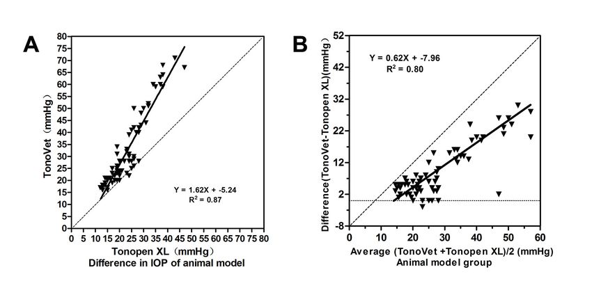
Molecular Vision 2019; 25:391-399                                              © 2019 Molecular Vision

and 8 kg) were purchased from Landao Biotechnology Co.,                    Identification of a chronic ocular hypertension monkey
Ltd. (Guangdong, China) and used to evaluate consistency                   model:
between the two tonometers. The monkeys were raised in                          Color photography—A color fundus photograph of 35°
large cages with adequate room for activities and fed nutri-               of the eye was obtained weekly in anesthetized animals in
tious food and water. The rooms were illuminated on a 12                   the prone position (with the body placed in the prone posi-
h:12 h light-dark cycle (daytime light intensity of approxi-               tion and the head then held and adjusted to a nearly upright
mately 200 lux) under controlled conditions, including                     position suitable for measurement) using a retinal camera
humidity (50–55%) and temperature (24–25 °C). The health                   (TRC-50DX Retinal Camera, Topcon, Tokyo, Japan) with a
of the monkeys was monitored daily by animal care staff                    Nikon 200 D digital camera. Surface topical anesthesia was
and veterinary personnel. All experimental procedures were                 applied after the eye was opened. The pupils were sufficiently
performed under deep general anesthesia via an intramus-                   dilated (≥8 mm) with a 0.25% tropicamide ophthalmic solu-
cular injection of ketamine hydrochloride (5 mg/kg, Ketalar                tion (Mydrin®, Santen Pharmaceutical, Osaka, Japan). The
50®, GuTian Pharmaceuticals Ltd., Fujian, China) plus chlor-               clarity of the corneas was preserved with an ocular lubri-
promazine hydrochloride (2.5 mg/kg, chlorpromazine 50®,                    cating agent (Artificial tears®, Zhongshan Ophthalmic Center,
JiaoZuo Pharmaceuticals Ltd., Tianjin, China).                             Guangdong, China) during the examination. The narrowed
                                                                           neuroretinal rim and the enlarged optic cup were observed
Establishment of a chronic ocular hypertension monkey
                                                                           in the animal model.
model: Eight out of ten monkeys underwent successful
induction of COHT (we randomly selected one eye in each                        Optical coherence tomography measurements of
animal for measurements). All of the animals were deeply                   RNFL thickness—Cross-sectional images of the RNFL
anesthetized. The body was placed in the prone position, and               were scanned weekly with a circular scan (3.4 mm diameter)
the head was then held and adjusted to a nearly upright posi-              procedure using a Stratus OCT Instrument (Model 3000, Carl
tion suitable for measurement. The important steps of this                 Zeiss Meditec, Dublin, CA). Deeply anesthetized animals
procedure are described below. Tests performed before laser                were immobilized using a board to maintain the spine’s
photocoagulation (to exclude existing ocular disease) included             posture and gently held in place by an operator. Surface
an IOP measurement, a slit-lamp examination, color fundus                  topical anesthesia was applied after the eye was opened.
photography, and optical coherence tomography (OCT) of                     The pupils were appropriately dilated, and the corneas were
the retinal nerve fiber layer (RNFL) thickness. The pupils                 kept clear using a method similar to that used during color
were sufficiently contracted (to 1 mm) with a 1% pilocarpine               photography measurements. The RNFL thickness was then
ophthalmic solution (Pilocarpine®; Zhongshan Ophthalmic                    examined in the animal model.
Center, Guangdong, China), and the corneas were anesthe-                   Central corneal thickness assessment: Anterior segment
tized with 0.5% proparacaine hydrochloride (Alcaine®, Alcon                optical coherence tomography (AS-OCT; Fourier OCT, Carl
Pharma, Belgium). The entire circumference of the trabecular               Zeiss Meditec, Dublin, CA) was used to measure the central
meshwork (TM) was ablated with the VISULAS Trion (Carl                     corneal thickness (CCT) before the first laser photocoagula-
Zeiss Meditec AG, Jena, Germany) using a slit-lamp delivery                tion to exclude the effect of corneal thickness the on IOP
system and laser gonioscope as previously described [16,17].               measurements in each animal. The posture of the spine was
The laser parameters were slightly modulated as follows:                   maintained in a manner similar to that used for color photog-
50 μm spot size, 0.5 s duration, 1,000 mW laser power, and                 raphy during the examination in all anesthetized animals.
150–250 spots. The laser treatments were performed repeat-                 Surface topical anesthesia and an ocular lubricating agent
edly to obtain a stable high IOP as previously described.                  were also used. The procedure was performed using a four-
Care was taken to indicate that the middle TM was photo-                   line scan model of the cornea. The line was centered on the
coagulated. The presence of a vapor bubble signified that                  corneal vertex, and the scans were performed as horizontal,
the TM was effectively ablated. Tobramycin dexamethasone                   vertical, 45°, and 135° scans. Particular attention was paid to
and tropicamide eye drops (Alcon Laboratories Inc.) were                   the eye position and scan lines through the central cornea.
used to alleviate any noninfectious inflammation during the                Measurement of intraocular pressure: IOP was measured in
immediate postlaser photocoagulation period. A single IOP                  ten healthy monkeys (one eye was randomly selected in each
measurement was obtained every week. If the IOP was not                    animal) and eight monkeys with COHT (eyes with high IOP)
consistently higher than 21 mmHg, additional laser photoco-                using the TonoVet® rebound tonometer (Finland, TV01) and
agulation was performed at 3-week intervals until stable IOP               the Tonopen XL® applanation tonometer (Reichert, Depew,
was obtained.                                                              NY) in compliance with the manufacturers’ recommended

                                                                       392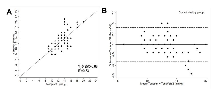
Molecular Vision 2019; 25:391-399                                             © 2019 Molecular Vision

procedures. All experimental monkeys were placed under                     IOP measurements. Bland–Altman plots constructed to assess
deep general anesthesia, but topical eye anesthetics were                  consistency for both tonometers. The deviations between the
avoided during this measurement. The animals were seated                   Tonopen applanation tonometer and the TonoVet rebound
and gently held in place at a desk by one operator while                   tonometer were analyzed with linear regression and two-
another experienced operator obtained all IOP measurements                 tailed Student t tests in the model. Data are expressed as the
with the two tonometers. Both tonometers were calibrated                   means ± standard deviation (SD). Deviation were considered
before each measurement. The TonoVet tonometer was                         statistically significant when the p value was less than or
used first and was followed by the Tonopen tonometer1 min                  equal to 0.05.
later. The reasoning behind this order was that the TonoVet
tonometer requires a smaller area of contact with the cornea;                                       RESULTS
however, measurement error can be reduced by recording the
                                                                           Corneal thickness and IOP measurements: The average CCT
measurements with both tonometers 1 min apart.
                                                                           values obtained in the ten healthy monkeys and the eight
     Due to the limited number of subjects, nine IOP measure-              monkeys with high IOP were 500.00 ± 10.95 μm and 496.25
ments were obtained in each animal; to obtain more digital                 ± 19.23 μm (mean ± SD), respectively. The correlation coef-
differential pressure data, these times were based on a 24 h               ficients between the IOPs obtained using the Tonopen and
pattern observed in IOP fluctuations [18]. According to our                TonoVet tonometers and the CCT values in the healthy control
previous study, the monkeys’ high IOP was sustained for 3–6                group were 0.15 (p = 0.66) and −0.30 (p = 0.37), respectively.
weeks [19]. Some problems, such as transient IOP increases                 Similarly, there was no statistically significant correlation
and corneal edema, were observed during the previous post-                 between the CCT and IOP values in the animal model group
laser photocoagulation period. To reduce measurement bias                  (Spearman correlation coefficient: −0.33 and −0.30, respec-
caused by these problems as much as possible, 24 h IOP levels              tively, and p = 0.42 and p = 0.47, respectively).
were measured after the high IOP and ocular conditions were
                                                                           Comparison of Tonopen and TonoVet measurements in
stabilized. To maintain a high IOP, every monkey underwent
                                                                           healthy monkeys: The average IOP measurements obtained
multiple laser procedures, and some monkeys experienced
                                                                           using the TonoVet and Tonopen devices were 12.99 ±
severe spikes in IOP. Extra laser photocoagulation was not
                                                                           3.04 mmHg (range, 8 to 21 mmHg) and 12.92 ± 2.31 mmHg
performed in the COHT animal models once a stable high
                                                                           (range, 6 to 20 mmHg), respectively. There was no statis-
IOP was achieved, and laser-induced ocular noninfectious
                                                                           tically significant difference between the measurements
inflammation and corneal edema then gradually disappeared.
                                                                           obtained for the average IOP between the two instruments
The fifth week after the first photocoagulation was chosen
                                                                           (two-tailed Student t test: 0.06 ± 2.08 mmHg, p = 0.761). The
as the time point for IOP measurement; at this time, single
                                                                           Tonopen and TonoVet readings were strongly and linearly
IOP measurements had gradually dropped to approximately
                                                                           correlated in the normal IOP monkey eyes, as shown in
21 mmHg, and different IOP levels were observed during the
                                                                           Figure 1A. The Bland–Altman analysis with 95% limit of
24 h span because of fluctuations in the model animals’ IOP.
                                                                           agreements (LoAs) is shown in Figure 1B. The 95% LoA
The 24 h IOP measurement points were obtained at 1:30, 3:30,
                                                                           width was between −4.13 and 4.00 mmHg. Table 1 indicate
6:30, 9:30, 12:30, 15:30, 18:30, 21:30, and 23:30, because of
                                                                           that there were small deviations in IOP for both tonometers.
the general anesthesia used on the monkeys [20].
                                                                           Furthermore, 56.67% of the deviations were concentrated
    According to the individual properties of both tonom-                  between −1.00 and +1.00 mmHg, and 91.12% were located
eters, six consecutive IOP readings were obtained using                    between −3.00 and +3.00 mmHg. There was good consistency
the TonoVet rebound tonometer, and ten consecutive IOP                     in the IOP measurements obtained using both tonometers in
readings by the Tonopen tonometer were acquired for each                   healthy monkeys with normal IOPs.
measurement. The average IOP value of each tonometer was
                                                                           Comparison of Tonopen and TonoVet measurements obtained
automatically calculated for each measurement. An average
                                                                           in monkeys with COHT: A COHT monkey model was estab-
IOP value for several measurements obtained at each time
                                                                           lished according to the ocular manifestations (Figure 2). The
point was used for the statistical analyses. Only consecutive
                                                                           average IOP measurements obtained using the Tonopen and
readings that showed little value variation (
Molecular Vision 2019; 25:391-399 © 2019 Molecular Vision
Table 1. Evaluation of the consistency between the Tonopen and
the TonoVet measurement in control healthy monkeys.
IOP ≤21 mmHg (n=90 readings)
Tonometers
Tonopen TonoVet
Mean ± SD 12.92±2.31 12.99±3.04
Range 6-20 8-21
Δ Mean ± SD 0.06±2.08
Δ Range -5-7
95% CI -4.13 – 4.00
Δ < ±1 mmHg* 56.67%
Δ < ±3 mmHg† 91.12%
P 0.761
Δ Mean ± SD: mean and standard deviation of difference (TonoVet minus Tonopen) * IOP difference
smaller than ± 1 mmHg † IOP difference smaller than ± 3 mmHg
1.62X + −5.24), as shown in Figure 3A. The mean deviation 7.0–16 mmHg), 22 were placed in the medium-pressure
in IOP measurements between the TonoVet and Tonopen group (range, 17–22 mmHg), and 37 were placed in the high-
devices was calculated as the TonoVet reading minus the pressure group (range, 23–60 mmHg). In the low-pressure
Tonopen reading, and was found to be 9.39 ± 8.22 mmHg group (IOP: 7–16 mmHg), the IOP readings deviated by
(range, −1.00 to +30.0 mmHg). The same strong linear regres- 4.46 ± 1.66 mmHg, as shown in Figure 4A; 23.08% of the
sion was observed in the mean IOP deviations between the IOP measurements acquired using the TonoVet tonometer
two tonometers (linear regression: Y = 0.62X + −7.96), with deviated by ± 5.00 mmHg from the Tonopen measurements,
higher IOP values associated with larger deviations between and this difference was statistically significant (two-tailed
the two tonometer measurements (Figure 3B). Student t test: pMolecular Vision 2019; 25:391-399 © 2019 Molecular Vision
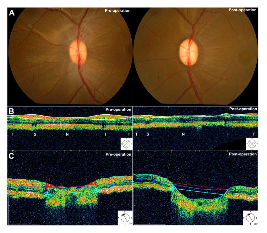
Figure 2. A monkey model of chronic ocular hypertension was established. A: Fundus: The enlarged optic cup. B: Optical coherence
tomography (OCT): The damaged retinal nerve fiber layer. C: OCT: The enlarged optic cup.
the deviation in IOP readings between the two devices was IOP [9-12]. However, under high IOP conditions, not enough
13.76 ± 9.19 mmHg, and 75.68% of the measurements devi- evidence is available to support the notion that measure-
ated by ± 5.00 mmHg; indicating instrumental systemic error ments are consistent between the two tonometers. The most
(two-tailed Student t test: p< 0.005) (Table 2 and Figure 4C). important finding of this study is that the TonoVet measure-
ments were consistent with the Tonopen measurements in
DISCUSSION monkey eyes with normal IOP; however, as the IOP gradually
increased, the readings produced by the TonoVet tonometer
The TonoVet rebound tonometer and the Tonopen applana-
tion tonometer are the most commonly used IOP measuring were larger than those produced by the Tonopen tonometer
instruments in experimental research studies. At present, Additionally, there was a quantitative relation between the
most published studies acknowledge that there is some level tonometer measurements when the deviations and the IOP
of consistency between these devices in eyes with normal levels were investigated.
395Molecular Vision 2019; 25:391-399 © 2019 Molecular Vision
Figure 3. Consistency analysis in
COHT monkey models. A: Regres-
sion analysis between TonoVet
rebound and Tonopen applanation
tonometer readings (n= 72 read-
ings); B: Deviations in IOP values
(calculated as TonoVet minus
Tonopen) are shown plotted against
the average IOP ((TonoVet plus
Tonopen)/2). The solid line repre-
sents the regression line, and the
dotted lines represent 95% confi-
dence interval limits. Duplicates
were distributed in the graph.
We reviewed previously published clinical papers that normal IOP, with 56.67% of the deviations between −1 and
explored consistency in IOP measurements. Munkwitz +1 mmHg, and 91.12% of the deviations below 3 mmHg (p
compared the rebound tonometer and the GAT in 75 eyes = 0.761). In the normal IOP group, therefore, the range of
of 75 patients, and reported that the deviations in IOP deviations was “acceptable.” Subsequently, the deviations
values were similar between these two tonometers (range, in measurements obtained using the TonoVet and Tonopen
7.0–22 mmHg). However, the deviations were almost twice devices were investigated in animals with different IOP levels.
as large in the higher IOP range (23–60 mmHg), where they
When the IOP was above 23 mmHg, 75.68% of the TonoVet
deviated by 12.56 ± 11.98 mmHg [14]. Martinez-de-la-Casa
measurements deviated by ± 5.00 mmHg from the Tonopen
also found that compared to the GAT, the rebound tonometer
measurements, and the deviation between the tonometers
tended to overestimate IOPs, and the deviation between the
reached as high as 13.76 ± 9.19 mmHg. The standard devia-
two instruments reached as high as 7.7 mmHg in eyes with
high IOP [15]. These reports led us to question the consis- tion for the TonoVet measurements was higher for eyes in the
tency between TonoVet and Tonopen measurements. In this high IOP range (23–60 mmHg). These results suggest that in
study, we first compared measurements obtained using both monkeys with normal IOP, the IOP readings obtained using
handheld devices in a healthy control group. As previously the TonoVet tonometer agreed with those obtained using the
reported [22], in monkeys, the IOP readings were similar Tonopen device, whereas the IOP readings were not always
between the Tonopen and TonoVet tonometers in eyes with in agreement in the high IOP range.
Figure 4. Consistency between TonoVet and Tonopen measurements was evaluated using the Bland–Altman analysis in animals with different
IOP levels in the COHT monkey group. The averages are shown plotted against the deviations. Higher intraocular pressure (IOP) values
were associated with worse consistency for both tonometers. The solid line represents the average deviation, and the dotted lines indicate
the 95% limits of the confidence intervals.
396Molecular Vision 2019; 25:391-399 © 2019 Molecular Vision
Thus far, few publications have explored the consis- dropped to approximately 21 mmHg, there were large fluc-
tency in evaluations obtained using these two tonometers in tuations in the 24 h IOP values. Thus, different IOP levels
monkeys with chronic high IOP over a wide range of IOP were obtained to guarantee proper data analysis. Moreover,
values. Some researchers have reported that these two conve- laser-induced transient IOP increases and corneal edema were
nient handheld tonometers have excellent consistency when avoided, and did not influence the IOP measurements. The
used in monkey eyes with normal IOP and in monkey eyes total number of IOP readings was in line with the statistical
with regulated IOP in which IOP was adjusted by modifying sample size estimation.
the height of a connected perfusate reservoir [12,23,24].
The GAT is the international gold standard for IOP
The novel finding presented in this study appears to be the
measurement. It is difficult for monkey to complete the GAT
comparison of monkeys with chronic experimentally induced
measurement because the position of the monkey’s eye (a mild
high IOP. These models closely mimic open angle glaucoma,
upshift) causes the corneal center to shift under general anes-
in which IOP is gradually and chronically elevated, and
thesia. It cause the two semicircles of the flattened corneal
does not sharply increase. The animal model used in this
surface to be unequal or, in some cases, two semicircles do
study presented with stable high IOP and an anterior eye
not form, affecting IOP measurements. Although it is regret-
segment condition. This model allowed us to avoid acute
table that the GAT could not be used as a control, in this study,
corneal edema, anterior chamber inflammation, and ciliary
we focused on the comparison between the TonoVet rebound
body dysfunction, which can be caused by acute high IOP.
tonometer and the Tonopen applanation tonometer, as these
It has been suggested that a laser-induced COHT monkey
two tonometers are the instruments most commonly used to
model could be useful for comparing deviations in tonometer
measure IOP in experimental research studies. It remains to
measurements.
be determined which tonometer is the most precise and reli-
Additionally, because the number of animals was limited, able. The quantitative relation between the IOP levels and the
and it is difficult to establish a chronic high IOP model, we deviations observed for both IOP measurement apparatuses
chose to use 24 h IOP measurements as a solution, to obtain was investigated. The present results could play a role in glau-
more differential digital pressure data for this experimental coma studies that use experimental animals. Additionally, the
study, although this decision represents a f law. To our Tonopen and TonoVet tonometers need only a small area of
knowledge, higher IOP values are associated with higher IOP contact with the cornea; the fact that it is not necessary to
fluctuations. There is some evidence indicating that monkeys form a semicircular ring to obtain an IOP value during the
exhibit large variations in IOP values from day to day and measurement process represents an obvious advantage for
hour to hour, especially under high IOP conditions [18,25]. measuring IOP in animals. The TonoVet tonometer might be
Thus, in this study, we used 18 monkeys (ten with normal IOP more useful in small animals that have normal or slightly
and eight with COHT) to measure 24 h IOP values (nine times high IOP, such as rats and other animals used to study corneal
for each eye). The 24 h IOP measurements were measured disease, because this device requires only a small contact
during the 15th week after the first photocoagulation. At that area. The Tonopen tonometer might be more suitable than the
time, although the single IOP measurements had gradually TonoVet tonometer in cases in which moderate to advanced
Table 2. Evaluation of the consistency between the Tonopen and the TonoVet measurement in COHT monkey models.
IOP 7–16 mmHg IOP 17–22 mmHg IOP 23–60 mmHg
Tonometers (n=13 readings) (n=22 readings) (n=37 readings)
Tonopen TonoVet Tonopen TonoVet Tonopen TonoVet
Mean ± SD 14.15 18.62 19.23 24.18 29.3 43.05
± 1.14 ± 1.98 ± 1.31 ± 3.62 ± 6.12 ± 14.29
Range 12-16 16 - 21 17 - 22 19 - 34 23 - 47 22 - 71
Δ Mean ± SD 4.46±1.66 4.95±3.57 13.76±9.19
Δ Range 1-7 0 - 15 -32
95% CI 3.46 - 5.47 3.37 - 6.54 10.69 - 16.82
Δ > ±5 mmHg* 23.08% 36.36% 75.68%
P 0 0 0.003
Δ Mean ± SD: mean and standard deviation of difference (TonoVet minus Tonopen) * IOP difference bigger than ± 5 mmHg
397Molecular Vision 2019; 25:391-399 © 2019 Molecular Vision
high IOP values must be measured, because the Tonopen 4. European Glaucoma Society. Terminology and Guidelines for
tonometer has a more stable standard deviation. Glaucoma. 4th ed. European Union, EU; 2014. http://www.
eugs.org/eng/SIGhedefinitions.asp; 2014 Accessed.
The use of anesthetized animals as experimental subjects 5. Goldmann H, Schmidt T. [Applanation tonometry]. Ophthal-
is one flaw of this study. In a clinical setting, IOP is likely to mologica Journal international d’ophtalmologie International
be influenced by anesthetics and anesthetic drugs [26-28]. In journal of ophthalmology Z Augenheilkd 1957; 134:221-42.
addition, anesthesia causes IOP to increase in rabbits, cats, Epub 1957/10/01
and Syrian hamsters [29-31]. However, no previous study 6. Wessels IF, Oh Y. Tonometer utilization, accuracy, and cali-
explored how anesthesia acts on IOP in nonhuman primates. bration under field conditions. Arch Ophthalmol 1990;
Anesthesia is difficult to avoid in animal research. In this 108:1709-12. [PMID: 2256841].
study, IOP measurements were obtained in anesthetized 7. Kontiola AI. A new induction-based impact method for
monkeys without topical anesthesia. The animals were measuring intraocular pressure. Acta Ophthalmol Scand
2000; 78:142-5. [PMID: 10794245].
reanesthetized before each IOP measurement obtained during
the study. Thus, all of the IOP measurement results were 8. Kontiola A, Puska P. Measuring intraocular pressure with the
Pulsair 3000 and Rebound tonometers in elderly patients
affected by anesthesia, and it may have had a small effect on
without an anesthetic. Graefe’s archive for clinical and
the conclusions regarding the IOP measurements obtained in experimental ophthalmology = Albrecht Von Graefes Arch
this study. Klin Exp Ophthalmol 2004; 242:3-7. Epub 2003/11/25
In summary, the results of this study suggest that TonoVet 9. Goldblum D, Kontiola AI, Mittag T, Chen B, Danias J. Non-
invasive determination of intraocular pressure in the rat
measurements are consistent with Tonopen measurements
eye. Comparison of an electronic tonometer (TonoPen), and
for normal IOP samples. However, in monkey models with a rebound (impact probe) tonometer. Graefe’s archive for
chronic high IOP, inconsistencies were observed between clinical and experimental ophthalmology = Albrecht Von
results obtained using these two tonometers. We found that Graefes Arch Klin Exp Ophthalmol 2002; 240:942-6. Epub
higher IOP values could be associated with larger measure- 2002/12/18[PMID: 12486518].
ment error. Therefore, it is necessary to be aware of the 10. Leiva M, Naranjo C, Pena MT. Comparison of the rebound
systemic error that can be observed in IOP measurements, tonometer (ICare) to the applanation tonometer (Tonopen
and the deviations among different tonometers, to facilitate XL) in normotensive dogs. Vet Ophthalmol 2006; 9:17-21.
[PMID: 16409240].
accurate individualized diagnoses and prevent the utilization
of misleading IOP values. 11. Wang WH, Millar JC, Pang IH, Wax MB, Clark AF. Nonin-
vasive measurement of rodent intraocular pressure with
a rebound tonometer. Invest Ophthalmol Vis Sci 2005;
ACKNOWLEDGMENTS 46:4617-21. [PMID: 16303957].
12. . Yu W. Cao G, Qiu J, Liu X, Ma J, Li N, Yu M, Yan N, Chen
This study was supported by the National Natural Science
L, Pang IH. Evaluation of monkey intraocular pressure by
Foundation of China (grant number: 81430009) and the rebound tonometer. Mol Vis 2009; 15:2196-201. [PMID:
Medical Scientific Research Foundation of Guangdong Prov- 19898690].
ince, China (grant number: A2018070). 13. . García-Resúa C. González-Meijome JM, Gilino J, Yebra-
Pimentel E. Accuracy of the new ICare rebound tonometer
vs. other portable tonometers in healthy eyes. Optom Vis Sci
REFERENCES 2006; 83:102-7. [PMID: 16501412].
14. Munkwitz S, Elkarmouty A, Hoffmann EM, Pfeiffer N,
1. Grodum K, Heijl A, Bengtsson B. A comparison of glaucoma
Thieme H. Comparison of the iCare rebound tonometer and
patients identified through mass screening and in routine
the Goldmann applanation tonometer over a wide IOP range.
clinical practice. Acta Ophthalmol Scand 2002; 80:627-31.
Graefe’s archive for clinical and experimental ophthalmology
[PMID: 12485284].
= Albrecht Von Graefes Arch Klin Exp Ophthalmol 2008;
2. Kass MA, Gordon MO. Intraocular pressure and visual field 246:875-9. .
progression in open-angle glaucoma. Am J Ophthalmol 15. Martinez-de-la-Casa JM, Garcia-Feijoo J, Castillo A, Garcia-
2000; 130:490-1. [PMID: 11024422]. Sanchez J. Reproducibility and clinical evaluation of rebound
tonometry. Invest Ophthalmol Vis Sci 2005; 46:4578-80.
3. Heijl A, Leske MC, Bengtsson B, Hyman L, Bengtsson B,
[PMID: 16303951].
Hussein M. Early Manifest Glaucoma Trial Group. Reduc-
tion of intraocular pressure and glaucoma progression: results 16. Gaasterland D, Kupfer C. Experimental glaucoma in the
from the Early Manifest Glaucoma Trial. Arch Ophthalmol rhesus monkey. Invest Ophthalmol 1974; 13:455-7. [PMID:
2002; 120:1268-79. [PMID: 12365904]. 4208801].
398Molecular Vision 2019; 25:391-399 © 2019 Molecular Vision
17. Quigley HA, Hohman RM. Laser energy levels for trabecular Tono-Pen versus manometry. Invest Ophthalmol Vis Sci
meshwork damage in the primate eye. Invest Ophthalmol Vis 1996; 37:1197-9. [PMID: 8631634].
Sci 1983; 24:1305-7. [PMID: 6885314]. 25. Ollivier FJ, Brooks DE, Kallberg ME, Sapp HL, Komáromy
18. Downs JC, Burgoyne CF, Seigfreid WP, Reynaud JF, AM, Stevens GR, Dawson WW, Sherwood MB, Lambrou
Strouthidis NG, Sallee V. 24-hour IOP telemetry in the GN. Time-specific intraocular pressure curves in Rhesus
nonhuman primate: implant system performance and initial macaques (Macaca mulatta) with laser-induced ocular hyper-
characterization of IOP at multiple timescales. Invest tension. Vet Ophthalmol 2004; 7:23-7. [PMID: 14738503].
Ophthalmol Vis Sci 2011; 52:7365-75. [PMID: 21791586]. 26. Jantzen JP, Kleemann PP. Klin Monatsbl Augenheilkd
19. Shu T, Kang L, Huang J. Patterns of optic nerve head and 1988; 193:1-7. Effect of muscle relaxants on intraocular
retinal nerve fiber layer damage in the monkey chronic pressure[PMID: 2972873].
ocular hypertension model. Chinese Journal of Optometry 27. Duncalf D, Foldes FF. Effect of anesthetic drugs and muscle
Ophthalmology and Visual Science. 2014; 16:436-40. . relaxants on intraocular pressure. Int Ophthalmol Clin 1973;
20. Deokule SP, Doshi A, Vizzeri G, Medeiros FA, Liu JH, Bowd 13:21-33. [PMID: 4274097].
C, Zangwill L, Weinreb RN. Relationship of the 24-hour 28. van Aken H, Scherer R, Lawin P. Anasth Intensivther
pattern of intraocular pressure with optic disc appearance Notfallmed 1980; 15:293-302. Anaesthesia and intraocular
in primary open-angle glaucoma. Ophthalmology 2009; pressure (author's transl)[PMID: 7416447].
116:833-9. [PMID: 19195707].
29. Hahnenberger RW. Influence of various anesthetic drugs on
21. ISO 8612. Ophthalmic instruments-Tonometers. https://www. the intraocular pressure of cats. Albrecht Von Graefes Arch
iso.org/obp/ui/#iso:std:iso:8612:ed-2:v1:en; 2009 Accessed. Klin Exp Ophthalmol 1976; 199:179-86. [PMID: 1083696].
22. Rusanen E, Florin M, Hassig M, Spiess BM. Evaluation of a 30. Rajaei SM, Mood MA, Paryani MR, Williams DL. Effects of
rebound tonometer (Tonovet) in clinically normal cat eyes. diurnal variation and anesthetic agents on intraocular pres-
Vet Ophthalmol 2010; 13:31-6. [PMID: 20149173]. sure in Syrian hamsters (Mesocricetus auratus). Am J Vet
23. Elsmo EJ, Kiland JA, Kaufman PL, McLellan GJ. Evaluation Res 2017; 78:85-9. [PMID: 28029289].
of rebound tonometry in non-human primates. Exp Eye Res 31. Schutten WH, Van Horn DL. The effects of ketamine sedation
2011; 92:268-73. [PMID: 21315069]. and ketamine-pentobarbital anesthesia upon the intraocular
24. Peterson JA, Kiland JA, Croft MA, Kaufman PL. Intra- pressure of the rabbit. Invest Ophthalmol Vis Sci 1977;
ocular pressure measurement in cynomolgus monkeys. 16:531-4. [PMID: 863613].
Articles are provided courtesy of Emory University and the Zhongshan Ophthalmic Center, Sun Yat-sen University, P.R. China.
The print version of this article was created on 3 August 2019. This reflects all typographical corrections and errata to the
article through that date. Details of any changes may be found in the online version of the article.
399You can also read