RV-GAN: Segmenting Retinal Vascular Structure in Fundus Photographs using a Novel Multi-scale Generative Adversarial Network
←
→
Page content transcription
If your browser does not render page correctly, please read the page content below
RV-GAN: Segmenting Retinal Vascular Structure in
                                                  Fundus Photographs using a Novel Multi-scale
                                                       Generative Adversarial Network

                                               Sharif Amit Kamran1 , Khondker Fariha Hossain1 , Alireza Tavakkoli1 , Stewart Lee
                                                           Zuckerbrod2 , Kenton M. Sanders3 , and Salah A. Baker3
arXiv:2101.00535v2 [eess.IV] 14 May 2021

                                                  1
                                                      Dept. of Computer Science & Engineering, University of Nevada, Reno, NV, USA
                                                                          2
                                                                            Houston Eye Associates, TX, USA
                                                               3
                                                                 School of Medicine, University of Nevada, Reno, NV, USA

                                                      Abstract. High fidelity segmentation of both macro and microvascular structure
                                                      of the retina plays a pivotal role in determining degenerative retinal diseases, yet
                                                      it is a difficult problem. Due to successive resolution loss in the encoding phase
                                                      combined with the inability to recover this lost information in the decoding phase,
                                                      autoencoding based segmentation approaches are limited in their ability to extract
                                                      retinal microvascular structure. We propose RV-GAN, a new multi-scale gener-
                                                      ative architecture for accurate retinal vessel segmentation to alleviate this. The
                                                      proposed architecture uses two generators and two multi-scale autoencoding dis-
                                                      criminators for better microvessel localization and segmentation. In order to avoid
                                                      the loss of fidelity suffered by traditional GAN-based segmentation systems, we
                                                      introduce a novel weighted feature matching loss. This new loss incorporates and
                                                      prioritizes features from the discriminator’s decoder over the encoder. Doing so
                                                      combined with the fact that the discriminator’s decoder attempts to determine
                                                      real or fake images at the pixel level better preserves macro and microvascular
                                                      structure. By combining reconstruction and weighted feature matching loss, the
                                                      proposed architecture achieves an area under the curve (AUC) of 0.9887, 0.9914,
                                                      and 0.9887 in pixel-wise segmentation of retinal vasculature from three publicly
                                                      available datasets, namely DRIVE, CHASE-DB1, and STARE, respectively. Ad-
                                                      ditionally, RV-GAN outperforms other architectures in two additional relevant
                                                      metrics, mean intersection-over-union (Mean-IOU) and structural similarity mea-
                                                      sure (SSIM).

                                                      Keywords: Retinal Vessel Segmentation · Generative Networks · Medical Imag-
                                                      ing · Opthalmology · Retinal Fundus

                                           1     Introduction
                                           The fundoscopic exam is a procedure that provides necessary information to diagnose
                                           different retinal degenerative diseases such as Diabetic Retinopathy, Macular Edema,
                                           Cytomegalovirus Retinitis[27]. A highly accurate system is required to segment retinal
                                           vessels and find abnormalities in the retinal subspace to diagnose these vascular dis-
                                           eases. Many image processing and machine learning-based approaches for retinal ves-
                                           sel segmentation have so far been proposed [26,7,23,13]. However, such methods fail to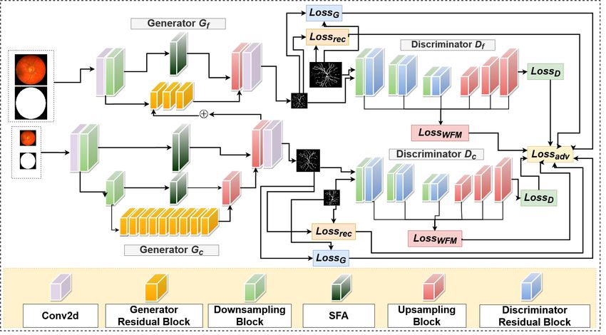
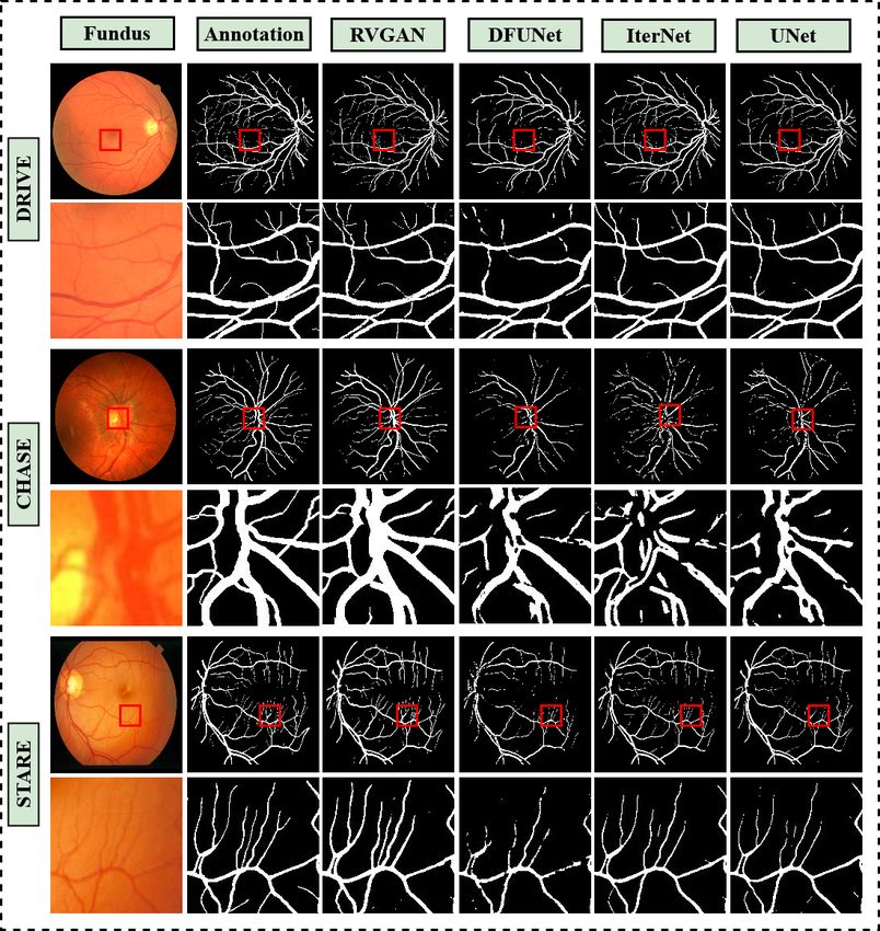
2       Kamran et al.

Fig. 1: RV-GAN segments vessel with better precision than other architectures. The 1st
row is the whole image, while 2nd row is specific zoomed-in area of the image. The Red
bounded box signifies the zoomed-in region. Here, the confidence score is, t > 0.5. The
row contains DRIVE, CHASE-DB1 and STARE data-set. Whereas the column contains
fundus images, ground-truths and segmentation maps for RV-GAN, DFUNet, IterNet
and UNet.

precisely pixel-wise segment blood vessels due to insufficient illumination and periodic
noises. Attributes like this present in the subspace can create false-positive segmen-
tation [7]. In recent times, UNet based deep learning architectures have become very
popular for retinal vessel segmentation. UNet consists of an encoder to capture con-
text information and a decoder for enabling precise localization[24]. Many derivative
works based on UNet have been proposed, such as Dense-UNet, Deformable UNet[10],
IterNet [16] etc. These models were able to achieve quite good results for macro ves-
sel segmentation. However, these architectures fail when segmenting microvessels with
higher certainty. One reason is successive resolution loss in the encoder, and failure
to capture those features in the decoder results in inferior microvessel segmentation.
Recent GAN-based architecture [21,32] tries to address this by incorporating discrim-
inative features from adversarial examples while training. However, the discriminator
being an encoder[9], only trains on patches of images rather than pixels, affecting the
true-positive-rate of the model. We need an architecture that can retain discriminative
manifold features and segment microvessels on pixel-level with higher confidence. Con-
RV-GAN          3

Fig. 2: RV-GAN consisting of Coarse and Fine generators Gf , Gc and discrimina-
tors Df , Dc . The generators incorporates reconstruction loss, Lossrec and Hinge loss
LossG . Whereas the discriminators uses weighted feature matching loss, Losswf m and
Hinge loss LossD . All of these losses are multiplied by weight multiplier and then
added in the final adversarial loss, Lossadv . The generators consists of Downsampling,
Upsampling, SFA and its distinct residual blocks. On the other hand, the discriminators
consists of Downsampling, Upsampling and counterpart residual blocks.

fidence signifies the probability distribution function of the segmented pixel falling un-
der vessel or background. By taking all of these into account, we propose Retinal-Vessel
GAN, consisting of coarse and fine generators and multi-scale autoencoder-based dis-
criminators for producing highly accurate segmentation of blood vessel with strong
confidence scores. Additionally, we come up with a new weighted feature matching
loss with inner and outer weights. And we combine it with reconstruction and hinge
loss for adversarial training of our architecture. From Fig. 1, it is apparent that our
architecture produces a segmentation map with a high confidence score.

2     Proposed Methodology
2.1   Multi-scale Generators
Pairing multi-scale coarse and fine generators produces high-quality domain-specific
retinal image synthesis, as observed in recent generative networks, such as Fundus2Angio
[12], V-GAN [27] and [29]. Inspired by this, we also adopt this feature in our architec-
ture by using two generators (Gf ine and Gcoarse ), as visualized in Fig. 2. The gen-
erator Gf ine synthesizes fine-grained vessel segmentation images by extracting local
features such as micro branches, connections, blockages, etc. In contrast, the genera-
tor Gcoarse tries to learn and conserve global information, such as the structures of the
maco branches, while producing less detailed microvessel segmentation. The detailed
structure of these generators is illustrated in Fig. 2.
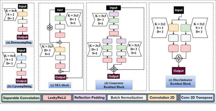
4       Kamran et al.

Fig. 3: Proposed Downsampling, Upsampling, Spatial Feature Aggregation block, Gen-
erator and Discriminator Residual blocks. Here, K=Kernel size, S=Stride, D=Dilation.

2.2   Residual Downsampling and Upsampling Blocks

Our generators and discriminators consist of both downsampling and upsampling blocks
to get the desired feature maps and output. The downsampling block comprises of a con-
volution layer, a batch-norm layer and a Leaky-ReLU activation function consecutively
and is illustrated in Fig. 3(a). Contrarily, the decoder block consists of a transposed
convolution layer, batch-norm, and Leaky-ReLU activation layer successively and can
be visualized in Fig.3(b).

2.3   Distinct Identity Blocks for Generator & Discriminator

For spatial and depth feature propagation, residual identity blocks have become go-
to building blocks for image style transfer, image inpainting, and image segmentation
tasks [25,30,4,3,22].Vanilla convolution layers are both computationally inefficient and
fail to retain accurate spatial and depth information, as opposed to separable convolu-
tion [5]. Separable convolution comprises of a depth-wise convolution and a point-wise
convolution successively. As a result, it extracts and preserves depth and spatial features
while forward propagating the network. Recent advancement in retinal image classifi-
cation has shown that combining separable convolutional layers with dilation allows for
more robust feature extraction [11]. We design two unique residual identity blocks, for
our generators and discriminators, as illustrated in Fig. 3(d) & Fig. 3(e).

2.4   Spatial Feature Aggregation

In this section, we discuss our proposed spatial feature aggregation (SFA) block, as
illustrated in Fig. 3(c). We use spatial feature aggregation block for combining spatial
RV-GAN          5

and depth features from the bottom layers of the network with the top layers of the
network, as illustrated in Fig. 2. The rationale behind employing the SFA block is to
extract and retain spatial and depth information, that is otherwise lost in deep networks.
Consequently, these features can be combined with the learned features of the deeper
layers of the network to get an accurate approximation, as observed in similar GAN
architectures. [33,2].

2.5   Auto-encoder as Discriminators
For better pixel-wise segmentation, we need an architecture that can extract both global
and local features from the image. To mitigate this underlying problem, we need a deep
and dense architecture with lots of computable parameters. It, in turn, might lead to
overfitting or vanishing gradient while training the model. To address this issue, rather
than having a single dense segmentation architecture, we adopt light-weight discrim-
inators as autoencoders. Additionally, we use multi-scale discriminators for both our
coarse and fine generators, as previously proposed in [15,30]. The arrangement consists
of two discriminators with variable sized input and can help with the overall adversarial
training.We define two discriminators as Df and Dc as illustrated in Fig. 2.

2.6   Proposed Weighted Feature Matching Loss
Feature matching loss [30] was incorporated by extracting features from discriminators
to do semantic segmentation. The feature-matching loss is given in Eq. 1 As the authors
used Patch-GAN as a discriminator, it only contains an encoding module. By contrast,
our work involves finding pixel-wise segmentation of retinal vessel and background and
thus requires an additional decoder. By successive downsampling and upsampling, we
lose essential spatial information and features; that’s why we need to give weightage
to different components in the overall architecture. We propose a new weighted feature
matching loss, as given in Eq. 2 that combines elements from both encoder and decoder
and prioritizes particular features to overcome this. For our case, we experiment and see
that giving more weightage to decoder feature map results in better vessel segmentation.
                                        k
                                     1 X     i             i
            Lf m (G, Denc ) = Ex,y         kDenc (x, y) − Denc (x, G(x))k             (1)
                                     N i=1

                             k
                          1 X i
  Lwf m (G, Dn ) = Ex,y        λ kDi (x, y)−Denc
                                             i
                                                 (x, G(x))k+λidec kDdec
                                                                    i
                                                                        (x, y)
                          N i=1 enc enc
                                                                    i
                                                                 − Ddec (x, G(x))k    (2)
Eq. 2 is calculated by taking the features from each of the downsampling blocks and
upsampling blocks of the discriminator’s encoder and decoder. We insert the real and the
synthesized segmentation maps consecutively. The N signifies the number of features.
Here, λenc and λdec is the inner weight multiplier for each of the extracted feature maps.
The weight values are between [0, 1], and the total sum of the weight is 1, and we use a
higher weight value for the decoder feature maps than the encoder feature maps.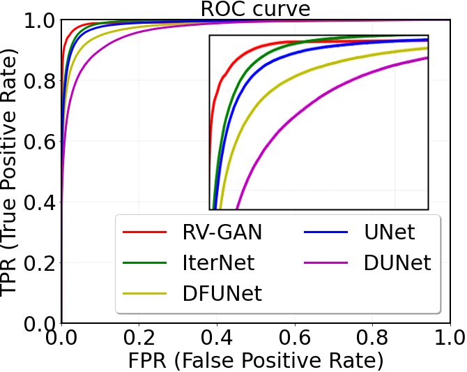
6           Kamran et al.

2.7    Weighted Objective and Adversarial Loss
For adversarial training, we use Hinge-Loss [33,18] as given in Eq. 3 and Eq. 4. Con-
clusively, all the fundus images and their corresponding segmentation map pairs are
normalized, to [−1, 1]. As a result, it broadens the difference between the pixel intensi-
ties of the real and synthesized segmentation maps. In Eq. 5, we multiply Ladv (G) with
λadv as weight multiplier. Next, we add Ladv (D) with the output of the multiplication.

  Ladv (D) = −Ex,y min(0, −1 + D(x, y)) − Ex min(0, −1 − D(x, G(x))) (3)

                           Ladv (G) = −Ex,y (D(G(x), y))                               (4)
                            Ladv (G, D) = Ladv (D) + λadv (Ladv (G))                  (5)
In Eq. 4 and Eq. 5, we first train the discriminators on the real fundus, x and real
segmentation map, y. After that, we train with the real fundus, x, and synthesized seg-
mentation map, G(x). We begin by batch-wise training the discriminators Df , and Dc
for a couple of iterations on the training data. Following that, we train the Gc while
keeping the weights of the discriminators frozen. In a similar fashion, we train Gf on a
batch training image while keeping weights of all the discriminators frozen.
    The generators also incorporate the reconstruction loss (Mean Squared Error) as
shown in Eq. 6. By utilizing the loss we ensure the synthesized images contain more
realistic microvessel, arteries, and vascular structure.
                                  Lrec (G) = Ex,y kG(x) − yk2                         (6)
    By incorporating Eq. 2, 5 and 6, we can formulate our final objective function as
given in Eq. 7.

      min      max (Ladv (Gf , Gc , Df , Dc ))+
    Gf ,Gc    Df ,Dc

                       λrec Lrec (Gf , Gc ) + λwf m Lwf m (Gf , Gc , Df , Dc )        (7)
Here, λadv , λrec , and λwf m implies different weights, that is multiplied with their re-
spective losses. The loss weighting decides which architecture to prioritize while train-
ing. For our system, more weights are given to the Ladv (G), Lrec , Lwf m , and thus we
select bigger λ values for those.

3     Experiments
3.1    Dataset
For benchmarking, we use three retinal segmentation datasets, namely, DRIVE [28],
CHASE-DB1 [20], and STARE [8]. The images are respectively in .tif (565 × 584),
.jpg(999 × 960), and .ppm(700 × 605). We train three different RV-GAN networks
with each of these datasets using 5-fold cross-validation. We use overlapping image
patches with a stride of 32 and an image size of 128 × 128 for training and validation.
So we end up having 4320 for STARE, 15120 for CHASE-DB1, and 4200 for DRIVE
from 20, 20, and 16 images. DRIVE dataset comes with official FoV masks for the test
images. For CHASE-DB1 and STARE dataset, we also generate FoV masks similar to
Li et al [16]. For testing, overlapping image patches with a stride of 3 were extracted
and averaged by taking 20, 8 and 4 images from DRIVE, CHASE-DB1, and STARE.
RV-GAN 7
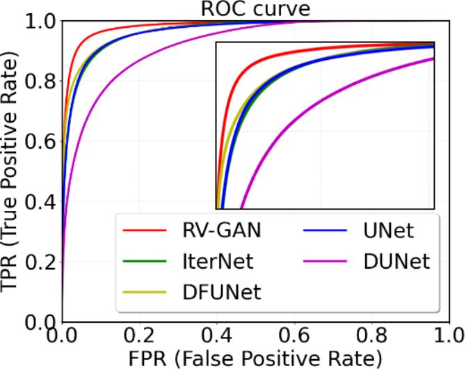
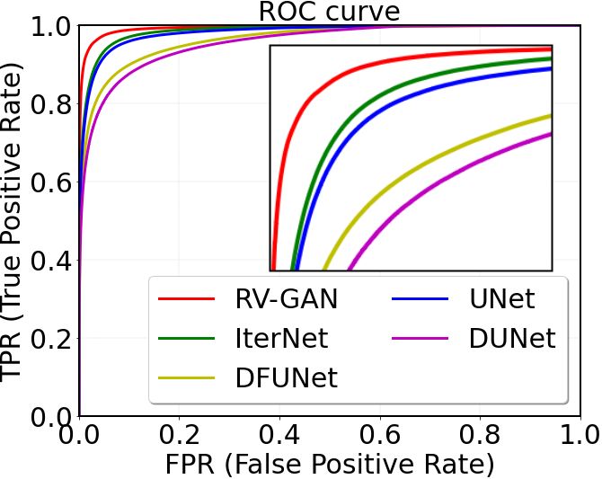
(a) DRIVE (b) STARE (c) CHASE-DB1
Fig. 4: ROC Curves on (a) DRIVE (b) STARE (c) CHASE-DB1.
3.2 Hyper-parameter Initialization
For adversarial training, we used hinge loss [33,18]. We picked λenc = 0.4 (Eq. 1),
λdec = 0.6 (Eq. 2), λadv = 10 (Eq. 5), λrec = 10 (Eq. 6) and λwf m = 10 (Eq. 7). We
used Adam optimizer [14], with learning rate α = 0.0002, β1 = 0.5 and β2 = 0.999.
We train with mini-batches with batch size, b = 24 for 100 epochs in three stages using
Tensorflow. It took between 24-48 hours to train our model on NVIDIA P100 GPU
depending on data-set. Because DRIVE and STARE have lower number of patches
compared to CHASE-DB1, it takes less amount to train. The inference time is 0.025
second per image.
3.3 Threshold vs. Confidence Score
Confidence score signifies the per-pixel probability density value of the segmentation
map. U-Net-derived models incorporate binary cross-entropy loss with a threshold of
0.5 to predict if the pixel is a vessel or background. So a pixel, predicted with a 0.5001
probability, will be classified as a vessel. As a result, the model suffers from Type I error
or a high false-positive rate (FPR). In contrast, we use the generators to produce realistic
segmentation maps and utilize the weighted feature matching loss to combine inherent
manifold information to predict real and fake pixels with higher certainty. Consequently,
we can see in Fig. 4 that our model’s Receiver Operating (ROC) curves for three data-
sets are relatively better than other previous methods due to a high confidence score.
3.4 Quantitative Bench-marking
We compared our architecture with some best performing ones, including UNet [10],
DenseBlock-UNet [17], Deform-UNet [10] and IterNet [16] as illustrated in Fig. 1. We
trained and evaluated the first three architectures using their publicly available source
code by ourselves on the three datasets. For IterNet, the pre-trained weight was pro-
vided, so we used that to get the inference result. Next, we do a comparative analysis
with existing retinal vessel segmentation architectures, which includes both UNet and
GAN based models. The prediction results for DRIVE, CHASE-DB1 and STARE are
provided in Table. 1. We report traditional metrics such as F1-score, Sensitivity, Speci-
ficity, Accuracy, and AUC-ROC. Additionally, we use two other metrics for predicting8 Kamran et al.
Table 1: Performance comparison on DRIVE [28], CHASE-DB1 [20], & STARE [8].
Dataset Method Year F1 Score Sensitivity Specificity Accuracy AUC-ROC Mean-IOU SSIM
UNet [10] 2018 0.8174 0.7822 0.9808 0.9555 0.9752 0.9635 0.8868
Residual UNet [1] 2018 0.8149 0.7726 0.9820 0.9553 0.9779 - -
Recurrent UNet [1] 2018 0.8155 0.7751 0.9816 0.9556 0.9782 - -
R2UNet [1] 2018 0.8171 0.7792 0.9813 0.9556 0.9784 - -
DRIVE DFUNet [10] 2019 0.8190 0.7863 0.9805 0.9558 0.9778 0.9605 0.8789
IterNet [16] 2019 0.8205 0.7735 0.9838 0.9573 0.9816 0.9692 0.9008
SUD-GAN [32] 2020 - 0.8340 0.9820 0.9560 0.9786 - -
M-GAN [21] 2020 0.8324 0.8346 0.9836 0.9706 0.9868 - -
RV-GAN (Ours) 2020 0.8690 0.7927 0.9969 0.9790 0.9887 0.9762 0.9237
UNet [10] 2018 0.7993 0.7841 0.9823 0.9643 0.9812 0.9536 0.9029
DenseBlock-UNet [17] 2018 0.8006 0.8178 0.9775 0.9631 0.9826 0.9454 0.8867
DFUNet [10] 2019 0.8001 0.7859 0.9822 0.9644 0.9834 0.9609 0.9175
CHASE-DB1
IterNet [16] 2019 0.8073 0.7970 0.9823 0.9655 0.9851 0.9584 0.9123
M-GAN [21] 2020 0.8110 0.8234 0.9938 0.9736 0.9859 - -
RV-GAN (Ours) 2020 0.8957 0.8199 0.9806 0.9697 0.9914 0.9705 0.9266
UNet [10] 2018 0.7595 0.6681 0.9915 0.9639 0.9710 0.9744 0.9271
DenseBlock-UNet [17] 2018 0.7691 0.6807 0.9916 0.9651 0.9755 0.9604 0.9034
DFUNet [10] 2019 0.7629 0.6810 0.9903 0.9639 0.9758 0.9701 0.9169
STARE IterNet [16] 2019 0.8146 0.7715 0.9886 0.9701 0.9881 0.9752 0.9219
SUD-GAN [32] 2020 - 0.8334 0.9897 0.9663 0.9734 - -
M-GAN [21] 2020 0.8370 0.8234 0.9938 0.9876 0.9873 - -
RV-GAN (Ours) 2020 0.8323 0.8356 0.9864 0.9754 0.9887 0.9754 0.9292
accurate segmentation and structural similarity of the retinal vessels, namely Mean-IOU
(Jaccard Similarity Coefficient) and Structural Similarity Index[31]. We chose Mean-
IOU because its the gold standard for measuring segmentation results for many Se-
mantic Segmentation Challenges such as Pascal-VOC2012 [6], MS-COCO [19]. Con-
trarily, SSIM is a standard metric for evaluating GANs for image-to-image translation
tasks. As illustrated in all the tables, our model outperforms both UNet derived archi-
tectures and recent GAN based models in terms of AUC-ROC, Mean-IOU, and SSIM,
the three main metrics for this task. M-GAN achieves better Specificity and Accuracy in
CHASE-DB1 and STARE. However, higher Specificity means better background pixel
segmentation (True Negative), which is less essential than having better retinal vessel
segmentation (True Positive). We want both, better Sensitivity and AUC-ROC, which
equates to having a higher confidence score. In Fig. 4 we can see that our True positive
Rate is always better than other architectures for all three data-set. We couldn’t report
SSIM and Mean-IOU for some of the architectures as source codes and pre-trained,
weights weren’t provided.
4 Conclusion
In this paper, we proposed a new multi-scale generative architecture called RV-GAN.
By combining our novel featuring matching loss, the architecture synthesizes precise
venular structure segmentation with high confidence scores for two relevant metrics.
As a result, we can efficiently employ this architecture in various applications of oph-
thalmology. The model is best suited for analyzing retinal degenerative diseases and
monitoring future prognosis. We hope to extend this work to other data modalities.RV-GAN 9
References
1. Alom, M.Z., Hasan, M., Yakopcic, C., Taha, T.M., Asari, V.K.: Recurrent residual convo-
lutional neural network based on u-net (r2u-net) for medical image segmentation. arXiv
preprint arXiv:1802.06955 (2018)
2. Chen, X., Xu, C., Yang, X., Tao, D.: Attention-gan for object transfiguration in wild images.
In: Proceedings of the European Conference on Computer Vision (ECCV). pp. 164–180
(2018)
3. Choi, Y., Choi, M., Kim, M., Ha, J.W., Kim, S., Choo, J.: Stargan: Unified generative adver-
sarial networks for multi-domain image-to-image translation. In: Proceedings of the IEEE
conference on computer vision and pattern recognition. pp. 8789–8797 (2018)
4. Choi, Y., Uh, Y., Yoo, J., Ha, J.W.: Stargan v2: Diverse image synthesis for multiple domains.
In: Proceedings of the IEEE/CVF Conference on Computer Vision and Pattern Recognition.
pp. 8188–8197 (2020)
5. Chollet, F.: Xception: Deep learning with depthwise separable convolutions. In: Proceedings
of the IEEE conference on computer vision and pattern recognition. pp. 1251–1258 (2017)
6. Everingham, M., Eslami, S.A., Van Gool, L., Williams, C.K., Winn, J., Zisserman, A.: The
pascal visual object classes challenge: A retrospective. International journal of computer
vision 111(1), 98–136 (2015)
7. Fraz, M.M., Remagnino, P., Hoppe, A., Uyyanonvara, B., Rudnicka, A.R., Owen, C.G., Bar-
man, S.A.: Blood vessel segmentation methodologies in retinal images–a survey. Computer
methods and programs in biomedicine 108(1), 407–433 (2012)
8. Hoover, A., Kouznetsova, V., Goldbaum, M.: Locating blood vessels in retinal images by
piecewise threshold probing of a matched filter response. IEEE Transactions on Medical
imaging 19(3), 203–210 (2000)
9. Isola, P., Zhu, J.Y., Zhou, T., Efros, A.A.: Image-to-image translation with conditional ad-
versarial networks. In: Proceedings of the IEEE conference on computer vision and pattern
recognition. pp. 1125–1134 (2017)
10. Jin, Q., Meng, Z., Pham, T.D., Chen, Q., Wei, L., Su, R.: Dunet: A deformable network for
retinal vessel segmentation. Knowledge-Based Systems 178, 149–162 (2019)
11. Kamran, S.A., Saha, S., Sabbir, A.S., Tavakkoli, A.: Optic-net: A novel convolutional neural
network for diagnosis of retinal diseases from optical tomography images. In: 2019 18th
IEEE International Conference On Machine Learning And Applications (ICMLA). pp. 964–
971 (2019)
12. Kamran, S.A., Hossain, K.F., Tavakkoli, A., Zuckerbrod, S., Baker, S.A., Sanders, K.M.:
Fundus2angio: A conditional gan architecture for generating fluorescein angiography images
from retinal fundus photography. In: International Symposium on Visual Computing. pp.
125–138. Springer (2020)
13. Kamran, S.A., Tavakkoli, A., Zuckerbrod, S.L.: Improving robustness using joint attention
network for detecting retinal degeneration from optical coherence tomography images. arXiv
preprint arXiv:2005.08094 (2020)
14. Kingma, D.P., Ba, J.: Adam: A method for stochastic optimization. arXiv preprint
arXiv:1412.6980 (2014)
15. Li, C., Wand, M.: Precomputed real-time texture synthesis with markovian generative adver-
sarial networks. In: European conference on computer vision. pp. 702–716. Springer (2016)
16. Li, L., Verma, M., Nakashima, Y., Nagahara, H., Kawasaki, R.: Iternet: Retinal image seg-
mentation utilizing structural redundancy in vessel networks. In: The IEEE Winter Confer-
ence on Applications of Computer Vision. pp. 3656–3665 (2020)
17. Li, X., Chen, H., Qi, X., Dou, Q., Fu, C.W., Heng, P.A.: H-denseunet: hybrid densely con-
nected unet for liver and tumor segmentation from ct volumes. IEEE transactions on medical
imaging 37(12), 2663–2674 (2018)10 Kamran et al.
18. Lim, J.H., Ye, J.C.: Geometric gan. arXiv preprint arXiv:1705.02894 (2017)
19. Lin, T.Y., Maire, M., Belongie, S., Hays, J., Perona, P., Ramanan, D., Dollár, P., Zitnick,
C.L.: Microsoft coco: Common objects in context. In: European conference on computer
vision. pp. 740–755. Springer (2014)
20. Owen, C.G., Rudnicka, A.R., Mullen, R., Barman, S.A., Monekosso, D., Whincup, P.H., Ng,
J., Paterson, C.: Measuring retinal vessel tortuosity in 10-year-old children: validation of the
computer-assisted image analysis of the retina (caiar) program. Investigative ophthalmology
& visual science 50(5), 2004–2010 (2009)
21. Park, K.B., Choi, S.H., Lee, J.Y.: M-gan: Retinal blood vessel segmentation by balancing
losses through stacked deep fully convolutional networks. IEEE Access (2020)
22. Park, T., Liu, M.Y., Wang, T.C., Zhu, J.Y.: Semantic image synthesis with spatially-adaptive
normalization. In: Proceedings of the IEEE Conference on Computer Vision and Pattern
Recognition. pp. 2337–2346 (2019)
23. Ricci, E., Perfetti, R.: Retinal blood vessel segmentation using line operators and support
vector classification. IEEE transactions on medical imaging 26(10), 1357–1365 (2007)
24. Ronneberger, O., Fischer, P., Brox, T.: U-net: Convolutional networks for biomedical im-
age segmentation. In: International Conference on Medical image computing and computer-
assisted intervention. pp. 234–241. Springer (2015)
25. Shaham, T.R., Dekel, T., Michaeli, T.: Singan: Learning a generative model from a single
natural image. In: Proceedings of the IEEE International Conference on Computer Vision.
pp. 4570–4580 (2019)
26. Soares, J.V., Leandro, J.J., Cesar, R.M., Jelinek, H.F., Cree, M.J.: Retinal vessel segmentation
using the 2-d gabor wavelet and supervised classification. IEEE Transactions on medical
Imaging 25(9), 1214–1222 (2006)
27. Son, J., Park, S.J., Jung, K.H.: Retinal vessel segmentation in fundoscopic images with gen-
erative adversarial networks. arXiv preprint arXiv:1706.09318 (2017)
28. Staal, J., Abràmoff, M.D., Niemeijer, M., Viergever, M.A., Van Ginneken, B.: Ridge-based
vessel segmentation in color images of the retina. IEEE transactions on medical imaging
23(4), 501–509 (2004)
29. Tavakkoli, A., Kamran, S.A., Hossain, K.F., Zuckerbrod, S.L.: A novel deep learning condi-
tional generative adversarial network for producing angiography images from retinal fundus
photographs. Scientific Reports 10(1), 1–15 (2020)
30. Wang, T.C., Liu, M.Y., Zhu, J.Y., Tao, A., Kautz, J., Catanzaro, B.: High-resolution image
synthesis and semantic manipulation with conditional gans. In: Proceedings of the IEEE
conference on computer vision and pattern recognition. pp. 8798–8807 (2018)
31. Wang, Z., Bovik, A.C., Sheikh, H.R., Simoncelli, E.P.: Image quality assessment: from er-
ror visibility to structural similarity. IEEE transactions on image processing 13(4), 600–612
(2004)
32. Yang, T., Wu, T., Li, L., Zhu, C.: Sud-gan: Deep convolution generative adversarial network
combined with short connection and dense block for retinal vessel segmentation. Journal of
Digital Imaging pp. 1–12 (2020)
33. Zhang, H., Goodfellow, I., Metaxas, D., Odena, A.: Self-attention generative adversarial net-
works. In: International Conference on Machine Learning. pp. 7354–7363 (2019)You can also read