Safety profiles of terahertz scanning in ophthalmology - Nature
←
→
Page content transcription
If your browser does not render page correctly, please read the page content below
www.nature.com/scientificreports
OPEN Safety profiles of terahertz
scanning in ophthalmology
Yu‑Chi Liu1,2,3*, Lin Ke4, Steve Wu Qing Yang4, Zhang Nan4, Ericia Pei Wen Teo1,
Nyein Chan Lwin1, Molly Tzu‑Yu Lin1, Isabelle Xin Yu Lee1, Anita Sook‑Yee Chan1,2,3,
Leopold Schmetterer1,3,5,6,7,8 & Jodhbir S. Mehta1,2,3
Terahertz (THz) technology has emerged recently as a potential novel imaging modality in biomedical
fields, including ophthalmology. However, the ocular biological responses after THz electromagnetic
exposure have not been investigated. We conducted a rabbit study to evaluate the safety profiles of
THz scanning on eyes, at a tissue, cellular, structural and functional level. Eight animals (16 eyes) were
analysed after excessive THz exposure (control, 1 h, 4 h, and 1 week after continuous 4-h exposure;
THz frequency = 0.3 THz with continuous pulse generated at 40 µW). We found that at all the time
points, the corneas and lens remained clear with no corneal haze or lens opacity formation clinically
and histopathologically. No thermal effect, assessed by thermographer, was observed. The rod and
cone cell-mediated electroretinography responses were not significantly altered, and the corneal
keratocytes activity as well as endothelial viability, assessed by in-vivo confocal microscopy, was
not affected. Post-exposed corneas, lens and retinas exhibited no significant changes in the mRNA
expression of heat shock protein (HSP)90AB1), DNA damage inducible transcript 3 (DDIT3), and early
growth response (EGR)1. These tissues were also negative for the inflammatory (CD11b), fibrotic
(fibronectin and α-smooth muscle actin), stress (HSP-47) and apoptotic (TUNEL assay) responses on
the immunohistochemical analyses. The optical transmittance of corneas did not change significantly,
and the inter-fibrillar distances of the corneal stroma evaluated with transmission electron microscopy
were not significantly altered after THz exposure. These results provide the basis for future research
work on the development of THz imaging system for its application in ophthalmology.
Terahertz (THz, 1012 Hz) waves are electromagnetic radiation. Recently, THz technology has emerged as a novel,
noninvasive and noncontact imaging modality in biomedical fields. As water has a very high dielectric constant
and is highly absorptive to THz spectrum, THz waves are therefore extremely sensitive to water content in tissue,
and THz scanning systems have been applied to a variety of hydration-related diseases or conditions1. By sensing
the changes of water content in the tissue, THz technology has been used to predict skin flap failure earlier than
clinical diagnosis2, to early detect the teeth that are intact but present with caries lesion3, to early identify the
features of skin cancer in the skin biopsy samples4, to help in the diagnosis of otitis media by early detection of
the presence of pus5, and to potentially assess the physiological dynamics of the tear film in dry eye syndrome6,7
The cornea is a transparent and avascular tissue that consists of 78% water by volume8. The high water con-
tent of the cornea, the homogeneity of corneal stromal tissue, and the relative lack of physiological variations
compared to other structures in the body, allow THz imaging to be a promising method for sensing corneal
hydration level as THz waves are very sensitive to w ater9. Corneal edema results from various etiologies, including
endothelial dystrophy, previous surgery such as cataract surgery or glaucoma filtrating surgery, trauma, toxic-
ity or hypoxia8,10. For patients with corneal edema, measurement of the corneal hydrodynamics is important
in monitoring the disease progression. However, the current imaging tools, such as anterior segment optical
coherence tomography (ASOCT) or ultrasound pachymetry, can only measure corneal thickness as a surrogate
evaluation for corneal e dema11–13. By contrast, an absolute corneal stromal water content can be measured by
THz scanning14. Brown and his colleagues reported that in a porcine eye model, there was an approximately
1
Singapore Eye Research Institute, The Academia, 20 College Road, Discovery Tower, Level 12, Singapore 169856,
Singapore. 2Singapore National Eye Centre, Singapore, Singapore. 3Ophthalmology and Visual Science Academic
Clinical Research Program, Duke-NUS Medical School, Singapore, Singapore. 4Institute of Materials Research
and Engineering, Agency for Science, Technology and Research, Singapore, Singapore. 5School of Chemical and
Biomedcial Engineering, Nanyang Technological University, Singapore, Singapore. 6Center for Medical Physics
and Biomedical Engineering, Medical University of Vienna, Vienna, Austria. 7Department of Clinical Pharmacology,
Medical University of Vienna, Vienna, Austria. 8Institute of Molecular and Clinical Ophthalmology, Basel,
Switzerland. *email: liuchiy@gmail.com
Scientific Reports | (2021) 11:2448 | https://doi.org/10.1038/s41598-021-82103-9 1
Vol.:(0123456789)
www.nature.com/scientificreports/
Figure 1. Clinical observation with slit lamp biomicroscopy (A,B) and fundus photography (C) for the control,
1 h, 4 h and 1 week after 4 h-exposure groups, respectively. Spectrum-wide transmittance trends of the corneas
in different groups were also shown (D). The corneas remained clear without corneal haze formation (A1–A4),
the lens were clear with no opacity (B1–B4), and there were no signs of vitritis, retinitis or optic neuropathy
(C1–C4).
linear relationship between the THz reflectivity and water c oncentration1. The same group further conducted
a feasibility study on rabbits and found a positive correlation between the THz reflection point signal and the
corneal hydration, as well as the central corneal thickness (CCT) measured by a pachymetry d evice15,16. Of note,
although the changes in the corneal water concentration in the experiment was only a few percent, the THz sys-
tem was able to detect the difference within this interval with statistical significance15. These pre-clinical results
indicated the potential of the application of THz technique in assessing corneal edema in an objective manner.
As the wavelength of THz is non-ionizing, the scanning system has been considered biologically innocuous.
The safety of THz scans has been previously studied in in-vitro m odels17–19. In-vitro experiments demonstrated
that no significant effects on cell cycle kinetics and no discernable chromosomal DNA damage after THz exposure
up to 84.8 mW/cm2 20. Koyama et al. evaluated the in-vitro cellular effects on human corneal epithelial cells fol-
lowing exposure to 0.12 THz radiation at 5 mW/cm2 for 24 h, and no significant genotoxicity or morphological
changes were o bserved21. However, for ocular tissue in the field of ophthalmology, the safety profiles of THz
scanning have not been studied and have to be ascertained before its application in ophthalmology.
In this study, we aimed to investigate the safety profiles, including the ocular cellular and tissue responses,
as well as structural and functional changes, following continuous THz exposure, using a rabbit model. To our
knowledge, this is the first report comprehensively studying the biological effects of THz on eyes. The results
would provide the necessary evidence for further clinical application of THz system in ophthalmology.
Results
Clinical evaluation with slit lamp biomicroscopy and fundus photography. On the slit lamp
evaluation, there was neither development of corneal haze and lens opacity, nor clinical signs of vitritis, retinitis
and optic neuropathy, after 1-h and 4-h excessive THz exposure, or 1 week after 4-h exposure (Fig. 1A–C). The
LOCS grade remained at 0 for the nuclear, cortical and posterior subcapsular opalescence in all the eyes. The
mean spectral transmittance was 82.0 ± 1.9, 80.1 ± 2.0, 80.5 ± 1.7, and 79.5 ± 2.2, for the control, 1-h, 4-h, and
1-week groups, respectively (P = 0.51, P = 0.55, and P = 0.62 when comparing post-exposure values with controls;
Fig. 1D).
In vivo confocal micrographs analysis and ASOCT evaluation. On in-vivo confocal microscopy
(IVCM) analysis, the stromal keratocytes appeared quiescent, and the keratocyte density at all the time points
presented comparable to that in the controls (Fig. 2A). Semi-quantitative analysis of the intensity of the reflec-
tivity revealed that the keratocyte reflectivity was not significantly different from that of the controls (mean
gray value = 80.5 ± 2.8, 77.0 ± 3.7 and 85.2 ± 6.1, for the 1-h, 4-h, and 1-week time points, respectively; P = 0.79,
P = 0.85, and P = 0.84 when comparing post-exposure values with controls; Fig. 2B). The corneal endothelium
was healthy with no evidence of polymegathism and polymorphism (Fig. 2A). The corneal endothelial count was
not affected, with the cell density at 3016 ± 119, 2937 ± 46, and 3078 ± 94 cells/mm2 for the 1-h, 4-h, and 1-week
time points, respectively (P = 0.89, P = 0.77, and P = 0.83 when comparing post-exposure values with controls;
Fig. 2C).
On ASOCT evaluation, there was no abnormal hyper-reflectivity seen in the stroma. The CCT remained
unchanged (313.3 ± 8.7, 319.3 ± 21.0 and 320.3 ± 15.5 µm, for the 1-h, 4-h, and 1-week time points, respectively;
P = 0.89, P = 0.92, and P = 0.91 when comparing post-scanning values with control values; Fig. 3).
Scientific Reports | (2021) 11:2448 | https://doi.org/10.1038/s41598-021-82103-9 2
Vol:.(1234567890)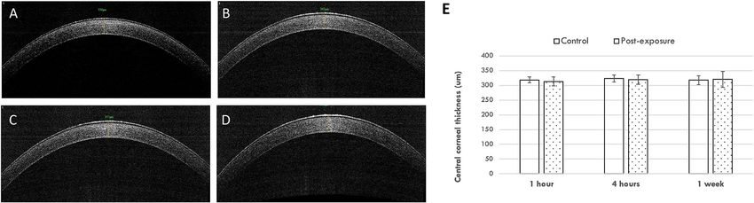
www.nature.com/scientificreports/

                                  Figure 2.  Representative IVCM micrographs at anterior stroma, posterior stroma and endothelial layer, after
                                  different periods of THz exposure (A). There were no significant changes in the mean intensity of stromal
                                  keratocytes reflectivity (B) and corneal endothelial density (C) when comparing to controls, at different time
                                  points. Error bars represent standard deviations.

                                  Figure 3.  Representative ASCOT pictures for the control (A), 1 h (B), 4 h (C) and 1 week after 4 h-exposure
                                  groups (D). The bar graph showed that the CCT was at a consistent level over time after exposure (E). Error bars
                                  represent standard deviations.

                                  Corneal temperature and ERG changes. On the thermographic maps, the ocular surface temperature
                                  was lower in the central area than in the peripheral area. There were no significant changes in the central and
                                  peripheral temperature over the THz continuous scanning period (P = 0.69 when comparing different exposure
                                  time points). The central and peripheral temperatures, at all the time points, were comparable to those in the
                                  control group (all P > 0.05; Fig. 4).
                                      Retinal function evaluated by electroretinography (ERG) showed that the average scotopic a- and b-wave
                                  amplitudes obtained at 200 cd s/m2 were not significantly reduced in all the experimental groups (all P > 0.05 for
                                  a- and b-wave amplitudes at all the time points, when compared to controls; Fig. 5).

                                  Histological analysis and immunohistochemistry assays. Histopathologically, no inflammatory
                                  cells or stromal fibrotic reaction were observed in corneas, and no signs of cell necrosis, gliosis, inflammation
                                  or degeneration of photoreceptors or neurons were seen in retinas in all eyes. The rings and parallel lens fibers
                                  were intact and parallel. No degeneration or vacuolation of the lens fibers, a characteristic of cortical cataract
                                  was seen. No accumulation of lens fibers or increased eosinophilic deposition of dense proteinaceous material, a

Scientific Reports |   (2021) 11:2448 |                https://doi.org/10.1038/s41598-021-82103-9                                                  3

                                                                                                                                             Vol.:(0123456789)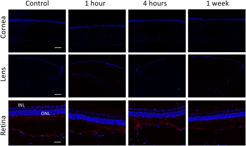
www.nature.com/scientificreports/

                                            Figure 4.  Representative thermographic maps showing the ocular surface temperature for the control (A), 1 h
                                            (B), 4 h (C) and 1 week after 4 h-exposure groups (D). The temperature was lower in the central area than that
                                            in the peripheral cornea, but no significant change was observed after different periods of exposure (E).

                                            Figure 5.  ERG responses after different duration of THz exposure. There were no significant changes in the
                                            scotopic a-wave (A) and b-wave (B) amplitudes after exposure compared to controls.

                                            pathological feature of nuclear cataract, was seen in the nucleus (Fig. 6). The histological sections were reviewed
                                            by an experienced ocular pathologist (A.S.Y.C) who had been masked to the experimental groups.
                                                There was no expression of CD11b and heat shock protein (HSP)-47, a macrophage marker in early inflamma-
                                            tory response and a collagen-specific stress protein marker, respectively, in corneas, lens epithelium and retinas
                                            in all eyes (Fig. 7). There was also no expression of fibronectin observed at all the time points. α-smooth muscle
                                            actin (α-SMA), a marker for myofibroblast transformation, was consistently present at Bruch’s membrane and
                                            sclera with no increase in the extent after THz exposure (Fig. 8). Apoptotic cells were not seen in corneas, lens
                                            epithelium, inner and outer nuclear layers of retinas at 1 h, 4 h and 1 week after THz radiation (Fig. 9).

                                            Corneal ultrastructure evaluated by TEM. The inter-fibillar distance was 60.7 ± 4.9, 50.6 ± 2.7, 66.5 ± 3.8,
                                            and 57.8 ± 3.1 pixel length, for the control, 1 h, 4 h and 1 week group respectively (P = 0.25, P = 0.29 and P = 0.33
                                            when comparing the experimental groups to controls). The chromatin in the keratocyte nucleus was condensed
                                            in all the corneas, and no signs of cell necrosis, such as swelling nuclei, cytoplasmic vacuoles or irregular clump-
                                            ings of chromatin, were observed (Fig. 10).

                                            mRNA expression and qRT‑PCR. The levels of mRNA expression of HSP90AB1, DDIT3, and EGFR1,
                                            which is a widespread heat-associated protein, a marker of cell stress response, and a transcriptional regulator,
                                            respectively, did not change significantly in the corneas, lens and scleral-retinal tissue, in the THz exposure
                                            groups, compared to the control group (all P > 0.05). The fold changes were at the range of 0.94–1.17, 0.79–1.11
                                            and 0.75–1.31 for HSP90AB1, DDIT3, and EGR1 levels, respectively (Fig. 11).

                                            Discussion
                                            In the present study, we demonstrated the biological responses, at tissue and cellular levels, after prolonged
                                            exposure to THz waves. With excessive exposure to THz up to 4 h, we did not observe detrimental effects on
                                            ocular tissue: the corneas and lens remained clear, the rod and cone cells- mediated ERG responses were not

          Scientific Reports |   (2021) 11:2448 |                https://doi.org/10.1038/s41598-021-82103-9                                                    4

Vol:.(1234567890)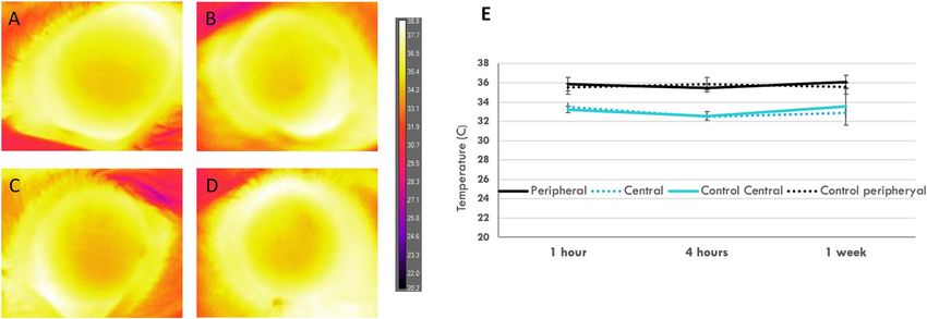
www.nature.com/scientificreports/

                                  Figure 6.  Histological sections with H&E staining presented that no inflammatory cell infiltrates or fibrotic
                                  reaction in the corneal stroma (A1–A4), and no retinal pathology such as gliosis, inflammation or degeneration
                                  of photoreceptors, were seen in the retinas (B1–B4), for the control, 1 h, 4 h and 1 week after 4 h-exposure
                                  groups, respectively. GCL: ganglion cell layer; IPL: inner plexiform layer; INL: inner nuclear layer; OPL;
                                  outer plexiform layer; ONL: outer nuclear layer; RPE: retinal pigment epithelium. After THz exposure,
                                  cataractogenesis was not detected. The crystalline lens fibers remained concentric and intact without disruption
                                  or liquefaction thus demonstrating no cortical cataract formation (note: artificial disruptions were seen due to
                                  processing artefacts). No increased coloration of the nuclear lens was seen thus excluding nuclear sclerosis (C1:
                                  control, C2: 1 week after 4-h exposure). Scale bar: 100 μm.

                                  Figure 7.  There was no expression of CD11b (A) and HSP-47 (B) in corneas, lens epithelium and retinas at
                                  different time points. IPL: inner plexiform layer; ONL: outer nuclear layer. Nuclei were counterstained with
                                  DAPI (blue). Scale bar 100 μm.

Scientific Reports |   (2021) 11:2448 |                https://doi.org/10.1038/s41598-021-82103-9                                                 5

                                                                                                                                             Vol.:(0123456789)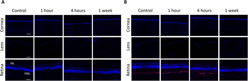
www.nature.com/scientificreports/
Figure 8. Immunohistochemical analysis on fibronectin showed negative staining in corneas, lens epithelium
and retinas at all time points (A). Staining for α-SMA was present at Bruch’s membrane and sclera with
a comparable extent in all eyes (B). IPL: inner plexiform layer; ONL: outer nuclear layer. Nuclei were
counterstained with DAPI (blue). Scale bar 100 μm.
Figure 9. There were no TUNEL-positive staining cells at all time points, in corneas, lens epithelium and
retinal layers. IPL: inner plexiform layer; ONL: outer nuclear layer. Nuclei were counterstained with DAPI
(blue). Scale bar 100 μm.
significantly altered, the thermal effect on ocular surface was not notable, and the corneal keratocytes activity, as
well as endothelial viability, was not affected. The tissue reaction, including the inflammatory, apoptotic, fibrotic
and stress responses, as well as the alternations in the corneal optical transmittance and ultrastructure, were not
observed. These biological safety results are essential to pave the path for further potential clinical applications
of THz scanning systems.
The application of THz sensing to the field of ophthalmology was first introduced in 201022. Due to a very
high dielectric constant and high sensitivity to changes of water content, THz imaging system has been reported
as a potential non-contact and non-invasive tool to quantify corneal hydration level22. In an ex-vivo rabbit
model, Elena et al. reported that an 1% decrease in the content of water mass in corneas led to a clearly detect-
able drop of the THz reflected signal by 13%, indicating good detection sensitivity23. Its high sensitivity to water
molecules allows it to be a potential novel device to diagnose early water gradient changes, before measurable
Scientific Reports | (2021) 11:2448 | https://doi.org/10.1038/s41598-021-82103-9 6
Vol:.(1234567890)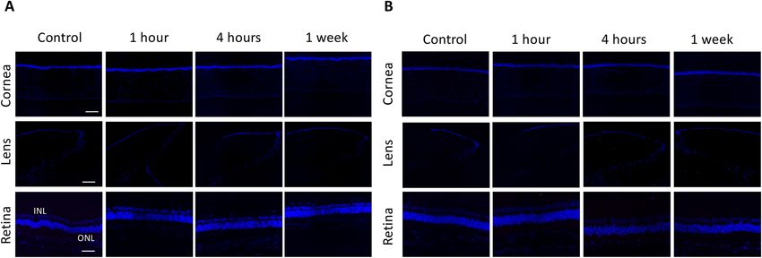
www.nature.com/scientificreports/
Figure 10. Transmission electron micrographs of the corneas showing transverse section of collagen fibrils (A,
scale bar 200 nm) and keratocytes (B, scale bar 1 μm) for the control , 1 h , 4 h and 1 week after 4 h-exposure
groups (A1–4 and B1–4, respectively).
Figure 11. Gene expression in corneas (A), lens (B) and sclero-retinal tissues (C) measured for different groups
using qRT-PCR. The mRNA expression fold values ( 2−ΔΔCT) were measured and normalized to that of the
control group. There were no significant changes in the expression of HSP90AB1, DDIT3 and EGR1 in all the
exposure groups.
changes in corneal thickness by conventional devices such as ASOCT or ultrasonic pachymetry. The idea of
using THz technology to generate hydration maps for an entire cornea was also proposed and tested, although
many technical challenges related to the imaging resolution and imaging field have to be a ddressed15,24. These
pre-clinical studies and pilot clinical study suggest that the THz-based system could be utilized as an independent
or adjuvant diagnostic and monitoring tool, for patients with corneal edema or hydration-related eye diseases.
As THz applications have been increasingly appearing, it is imperative to investigate the biological effects of THz
exposure at standard-scanning usage levels.
The biological effects resulting from radiation depend on three factors: the type of radiation, the amount and
the frequency of radiation, and the type of cells affected25,26. This can explain why there are some contradictory
results concerning the biological effects of THz on different cells types. There was no genomic damage observed
in in-vitro human skin cells after 2–8 h-THz exposure with the THz frequency up to 2.52 THz (power intensity
0.03–0.4 mW/cm2)27. An exposure of 0.12 THz radiation at 5 mW/cm2 for 24 h also did not induce genotoxicity,
morphological changes, and HSP protein expression in in-vitro human corneal epithelial cells21. Similar safety
was reported in blood samples from 9 healthy donors: no direct chromosomal damage and alteration of cell cycle
kinetics on blood cells following 20 min and 0.12 THz exposure (intensity 1 mW/cm2)20. On the other hand,
Scientific Reports | (2021) 11:2448 | https://doi.org/10.1038/s41598-021-82103-9 7
Vol.:(0123456789)www.nature.com/scientificreports/
Alexandrov et al. reported that the expression of certain genes was affected in mouse mesenchymal stem cells after
prolonged (9 h) and broad-band (10 THz) e xposure28. This might be due to the nature of high radiosensitivity of
stem cells29 and high-intensity exposure. The authors also concluded that the alteration of genes did not result
from thermal effects as the increase in temperature was minimal, which is in agreement with the present study.
The present study used a commercialized THz system in which the continuous pulse generated was at 40 µW
and 0.3 THz. The exposure period in the experiments was prolonged, up to 4 h, which is much longer than the
actual image acquisition time (within a minute). Moreover, as the biological expression after radiation can be
immediate if the exposure is vast, or can be few days after the molecular absorption event (delayed onset)30, we
also included a delayed time point at 1 week after exposure. On the molecular basis, we did not observe signifi-
cant changes in the mRNA expression of HSP90AB1, DDIT3, and EGFR1 in our in-vivo experiments. Of note,
previous studies used in-vivo e xperiment20,21,27,28, which were not taken into account physical barrier and buffer
of the tissues. In normal eyes, the presence of tear film, aqueous humor and vitreous body protects the direct
exposure of radiation.
The wavelength of THz is approximately 30 μm, which is short enough to provide reasonable resolution but
long enough to prevent serious loss of signal due to scattering31. The wavelength is longer than that of infrared
and ultraviolet (UV) light but is shorter than that of microwaves. It has been shown that an UV irradiation, at a
wavelength of 300 nm, has a penetration depth of around 0.5 m m32, and therefore the penetration of THz waves
are expected to be deeper than 0.5 mm. Hence we examined the biological effects down to the retinal level.
Furthermore, THz waves belong to non-ionizing radiation, defined as the electromagnetic radiation that does
not carry enough photon energy to ionize atoms or molecules. In contrast, ionizing radiation, such as X-rays or
gamma-rays, has a higher frequency and shorter wavelength than non-ionizing radiation and can cause health
hazards. These physical characteristics provide the background knowledge on the evaluation of its clinical appli-
cations in the aspect of safety.
The lens is one of the radiosensitive tissue in the human b ody33, and radiation is a well-known risk factor
10
for cataract . UV and infrared light are absorbed in the lens, and lens epithelial cell DNA is easily damaged by
oxidative stress, direct photochemical action of radiation, or thermal damage resulting from high-frequency
vibration of radiation, causing cataract30,33. Ionizing radiation, compared to non-ionizing radiation, has an even
greater detrimental impact on the lens, hence radiological protection measures are required33. We did not observe
any lens opacity, clinically or pathologically, after the THz exposure. The apoptotic cell death, the pathologic
mechanism of c ataractogenesis34, was not observed in the lens epithelium.
The cornea is at the anterior aspect of the eye and is highly exposed to irradiation. The wavelength of THz is
close to that of infrared, and infrared devices have been safely applied in the diagnosis and treatment of several
ocular surface diseases. Infrared meibography-ASOCT technology has been used to obtain the glandular archi-
tecture of Meibomian g lands35. Infrared warm compression has been an effective treatment for patients with
Meibomian gland dysfunction, by improving the release of meibum and tear stability36. With similar safety, we
did not observe any side effects, such as stromal haze and negative impact on keratocytes or endothelium, evalu-
ated clinically, optically, histologically and immunochemically, on the corneas after excessive THz exposure. On
the other hand, radiation with longer wavelengths, such as microwaves, is associated with thermal effects, which
can disrupt the extracellular matrix, change the inter-fibrillar distance, biomechanical property and transparency
of the corneas37. Ionizing radiation, like gamma-rays, induce more detrimental effects, killing keratocytes and
endothelium38. In the present study, the inter-fibrillar distances were not significantly altered after THz exposure,
and no necrotic keratocytes were observed.
The irradiance transmitted to the retina is minimized by the radiation absorbance of the cornea, aqueous
humor, lens and vitreous. The photoreceptors and retinal pigment epithelium (RPE) located in the posterior
pole are more susceptible to radiation. Unprotected or prolonged exposure of UV light (UV-B particularly)
results in photochemical damage in RPE and outer segment of photoreceptor39. Talebnejad et al. evaluated the
effects of microwaves on rabbit’s retinas: there were no pathological changes on the histopathological sections,
but the changes in the ERG responses were greater in the microwave groups than the sham group although
not significant40. Similarly, our results revealed that no apoptotic, inflammatory, fibrotic or stress reaction was
observed following prolonged THz exposure. However, the ERG evaluation did not have a consistent trend. The
a- and b-waves were slightly and insignificantly lower in the exposure group than control group at 1 h. The waves
were then returned to the control level at 4 h and 1 week.
There are several limitations in the present study. Firstly, as the wound healing process would be distinct
1 week after i nsult41, and the formation of cataract was reported a few days after radiation30, we set the last sacri-
ficial time point at 1 week. Delayed onset consequences beyond one week will be investigated in future studies.
Secondly, the ERG results were inconclusive, and this might be due to the inherently limited sensitivity of ERG
on rabbits or small sample size.
In conclusion, we evaluated the biological responses of ocular tissue, ranging from the anterior segment
to posterior segment, following excessive THz exposure. No adverse responses were observed from the tissue,
cellular, structural or functional levels. These safety profiles provide favourable evidence and basis for further
research work on the development and refinement of THz imaging system for its application in ophthalmology.
Methods
THz spectroscopy system. TERA K15 (Menlo Systems, GmbH, Germany) was used in this study. In the
system, two femtosecond fibre lasers with 250 MHz repetition rate, 90 femtosecond laser pulses and approxi-
mately 1.56 μm central wavelength, were used to excite two photoconductive antennas: one was used as emitter
and the other was used as a receiver. The THz emitter and receiver were based on the principle of a photoconduc-
tive switch. The THz pulse was generated with the coverage of bandwidth of 0.3–3 THz and 40 µW continuous
Scientific Reports | (2021) 11:2448 | https://doi.org/10.1038/s41598-021-82103-9 8
Vol:.(1234567890)www.nature.com/scientificreports/
power. The optical power of the lasers was approximately 30 mW with a pulse energy of approximately 0.3 nJ,
which corresponded to 5 × 10⁻¹³ J for the THz radiation. The THz beam was focused with TPX lenses, and the
wrist of focus beam was approximately 2 mm with maximum THz output at 0.3 THz. The repetition rates of both
lasers were locked and stabilized by two synchronization electronic devices.
Study animals and experimental groups. Sixteen 12- to 15-week-old New Zealand White rabbits (32
eyes) with 3–4 kg body weight were obtained from National University of Singapore and housed under standard
laboratory conditions. All animals were treated according to the guidelines of the Animal Research: Reporting
of In Vivo Experiments (ARRIVE guidelines, reference number 2017/SHS/1325). The protocol was approved
by the Institutional Animal Care and Use Committee of SingHealth. The animals were randomly divided into
4 groups (n = 8 eyes of 4 animals for each): control, 1-h, 4-h and 1-week groups. In the 1-h and 4-h groups, the
animals received prolonged THz radiation for 1 and 4 h, respectively, and then were euthanized under general
anaesthesia by intracardiac injection of overdosed sodium pentobarbitone (Jurox, Rutherford, Australia). In the
1-week group, the rabbits were euthanized at 1 week after 4-h continuous THz exposure, in order to study any
delayed tissue responses.
Clinical evaluation. All the eyes underwent clinical evaluation by slit lamp biomicroscopy (Nikon FS-3V;
Nikon, Japan), ASOCT (RTVue; Optovue, USA), IVCM (HRT3; Heidelberg Engineering GmbH, Germany),
fundus photography (Micron IV fundus camera; Phoenix Research Laboratories, USA), ERG (E3; Diagnosys
LLC, USA), and infrared ocular surface thermography (TG-1000; Tomey Corporation, Japan) under general
anaesthesia at baseline and 1 h and 4 h after THz exposure, and 1 week after 4-h exposure. The lens opacity was
graded with the Lens Opacities Classification system III (LOCS III)42.
For ASOCT evaluation, three high-resolution corneal cross-sectional scans (8 mm scan length, single scan
mode) were obtained for each eye at each time point. The CCT was measured by an independent observer (NCL),
and the average value was taken. For IVCM evaluation, the central aspect of the corneas was examined with a
minimum of three z-axis scans, consisting of the entire corneal thickness. For each eye, three micrographs from
anterior stroma (IVCM scanning depth < 160 μm), posterior stroma (scanning depth > 160 μm), and corneal
endothelium, respectively, were selected. The gray values of reflectivity of six stromal scans were semi-quantified
as described p reviously43, and the endothelial cells of 3 micrographs (frame area 400 μm × 400 μm) were counted,
using Image J (National Institutes of Health, USA). The IVCM assessment was performed by two independent
observers (YCL, NCL), and then the average value of micrographs was used. The fundus photographs were taken
30 min after instillation of dilation eye drops (1% tropicamide (Alcon, USA) and 2.5% phenylephrine (Bausch
and Lomb, USA)). All the ERG recordings were performed in a dark room under dim red light illumination, as
previously described44. Scotopic ERG responses were recorded across increasing light intensities from − 3.3 to
1.5 log cd s m−2 in 0.3-log-unit increments. Full-field ERG was recorded, and each response was the average of
3 trials. In addition, corneal temperature was evaluated with a contactless thermographer: the temperature of
corneal surface was quantified with 28 equidistance test locations (each grid corresponded to 1.8 mm distance
approximately)45, and we recorded the readings at the central cornea as well as 3.6 mm away from the center at
each side (periphery).
Histology and immunohistochemistry. The histologic and immunohistochemical analyses were per-
formed as previously d escribed46,47. In brief, sections of paraffin (5 µm thickness) embedded corneas, lens and
retinas were stained with hematoxylin and eosin histochemistry and visualized under light microscopy (Axi-
oplan 2; Carl Zeiss, Germany). For immunohistochemistry staining, sections were subjected to antigen retrieval
in citrate buffer (pH = 6.0) for 20 min, and the slides were rinsed with phosphate-buffered saline (PBS). The slides
were quenched with 10 mM ammonia chloride, followed by a blocking step for 1 h. They are then stained using
the following primary antibodies: cellular fibronectin (Millipore, USA) diluted 1∶100; α-SMA (Dako Cytoma-
tion, Denmark) diluted 1∶50; HSP-47, Enzolife Sciences, Switzerland) diluted 1:200; CD11b (BD Pharmin-
gen, USA) diluted 1:50, in the blocking solution. The secondary antibody was goat anti-mouse Alexa Fluor
488-conjugated (Invitrogen, USA). Slides were then mounted with UltraCruz mounting medium containing
DAPI (Santa Cruz Biotechnology, USA) and were observed and imaged with a fluorescence microscope (Axi-
oplan 2). To detect apoptotic cells, a fluorescence-based terminal deoxynucleotidyl transferase dUTP nick end
labelling (TUNEL) assay (Roche Applied Science, USA) was used according to the manufacturer’s instructions.
Optical transmittance measurements. Corneas from each group were placed in a 96-well plate, and
100 μL of wash buffer was added in to each well, together with an optical blank. Absorbance measurements were
obtained using a Tecan Infinite M200 (Tecan, Männedorf, Switzerland). Absorbance, A, was measured over the
wavelength range 380–780 nm at 1 nm intervals. Transmittance was calculated as T = 10−A, yielding transmit-
tance of either the blank solution (TB) or of the samples (TB+S). Transmittance of the samples itself, TS, was then
calculated as TS = TB+S + (1 − TB)48.
Transmission electron microscopy (TEM) and quantitative real‑time reverse transcription pol-
ymerase chain reaction (qRT‑PCR). TEM was performed with the protocol as we previously d escribed48.
In brief, corneas were fixed in 2% paraformaldehyde and 2% glutaraldehyde in PBS for one hour at room tem-
perature and then cut into 1 m m2 small pieces, before being fixed for another 1 h and then washed 3 times for
5 min with PBS. Corneas were then fixed with 1% potassium ferrocyanide and 1% osmium tetroxide for 1 h and
rinsed with distilled water. Subsequently, samples were dehydrated in a graded series of ethanol, and embedded
in Araldite (Electron Microscopy Sciences, Pennsylvania, USA). The 70–90 nm ultra-thin sections were cut
Scientific Reports | (2021) 11:2448 | https://doi.org/10.1038/s41598-021-82103-9 9
Vol.:(0123456789)www.nature.com/scientificreports/
with an Ultramicrotome (C. Reichert Optische Werke AG, Austria) and were collected on copper grids, double
stained with uranyl acetate and lead citrate for 8 min each, and then imaged on a JEM 1220 electron microscope
(JEOL, Tokyo, Japan) at 100 kV. To evaluate the effects of THz on the collagen ultrastructure of corneas, two
TEM images of transverse collagen fibrils from each quadrant was selected, and the fibril spacing was measured
using Image J. The center-to-center interfibrillar distance (in terms of pixel length) was defined as the spacing
etween49.
between the reference fibril spot and its closest neighbors without other fibrils blocking in b
For qRT-PCR, the corneas, lens and sclero-retinal tissues were cut into small pieces and immediately trans-
ferred into chilled TRIzol reagent (Invitrogen, USA), followed by homogenization steps using sonication for 20 s
at 20% power. Total RNA was extracted by the homogenized tissues using the PureLink Mini Kit (Ambion, Life
Technologies, Carlsbad, CA, USA) according to manufacturer’s instructions. Total RNA was quantified using a
NanoDrop ND-1000 UV–Vis Spectrophotometer (Thermo Scientific, USA). Total RNA (1 µg) from each sample
was reverse transcribed into cDNAs using Superscript III (Invitrogen, USA) according to the manufacturer’s
protocol. qRT-PCR was performed in 384-well plate in a total volume of 10 µL containing LightCycler 480 SYBR
Green I Master (Roche, Switzerland), primers, PCR grade water, and cDNA.
Each pair of primers and samples were run in triplicate wells and were performed three times. The relative
fold change was analyzed by the ΔΔCT method. The expression level of each gene in the control samples was used
for calibration. Threshold cycles ( CT) were normalized to expression of the housekeeping gene glyceraldehyde-
3-phosphate dehydrogenase (GAPDH)50: forward, 5′-GGG TGG TGG ACC TCA TGG T-3′, and reverse, 5′-CGG
TGG TTT GAG GGC TCT TA-3′). ΔCT in each sample was obtained by subtracting the CT of GAPDH from the
CT of the targeted gene. ΔΔCT of the samples for the 1-h, 4-h and 1-week groups was then calculated respectively
by subtracting the ΔCT of control samples from the ΔCT of each THz-exposed samples. The fold change of the
targeted gene in the THz-exposed samples compared with the controls was determined as 2−ΔΔCT. Gene specific
primers were selected (PRIMER BLAST, NIH) as follows: HSP90AB1 (Heat shock protein 90 kDa alpha family
class B member 1): forward, 5′-ATG ACT GGG AGG ACC ACT TG-3′, and reverse, 5′-GGG ATG AAA AGC
AAA GCC CTG-3′; DDIT3 (DNA damage inducible transcript 3): forward, 5′-CTG TCC GTG TCC CCC AAG
AT-3′, reverse, 5′-GGA GAG AGC GGT GCT TGC TA-3′; EGR1 (Early growth response 1): forward, 5′-CTA
CGA GCA CCT GAC CGC A-3′, reverse, 5′-AGG GTG TTG CCA CTG TTG GG-3’.
Statistical analysis. All data were expressed as mean ± standard deviation. Statistical comparisons among
the data of different exposure time points were performed using a Friedman test. Comparisons between post-
exposure values and controls were carried out with a Mann–Whitney U test. Statistical analyses were performed
using STATA software (version 13, STATACrop, College Station, TX). P values less than 0.05 were considered
statistically significant.
Received: 28 January 2020; Accepted: 15 January 2021
References
1. Bennett, D. B. et al. Terahertz sensing in corneal tissues. J. Biomed. Opt. 16, 057003 (2011).
2. Bajwa, N. et al. Non-invasive terahertz imaging of tissue water content for flap viability assessment. Biomed. Opt. Express. 8, 460–474
(2017).
3. Hall, A. & Girkin, J. M. A review of potential new diagnostic modalities for caries lesions. J. Dent. Res. 83, C89-94 (2004).
4. Rahman, A., Rahman, A. K. & Rao, B. Early detection of skin cancer via terahertz spectral profiling and 3D imaging. Biosens.
Bioelectron. 82, 64–70 (2016).
5. Ji, Y. B. et al. Terahertz otoscope and potential for diagnosing otitis media. Biomed. Opt. Express 7, 1201–1209 (2016).
6. Ozheredov, I. et al. Potential clinical applications of terahertz reflectometry for the assessment of the tear film stability. Opt. Eng.
59, 061622 (2020).
7. Ozheredov, I. et al. In vivo THz sensing of the cornea of the eye. Laser Phys. Lett 15, 055601 (2018).
8. Afshari, N. et al. Structure and dunction of the external eye and cornea. In External Disease and Cornea (ed. Thomas, J.) 8–9
(American Academy of Ophthalmology, San Francisco, 2016).
9. Sung, S. et al. Preliminary results of non-contact THz imaging of cornea. Proc. SPIE Int. Soc. Opt. Eng. https: //doi.
org/10.1117/12.2086866 (2015).
10. Liu, Y. C., Wilkins, M., Kim, T., Malyugin, B. & Mehta, J. S. Cataracts. Lancet 390, 600–612 (2017).
11. Han, S. B., Liu, Y. C., Noriega, K. M. & Mehta, J. S. Applications of anterior segment optical coherence tomography in cornea and
ocular surface diseases. J. Ophthalmol. 2016, 4971572 (2016).
12. Liu, Y. C., Lwin, N. C., Chan, N. S. & Mehta, J. S. Use of anterior segment optical coherence tomography to predict corneal graft
rejection in small animal models. Investig. Ophthalmol. Vis. Sci. 55, 6736–6741 (2014).
13. Ang, M. et al. Anterior segment optical coherence tomography. Prog. Retin. Eye Res. 66, 132–156 (2018).
14. Taylor, Z. D. et al. THz and mm-wave sensing of corneal tissue water content: Electromagnetic modeling and analysis. IEEE Trans.
Terahertz Sci. Technol. 5, 170–183 (2015).
15. Bennett, D. et al. Assessment of corneal hydration sensing in the terahertz band: In vivo results at 100 GHz. J. Biomed. Opt. 17,
97008 (2012).
16. Taylor, Z. D. et al. THz and mm-wave sensing of corneal tissue water content: In vivo sensing and imaging results. IEEE Trans.
Terahertz Sci. Technol. 5, 184–196 (2015).
17. Taylor, Z. D. et al. THz medical imaging: In vivo hydration sensing. IEEE Trans. Terahertz Sci. Technol. 1, 201–219 (2011).
18. Wilmink, G. et al. In Quantitative Investigation of the Bioeffects Associated with Terahertz RADIATION 75620L-75610 (San Fran-
cisco, CA, 2010).
19. Wilmink, G. J. et al. In Quantitative Investigation of the Bioeffects Associated with Terahertz Radiation 75620L (San Francisco, CA,
2010).
20. Scarfi, M. R. et al. THz exposure of whole blood for the study of biological effects on human lymphocytes. J. Biol. Phys. 29, 171–176
(2003).
21. Koyama, S. et al. Twenty four-hour exposure to a 0.12 THz electromagnetic field does not affect the genotoxicity, morphological
changes, or expression of heat shock protein in HCE-T cells. Int. J. Environ. Res. Public Health. 13, 793 (2016).
Scientific Reports | (2021) 11:2448 | https://doi.org/10.1038/s41598-021-82103-9 10
Vol:.(1234567890)www.nature.com/scientificreports/
22. Singh, R. S. et al. Terahertz sensing of corneal hydration. Conf. Proc. IEEE Eng. Med. Biol. Soc. 2010, 3021–3024 (2010).
23. Iomdina, E. N. et al. Study of transmittance and reflectance spectra of the cornea and the sclera in the THz frequency range. J.
Biomed. Opt. 21, 97002 (2016).
24. Sung, S. et al. THz imaging system for in vivo human cornea. IEEE Trans. Terahertz Sci. Technol. 8, 27–37 (2018).
25. Valentin, J. Protection in recommendations of the international commission on radiological protection 2–4 (2007).
26. Fuest, M. et al. Femtosecond laser-assisted conjunctival autograft preparation for pterygium surgery. Ocul. Surf. 15, 211–217
(2017).
27. Hintzsche, H. et al. Terahertz radiation at 0.380 THz and 2.520 THz does not lead to DNA damage in skin cells in vitro. Radiat
Res. 179, 38–45 (2013).
28. Alexandrov, B. S. et al. Non-thermal effects of terahertz radiation on gene expression in mouse stem cells. Biomed. Opt. Express 2,
2679–2689 (2011).
29. Prise, K. M. & Saran, A. Concise review: Stem cell effects in radiation risk. Stem Cells. 29, 1315–1321 (2011).
30. Soderberg, P. G., Talebizadeh, N., Yu, Z. & Galichanin, K. Does infrared or ultraviolet light damage the lens?. Eye 30, 241–246
(2016).
31. Joo-Huik, S. Introduction to biomedical studies using terahertz waves. In Terahertz Biomedical Science and Technology (ed. Joo-
Huik, S.) 1–9 (CRC Press, Boca Raton, 2014).
32. Lofgren, S. & Soderberg, P. G. Lens lactate dehydrogenase inactivation after UV-B irradiation: An in vivo measure of UVR-B
penetration. Investig. Ophthalmol. Vis. Sci. 42, 1833–1836 (2001).
33. Kleiman, N. J. Radiation cataract. Ann. ICRP. 41, 80–97 (2012).
34. Lee, E. H. et al. Lens epithelial cell death and reduction of anti-apoptotic protein Bcl-2 in human anterior polar cataracts. Mol. Vis.
8, 235–240 (2002).
35. Napoli, P. E. et al. A simple novel technique of infrared meibography by means of spectral-domain optical coherence tomography:
A cross-sectional clinical study. PLoS ONE 11, e0165558 (2016).
36. Goto, E. et al. Treatment of non-inflamed obstructive meibomian gland dysfunction by an infrared warm compression device. Br.
J. Ophthalmol. 86, 1403–1407 (2002).
37. Morgan, S. R. et al. Microwave treatment of the cornea leads to localised disruption of the extracellular matrix. Sci. Rep. 8, 13742
(2018).
38. Yoshida, J. et al. Gamma-irradiated sterile cornea for use in corneal transplants in a rabbit model. Middle East Afr. J. Ophthalmol.
22, 346–351 (2015).
39. Begaj, T. & Schaal, S. Sunlight and ultraviolet radiation-pertinent retinal implications and current management. Surv. Ophthalmol.
63, 174–192 (2018).
40. Talebnejad, M. R. et al. The effects of microwave radiation on rabbit’s retina. J. Curr. Ophthalmol. 30, 74–79 (2018).
41. Liu, Y. C., Teo, E. P., Lwin, N. C., Yam, G. H. & Mehta, J. S. Early corneal wound healing and inflammatory responses after SMILE:
Comparison of the effects of different refractive corrections and surgical experiences. J. Refract. Surg. 32, 346–353 (2016).
42. Chylack, L. T. Jr. et al. The Lens Opacities Classification System III. The longitudinal study of cataract study group. Arch. Ophthal-
mol. 111, 831–836 (1993).
43. Liu, Y. C. et al. Biological corneal inlay for presbyopia derived from small incision lenticule extraction (SMILE). Sci. Rep. 8, 1831
(2018).
44. Chaurasia, S. S. et al. The NLRP3 inflammasome may contribute to pathologic neovascularization in the advanced stages of diabetic
retinopathy. Sci. Rep. 8, 2847 (2018).
45. Konieczka, K., Schoetzau, A., Koch, S., Hauenstein, D. & Flammer, J. Cornea thermography: Optimal evaluation of the outcome
and the resulting reproducibility. Transl. Vis. Sci. Technol. 7, 14 (2018).
46. Liu, Y. C. et al. A biodegradable, sustained-released, prednisolone acetate microfilm drug delivery system effectively prolongs
corneal allograft survival in the rat keratoplasty model. PLoS ONE 8, e70419 (2013).
47. Williams, G. P. et al. Hyperopic refractive correction by LASIK, SMILE or lenticule reimplantation in a non-human primate model.
PLoS ONE 13, e0194209 (2018).
48. Liu, Y. C. et al. Corneal lenticule storage before reimplantation. Mol. Vis. 23, 753–764 (2017).
49. Yam, G. H. et al. Decellularization of human stromal refractive lenticules for corneal tissue engineering. Sci. Rep. 6, 26339 (2016).
50. Seol, D. et al. Selection of reference genes for normalization of quantitative real-time PCR in organ culture of the rat and rabbit
intervertebral disc. BMC Res. Notes. 4, 162 (2011).
Acknowledgements
We thank Anor Tech, Singapore, for the support of THz spectroscopy system.
Author contributions
Y.C.L., K.L., and J.S.M. conceived and designed the study; Y.C.L., Y.S.W.Q, M.T.Y.L., I.X.Y.L, N.Z., T.E.P.W, and
L.N.C. conducted the experiments; Y.C.L., K.L., and C.A.S.Y. analysed and interpreted the data; Y.C.L., K.L.,
L.S., and J.S.M. wrote the manuscript.
Funding
This research was supported by the Transition Award, National Medical Research Council (R1482/65/2017),
Singapore.
Competing interests
The authors declare no competing interests.
Additional information
Correspondence and requests for materials should be addressed to Y.-C.L.
Reprints and permissions information is available at www.nature.com/reprints.
Publisher’s note Springer Nature remains neutral with regard to jurisdictional claims in published maps and
institutional affiliations.
Scientific Reports | (2021) 11:2448 | https://doi.org/10.1038/s41598-021-82103-9 11
Vol.:(0123456789)www.nature.com/scientificreports/
Open Access This article is licensed under a Creative Commons Attribution 4.0 International
License, which permits use, sharing, adaptation, distribution and reproduction in any medium or
format, as long as you give appropriate credit to the original author(s) and the source, provide a link to the
Creative Commons licence, and indicate if changes were made. The images or other third party material in this
article are included in the article’s Creative Commons licence, unless indicated otherwise in a credit line to the
material. If material is not included in the article’s Creative Commons licence and your intended use is not
permitted by statutory regulation or exceeds the permitted use, you will need to obtain permission directly from
the copyright holder. To view a copy of this licence, visit http://creativecommons.org/licenses/by/4.0/.
© The Author(s) 2021
Scientific Reports | (2021) 11:2448 | https://doi.org/10.1038/s41598-021-82103-9 12
Vol:.(1234567890)You can also read