SAS and all other SAS Institute Inc. product or service names are registered trademarks or trademarks of SAS Institute Inc. in the USA and other ...
←
→
Page content transcription
If your browser does not render page correctly, please read the page content below

SAS and all other SAS Institute Inc. product or service names are registered trademarks or trademarks of SAS Institute Inc. in the USA and other countries. ® indicates USA registration. Other brand and product names are trademarks of their respective companies.

Computer-aided diagnosis system for breast ultrasound images using deep learning
 Hiroki Tanaka1, Shih-Wei Chiu2, Takanori Watanabe3, Setsuko Kaoku4, Takuhiro Yamaguchi2
 1 Japan Tobacco Inc, Tokyo, Japan; 2 Tohoku University Graduate School of Medicine; 3 National Hospital Organization Sendai Medical Center, Sendai, Miyagi, Japan;
 4 National Hospital Organization Osaka National Hospital, Osaka, Japan

 Abstract Intro 2
 The purpose of this study was to develop a computer-aided diagnosis (CAD) system for the
 • A computer-aided diagnosis (CAD) system was developed to assist doctors
Abstract classification of malignant and benign breast masses using ultrasonography based on a convolutional
 neural network (CNN), a state-of-the-art deep learning technique.
 • A CAD system automatically classifies the breast lesions in ultrasound images into
Introduction From the clinical data obtained in a previously conducted large-scale clinical trial, we collected malignant or benign, which helps doctors in providing a more accurate diagnosis.
 images of 1536 breast masses (897 malignant and 639 benign) confirmed by pathological
Methods examinations, with each breast mass captured from various angles. We constructed an ensemble • Convolutional neural networks (CNNs), a deep learning technique, have attracted
 network by combining two CNN models (VGG19 and ResNet152) fine-tuned on training data and considerable attention as a powerful tool to extract and learn efficient features
Results used the mass-level classification method to enable the CNN to classify a given mass using all views. directly from a data set.
 To visualize the regions detected by the CNN models to classify breast masses, we performed a
Discussion heatmap analysis.

Conclusion For an independent test set consisting of 154 masses (77 malignant and 77 benign), our network
 showed outstanding classification performance with a sensitivity of 90.9% (95% CI 84.5–97.3), a
 specificity of 87.0% (79.5–94.5), and area under the curve of 0.951 (0.916–0.987) . In addition, our
 study indicated that not only the breast masses but also the surrounding tissues were important
 regions for correct classification.

 Collectively, this CNN-based CAD system is expected to assist doctors and improve the diagnosis of
 breast cancer in clinical practice.

 Introduction
 Intro 1
 • Ultrasonography has been recommended as an adjunctive modality to mammography
 Figure 2. Structure of a CNN
Main Author: • Ultrasonography has the disadvantage of being operator dependent and requiring
Hiroki Tanaka proficiency in reading ultrasound images. Objective
 • This study aimed to develop a CNN-based CAD system to automatically classify the
 breast masses using all related ultrasound images by using a large-scale dataset.

 (a) Benign (b) malignancy
 Figure 1. Examples of breast masses on ultrasound images
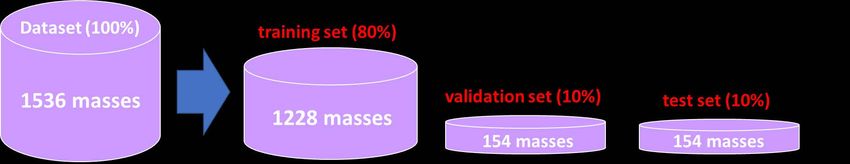
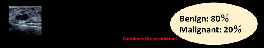
Computer-aided diagnosis system for breast ultrasound images using deep learning
 Hiroki Tanaka1, Shih-Wei Chiu2, Takanori Watanabe3, Setsuko Kaoku4, Takuhiro Yamaguchi2
 1 Division of Pharmaceutical, JAPAN TOBACCO INC, Tokyo, Japan; 2 Tohoku University Graduate School of Medicine; 3 National Hospital Organization Sendai Medical
 Center, Sendai, Miyagi, Japan; 4 National Hospital Organization Osaka National Hospital, Osaka, Japan

 Methods
 Method 1 : Image collection and datasets Method 2 : data argumentation
Abstract • In this study, we applied data augmentation for the training set.
 • 1536 breast masses (897 malignant and 639 benign) used in this study were
Introduction collected from a previously conducted large-scale clinical trial in Japan.
 • Data augmentation is a technique for synthetically generating new samples from an original
 training data.
Methods • Breast masses were identified by pathological examination.
 Original The number of training images
 Example)
Results
 increased from 6712 to 136,160
 • The dataset was randomly divided by mass in an 8:1:1 ratio into a training set, a
 images.
Discussion
 validation set and a test set.

Conclusion

 Horizontal flip Transformation of the contrast, brightness, or saturation
 Method 3 : CNN models

Main Author:
Hiroki Tanaka

 Figure 3. Ensemble model Figure 4. VGGNet19
 • We constructed an ensemble model by combining two CNN models called VGGNet19
 (Simonyan et al 2015) and ResNet152 (He et al 2016). This number indicates the
 number of layers.
 • This model combines the predictions of the trained VGGNet and ResNet to enhance
 the classification performance. Figure 5. ResNet152
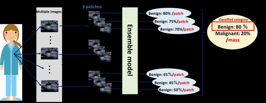
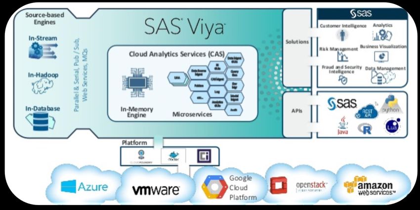
Computer-aided diagnosis system for breast ultrasound images using deep learning
 Hiroki Tanaka1, Shih-Wei Chiu2, Takanori Watanabe3, Setsuko Kaoku4, Takuhiro Yamaguchi2
 1 Division of Pharmaceutical, JAPAN TOBACCO INC, Tokyo, Japan; 2 Tohoku University Graduate School of Medicine; 3 National Hospital Organization Sendai Medical
 Center, Sendai, Miyagi, Japan; 4 National Hospital Organization Osaka National Hospital, Osaka, Japan

 Methods
 Method 3 : Procedure to classify masses
Abstract
Introduction
Methods
 • For each mass, there were multiple
Results ultrasound images because each breast
 mass was captured from various angles.
Discussion
 • In practice, doctors evaluate some views
Conclusion in ultrasound images and make a
 diagnosis per mass (patient) and not per
 view.

 • Therefore, it is desirable for a CNN to
 perform its diagnosis accordingly.

 Figure 6. Illustration of procedure to classify masses
Main Author: Method 4 : Software
Hiroki Tanaka
 • SAS® Visual Data Mining and Machine Learning 8.3 / SAS® Viya® 3.4

 • DLPy 0.7, the high-level Python APIs designed to efficiently apply the deep
 learning methods in SAS Visual Data Mining and Machine Learning

 ➢ Easy to code CNN models and Image processing such as data augmentation.

 ➢ Can visualize the process of the CNN models with heat map analysis.

Computer-aided diagnosis system for breast ultrasound images using deep learning
 Hiroki Tanaka1, Shih-Wei Chiu2, Takanori Watanabe3, Setsuko Kaoku4, Takuhiro Yamaguchi2
 1 Division of Pharmaceutical, JAPAN TOBACCO INC, Tokyo, Japan; 2 Tohoku University Graduate School of Medicine; 3 National Hospital Organization Sendai Medical
 Center, Sendai, Miyagi, Japan; 4 National Hospital Organization Osaka National Hospital, Osaka, Japan

 Result 1 : classification performance
 Table 1. Classification performance of the ensemble model
Abstract Accuracy (95%CI) Sensitivity (95%CI) Specificity (95%CI) AUC (95%CI *)
 • The classification performance of the ensemble model were evaluated
Introduction 89.0% 90.9% 87.0% 0.951 using the test set consisting of 154 masses (77 benign and 77 malignant).
Methods (84.0–93.9) (84.5–97.3) (79.5–94.5) (0.916–0.987)
 * 95% confidence interval
Results 1
 Result 2 : heat map analysis
Results 2
Discussion
Conclusion Table 2. Detection rate for overall masses in randomly selected test patches
 =
 Number of detected masses/number of randomly selected 50 test patches

 VGGNet 19 47.7 %
 ResNet 152 37.0 %
 Figure 7. Example where ResNet detected the mass Figure 8. Example where ResNet did not detect the mass

 Discussion Acknowledgments
 • In a large-scale clinical trial where screening was combined with mammography and We would like to thank SAS Institute Japan Ltd. for their technical support and provision of
Main Author: ultrasonography, sensitivity was shown to 91 % and specificity was 87 % (Ohuchi et the development environment for deep learning, which was founded by SAS Institute Inc.
Hiroki Tanaka al 2016).
 References
 • Our model provided equivalent results.
 • He K, Zhang X, Ren S and Sun J 2016 Deep residual learning for image recognition Proc. of the IEEE
 • Thus, we believe that our model might provide a second opinion to doctors and Conf. on Computer Vision and Pattern Recognition 770–778
 might assist doctors in decision making regarding diagnosis. • Ohuchi N et al 2016 Sensitivity and specificity of mammography and adjunctive ultrasonography to
 screen for breast cancer in the Japan Strategic Anti-cancer Randomized Trial (J-START): a randomised
 Conclusion controlled trial Lancet 387 341–348
 • In summary, it is expected that our system will be useful for doctors as a • Simonyan K and Zisserman A 2015 Very deep convolutional networks for large-scale image
 recognition Int. Conf. on Learning Representation (ICLR)
 supplemental modality for screening women with breast masses.

SAS and all other SAS Institute Inc. product or service names are registered trademarks or trademarks of SAS Institute Inc. in the USA and other countries. ® indicates USA registration. Other brand and product names are trademarks of their respective companies.

You can also read