Scaphoid screw dislocation
←
→
Page content transcription
If your browser does not render page correctly, please read the page content below
Scaphoid screw
dislocation
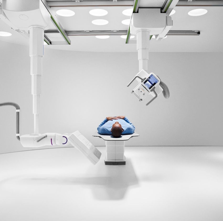
Multitom Rax Real 3D1 Hi-Res clinical case
University Hospital Wuerzburg, Germany
Study ID 5aab557
Image reprocessed on syngo.via with cinematic VRT. 1
1 Option Cinematic VRT is recommended for communication,
Unrestricted ©education,
Siemens Healthineers, 2021
and publication purposes and not intended for diagnostic reading.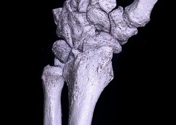
Clinical background and indication for
Multitom Rax Real 3D1 Hi-Res examination

Patient
Female |*1951 | BMI 24.4 kg/m²

Anamnesis
Patient fell on the left wrist three months before the
present examination and suffered a scaphoid waist
fracture (Herbert B2). Conventional screw
osteosynthesis was performed with subsequent cast
immobilization for six weeks.
Currently, the patient reports subtle pressure pain

                                                                                                                                                          Study ID 5aab935
over the radial side of the distal carpal row.

Indication for Real 3D1 examination
Radiography is unable to visualize proper screw                                  Lateral                      AP
placement and remains inconclusive with regard to                                Conventional X-ray examination
fracture healing.
The products/features (mentioned herein) are not commercially available in all
countries. Their future availability cannot be guaranteed.                                                                                   SHS DI XP      2
1 Option
                                                                                                                   Unrestricted ©  Siemens Healthineers, 2021
                                                                                                                   Unrestricted © Siemens Healthineers , 2021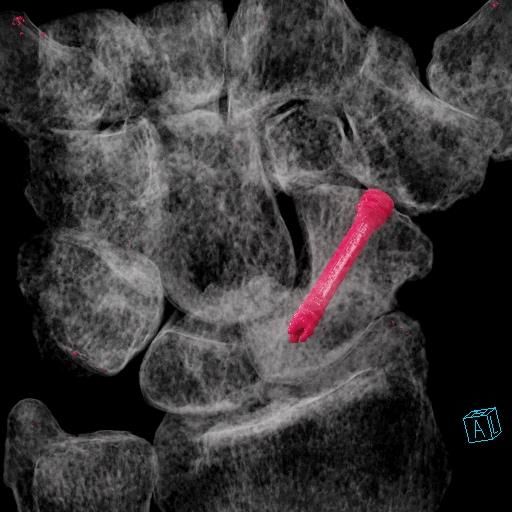
Multitom Rax Real 3D1 Hi-Res
Settings
Settings for tableside scan using dedicated metal protocol
Tube voltage 116 kV
Current time product 223 mAs
Dose area product 335 µGym²
Estimated value for CTDIvol,16 18 mGy
Scan time 14 sec
Number of projections 318
Reconstruction settings for sectional views
Pixel size 0.2 mm
Study ID 5aab557 Reconstruction kernel very sharp (equivalent to UR77)
Slice thickness 2 mm
The products/features (mentioned herein) are not commercially available in all
countries. Their future availability cannot be guaranteed. SHS DI XP 3
1 Option
Unrestricted © Siemens Healthineers, 2021
Multitom Rax Real 3D1 Hi-Res
Diagnostic findings

In addition to completed fracture healing,
Real 3D1 images with metal artefact reduction
reveal screw displacement into the
scaphotrapezial joint.
The proximal articular surface of the trapezium
displays a small notch (arrow) congruent to the
distal portion of the dislocated screw. Signs of
secondary osteoarthritis are visible (joint space                                           Coronal view   Sagittal view
narrowing, subchondral sclerosis).

                                                                                                                                                        Study ID 5aab557
The products/features (mentioned herein) are not commercially available in all countries.   Axial view     VRT view
Their future availability cannot be guaranteed.
Cinematic VRT is recommended for communication, education, and publication purposes and
not intended for diagnostic reading.                                                                                                                     4
1 Option
                                                                                                              Unrestricted
                                                                                                                  Unrestricted
                                                                                                                           © Siemens
                                                                                                                                © Siemens
                                                                                                                                     Healthcare
                                                                                                                                          Healthineers,
                                                                                                                                                GmbH, 2021
“VRT shows sufficient detail to be used for image
demonstration in interdisciplinary meetings with
orthopedic surgeons. After customization to
highlight metal implants in color, it is particularly
helpful for visualization of screw dislocation.” 1
Jan-Peter Grunz, MD
University Hospital Wuerzburg, Germany
1 Thestatements by Siemens Healthineers customers described herein are based on results that were achieved in the customer’s unique setting.
Since there is no “typical“ hospital and many variables exist (e.g., hospital size, case mix, level of IT adoption) there can be no guarantee that other customers will achieve the same results. SHS DI XP 5
Unrestricted © Siemens Healthineers, 2021The products/features (mentioned herein) are not
commercially available in all countries. Their future
availability cannot be guaranteed.
Results from case studies are not predictive of results
in other cases. Results in other cases may vary.
Dr. Jan-Peter Grunz is employed by an institution that
receives financial support from Siemens Healthineers
for collaborations.
Click to add footnote 6
second line Unrestricted © Siemens Healthineers, 2021You can also read