Steady streaming as a method for drug delivery to the inner ear
←
→
Page content transcription
If your browser does not render page correctly, please read the page content below
www.nature.com/scientificreports
OPEN Steady streaming as a method
for drug delivery to the inner ear
Laura Sumner1, Jonathan Mestel2 & Tobias Reichenbach1*
The inner ear, or cochlea, is a fluid-filled organ housing the mechanosensitive hair cells. Sound
stimulation is relayed to the hair cells through waves that propagate on the elastic basilar membrane.
Sensorineural hearing loss occurs from damage to the hair cells and cannot currently be cured.
Although drugs have been proposed to prevent damage or restore functionality to hair cells, a
difficulty with such treatments is ensuring adequate drug delivery to the cells. Because the cochlea is
encased in the temporal bone, it can only be accessed from its basal end. However, the hair cells that
are responsible for detecting speech-frequency sounds reside at the opposite, apical end. In this paper
we show that steady streaming can be used to transport drugs along the cochlea. Steady streaming
is a nonlinear process that accompanies many fluctuating fluid motions, including the sound-evoked
waves in the inner ear. We combine an analytical approximation for the waves in the cochlea with
computational fluid dynamic simulations to demonstrate that the combined steady streaming effects
of several different frequencies can transport drugs from the base of the cochlea further towards the
apex. Our results therefore show that multi-frequency sound stimulation can serve as a non-invasive
method to transport drugs efficiently along the cochlea.
Sensorineural hearing loss is caused by damage to the mechanosensitive hair cells in the inner ear, or cochlea,
and is one of the most common disabilities across the w orld1,2. Although research into medical treatments such
3–5
as gene therapy has advanced significantly , the progress of effective therapies is stunted by the fact that the
inner ear is a very complex part of the body and particularly difficult to access. The cochlea is encased in the tem-
poral bone, the hardest bone in the body. Artificial openings of the temporal bone can disturb the endocochlear
rocess6,7. Drug
potential, an electrical potential across the hair cells that is important for the inner ear’s active p
delivery through the cardiovascular system after oral or intravenous application of a drug is also difficult, due to
the blood-perilymph barrier segregating the cardiovascular system from the cochlear fluids8,9.
The cochlea can be accessed through the membrane-covered oval or the round window at its base that connect
to the middle ear. Drugs can be applied either through the middle ear and be deposited near the round window
(intratympanic delivery), relying on diffusion through the round window to reach the inner ear, or be injected
directly through the round window into the cochlea (intracochlear delivery). In the case of intratympanic deliv-
ery, a variety of methods are used in order to maintain contact with the membrane. Hydrogels10–12 and gelatin
foams13–16 can be used as carriers for drugs or micro/nanoparticles in order to extend contact time with the oval
or round windows17,18, as well as combinations of these in so-called ferrogels which can be manipulated through
the use of magnetic fi elds19. Osmotic pumps are also able to extend the delivery t imescales20. As an alternative
invasive approach, a small hole can be drilled into the side of the cochlea and drugs inserted through the open-
ing. However, this so-called cochleostomy alters the functioning of the cochlea and is therefore normally only
used for cochlear implants that replace key parts of the inner ear’s function21.
In many of these cases, the drugs are delivered at the basal end of the cochlea, where the highest audible
frequencies are detected, around 20 kHz in men6. However, the frequencies that matter most for speech range
from 300 Hz to 3 kHz and are sensed by hair cells further towards the apical end of the cochlea6,22. This leads
to the next stage of the delivery problem: distribution of the drug once it has been administered to the cochlea.
Whilst there are methods such as m icropumps23,24 which can inject and distribute a drug, most of the current
drug delivery methods rely on passive diffusion to reach the mid and apical region. This process is slow, inef-
ficient, and difficult to control or v erify25,26.
In the work which follows, we assume the drug in question is already present at the basal end of the cochlea,
either as a result of intracochlear injection through, or diffusion across, the round window. Once the drug has
been administered, active propulsion of drugs along the inner ear can potentially result from steady s treaming27,28.
Steady streaming describes the phenomenon of a net time-averaged fluid motion accompanying an oscillating
1
Department of Bioengineering and Centre for Neurotechnology, Imperial College London, South Kensington
Campus, London SW7 2AZ, UK. 2Department of Mathematics, Imperial College London, South Kensington
Campus, London SW7 2AZ, UK. *email: reichenbach@imperial.ac.uk
Scientific Reports | (2021) 11:57 | https://doi.org/10.1038/s41598-020-79946-z 1
Vol.:(0123456789)
www.nature.com/scientificreports/
Figure 1. The morphology of the inner ear. (a) The cochlea is a spiral-shaped duct filled with fluid and
segregated into two chambers by the elastic basilar membrane (dark blue), which can be accessed through the
round and the oval windows at the base. Mathematical descriptions of the fluid dynamics typically consider
the simplification of an uncoiled, linear cochlea. (b) Many aspects of cochlear fluid dynamics can be captured
in a two-dimensional representation, in which one dimension, x, represents the inner ear’s longitudinal extent
and the other dimension, y, denotes the vertical deviation from the basilar membrane. We further denote by
u(1) and p(1) the velocity and the pressure in the upper chamber. Analogously, u(2) and p(2) are the velocity
and the pressure in the lower chamber. Both chambers are connected at the apical end through the so-called
helicotrema.
primary flow field with zero m ean29. Such a net flow can, for instance, result from Reynolds stresses in boundary
layers near no-slip boundaries or from Stokes drift due to differences in the mean motion at a fixed point and the
mean motion of a fixed particle moving with the fluid. Both effects result in steady streaming in the c ochlea27,30.
For the purpose of describing the inner ear’s fluid dynamics, the organ can be viewed as consisting of two
fluid-filled elongated chambers that are separated by the elastic basilar membrane (Fig. 1). Sound stimulation
leads to a displacement of the oval window at the basal end of one chamber, and the induced pressure can be
released at the elastic round window at the basal end of the other chamber22,31. The sound then leads to a pressure
difference across the membrane and therefore to its displacement. The pressure difference and the corresponding
membrane displacement travel as waves along the longitudinal extent of the cochlea towards its apex. However,
because the impedance of the basilar membrane changes systematically from base to apex, the wave induced by
a single frequency exhibits a shortening wavelength and an increasing amplitude as it propagates apically, reach-
ing a peak in amplitude at a characteristic position, beyond which it decays rapidly. This characteristic place is
located near the base for high-frequency stimulation and shifts systematically further towards the apex for lower
frequencies. The cochlea thereby establishes a tonotopic mapping between frequency and longitudinal location
of the wave’s peak. The mapping is approximately logarithmic: frequencies that differ by a constant factor peak
at locations that are a constant distance a part6,22.
Steady streaming is quadratic in the wave amplitude and so is particularly pronounced near the peak of the
wave induced by a pure tone, since the amplitude of the evoked fluid motion there is largest and because of the
rapid attenuation beyond the peak location27,30. The steady-streaming induced by a single frequency is thus
localised in its effect, and can not provide efficient long range drug transport. However, a complex tone, com-
prising many frequencies could simultaneously excite different regions of the basilar membrane, and particles
might be relayed from the vicinity of one excited point to another. In this way, drugs might be transported deep
into the inner ear.
Here we combined an analytical approximation of the basilar-membrane wave with computational fluid
dynamics simulations and tracking of finite mass particles to investigate the steady streaming induced by a pure
tone as well as by complex tones, and to quantify the efficacy of this mechanism for drug transport.
Methods
Analytical approximation for the basilar‑membrane displacement. We employed an analytical
approximation to describe the wave on the basilar membrane. We simplify the spiral geometry of the cochlea by
considering a planar membrane with fluid above and below it. The resulting one-dimensional model describes
the longitudinal extent of the cochlea through the variable x and assumes that the variation in pressure and fluid
motion in the vertical dimension is negligible (Fig. 1). The difference p in the pressure between the lower and
the upper chamber, p = p(2) − p(1), then leads to an upward velocity V of the basilar membrane. In a passive
cochlea, the relationship between the basilar-membrane velocity and the pressure difference is a linear one, and
depends on the frequency of the vibration. For a pure tone of a frequency f, and angular frequency ω = 2πf ,
the pressure difference can be written as p = p̃eiωt + c.c. in which “c.c.” denotes the complex conjugate. The
resulting displacement of the basilar membrane can similarly be expressed as V = V eiωt + c.c.. We define the
complex impedance Z(x) relating the Fourier coefficients of the pressure and velocity of the basilar-membrane
vibration as
Scientific Reports | (2021) 11:57 | https://doi.org/10.1038/s41598-020-79946-z 2
Vol:.(1234567890)
www.nature.com/scientificreports/
= p.
V (1)
Z
reviously31 the pressure difference then satisfies the wave equation
As has been shown p
d 2
p 2iωρ0
=
p, (2)
dx 2 Zh
in which ρ0 denotes the density of the fluid, and h is the height of each chamber.
The main difficulty in solving Eq. (2) is the spatial variation of the impedance Z(x). However, the length
scale over which the impedance changes significantly is typically small compared to the wavelength of the
resulting wave. As first shown by Lighthill, we can therefore employ the Wentzel-Kramers-Brillouin (WKB)
approximation31,32. We introduce the complex, x-dependent wavenumber
2ωρ0
k(x) = , (3)
iZ(x)h
with the square root defined to have a negative imaginary part. The WKB approximation starts from the ansatz
x
k(x ′ )dx ′ (4)
p(x) =
p(x)e−i 0 ,
where the pressure amplitude p(x) is assumed to vary more slowly than 1/k, i.e. k ≫ p . Substituting this
p ′ /
expression into Eq. (2) and equating terms of O(k), requires
k0 Z(x) 1/4
p(x) = p0 = p0 , (5)
k(x) Z0
where p0 = p(0) is the pressure amplitude applied at the base of the inner ear, x = 0 , while k0 = k(0) and
Z0 = Z(0). The velocity of the basilar membrane follows as
Z0 3/4 −i
x k(x ′ )dx ′ +iωt
V (x, t) = V0 e 0 + c.c. (6)
Z(x)
where V0 = p0 /Z0 is the velocity amplitude at the basal end. It is possible to further analyse the WKB integral
asymptotically, and obtain a formula in terms of Bessel functions (see for example33), but we used Eq. (6) directly
in the numerics below.
Parameter values. We model the impedance of the basilar membrane in terms of its stiffness K, mass m,
and damping ξ per unit area. All of these can depend on the longitudinal location x and we allow for nonlinear
effects by permitting ξ to vary with amplitude
Z(x) = [iωm(x) + ξ(x, p0 ) − iK(x)/ω]/A. (7)
The cross-sectional area A represents a transverse strip of the basilar membrane with the width of one hair cell,
8 µm , and a breadth of 186 µm31. Stiffness, mass and damping refer henceforth to this narrow section of the
basilar membrane.
In the absence of damping, at each frequency there is a position of resonant response, where ω2 = K/m. The
small damping coefficient ensures the regularity of the solution, but maintains the link between frequency and
cochlear position. The damping derives from two sources; firstly, the viscous damping of the fluid, which is linear
at low Reynolds number, and secondly, the nonlinear cochleal response as described below.
Experimental data regarding the different contributions to the basilar-membrane impedance are not reliably
available in humans. We therefore based our computations on experimental data from the gerbil’s inner ear. The
hearing range of gerbils, about 200 Hz to 60 kHz, overlaps largely with that of humans, which extends from 20 Hz
to 20 kHz. Such data show that the stiffness of the basilar membrane decays exponentially from base to apex, at
least between the basal and the mid region of the inner e ar34. The main contribution to the basilar-membrane
mass m(x) derives from the organ of Corti on the membrane. Since the cross-section of the organ increases from
base to apex, the basilar-membrane mass also i ncreases35.
We assume exponential dependencies of both stiffness and mass on the longitudinal location, correspond-
ing to the cochlea’s logarithmic tonotopic mapping between frequency and spatial location: m = m0 ex/lm and
K = K0 e−x/lK . The parameters m0 and K0 denote the mass and stiffness at the basal end, x = 0, and lm and lK
are the length scales that correspond to the exponential variation. We obtained these parameters from experi-
mental measurements in the gerbil cochlea. The cross-sectional area of the organ of Corti, which sits on the
basilar membrane, at the basal end is about 8000 µm2, implying a moving mass of about m0 = 32 ng31,35. The
stiffness of the membrane at this location is about K0 = 1 N/m34. At a location of 7 mm apical from the basal
end, the cross-sectional area of the organ of Corti has increased to about 18,000 µm2, yielding a moving mass
of the basilar membrane of about 65 ng. The stiffness of the membrane at this cochlear location has decreased
to 0.03 N/m. These changes in stiffness and mass imply the length scales lm = 115 nm and lK = 501 nm. For the
height of each fluid filled chamber in the gerbil’s inner ear we have assumed h = 0.5 mm.
Scientific Reports | (2021) 11:57 | https://doi.org/10.1038/s41598-020-79946-z 3
Vol.:(0123456789)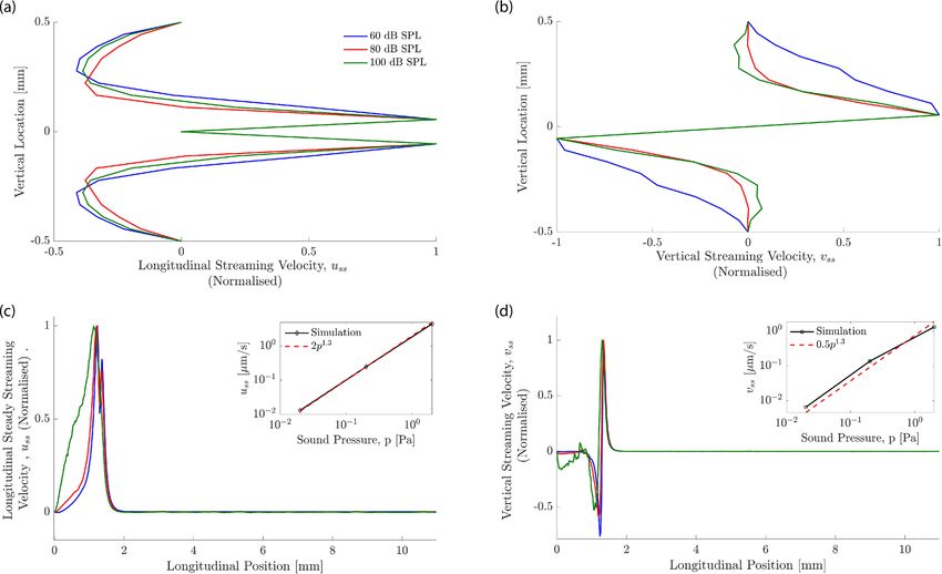
www.nature.com/scientificreports/
Figure 2. Amplitude dependent response of the basilar-membrane near the peak, modelled using a level-
dependent drag coefficient ξ in the membrane impedance. (a) The membrane displacement increases
linearly with the stimulation intensity basal and apical to the peak, but sublinearly at and near the peak. (b)
The nonlinear response emerges clearly in the sensitivity, defined as the ratio of the membrane’s vibrational
amplitude to the applied sound pressure. Where the sensitivity is independent of the sound-pressure level the
membrane response is linear, but nonlinear otherwise. The compressive nonlinearity near the wave’s peak shows
that the peak is sharper at lower stimulation intensities.
√
At each longitudinal location x, mass and stiffness define a resonance frequency f0 = K/m/(2π) at which
the contributions to the basilar membrane impedance from stiffness and mass cancel. The wave induced by
stimulation at frequency f peaks at that location, with an amplitude that is limited by the drag portion of the
basilar membrane impedance. A healthy inner ear has, however, an active process that provides mechanical
amplification of weak sound vibrations, effectively counteracting the viscous drag22,36,37. This amplification varies
with sound-pressure level: small vibrations are amplified most, and larger vibrations successively less. This leads
to a compressively nonlinear response of the basilar-membrane vibration that can, for instance, be explained
through the membrane’s segment being poised near a Hopf bifurcation38–40. As another consequence, the peak
of the basilar-membrane wave is sharper at low rather than high sound pressure levels.
Here, we modelled this level-dependent amplification through considering a spatially constant, amplitude
dependent damping ξ(p0 ). In particular, we considered three distinct sound-pressure levels, and adjusted the
damping parameter ξ so that the peak of the basilar-membrane displacement as predicted by the WKB approxi-
mation (6) had a width corresponding to experimental data41. We performed the analysis for a stimulation
frequency of 17 kHz and the three sound-pressure levels of 60, 80 and 100 dB SPL (Fig. 2). For these sound
levels we identified respectively the coefficients ξ = 10 nNs/m, ξ = 500 nNs/m, and ξ = 2 µNs/m for the cor-
responding total drag, including viscous and amplification effects. These drag coefficients reproduced both the
experimentally-observed nonlinear response of the basilar-membrane vibration at the peak and the broadening
of the peak for larger sound intensities.
Computational fluid dynamics (CFD). Computational fluid dynamic simulations were carried out in
OpenFOAM v 3.0.1., a finite volume, open source computational fluid dynamics toolkit. We simulated the fluid
flow in a two-dimensional model of the cochlea, in which two parallel chambers were separated by the basilar
membrane but were connected at the apical end (Fig. 1). Using the known solution for the basilar membrane
waves, the time-dependent displacement of the membrane was obtained from the numerical solution of the
WKB approximation, Eq. (6), using MATLAB. The output of the MATLAB analysis was a matrix whose rows
corresponded to the set of membrane coordinates at a given time step. This was then used as an input to the fluid
simulation, allowing the membrane’s boundary to be updated at each time step and the fluid to react accordingly.
OpenFOAM’s built in dynamic mesh capabilities were used to ensure mesh conformation. No-slip boundary
conditions were imposed at the membrane and all boundaries. Through this approach, the fluid-structure inter-
action between the cochlear fluids and the basilar membrane was reduced to a purely fluid-dynamic problem.
Fluid flow in the inner ear results from motion of the middle ear and resulting movement of the oval and
round windows. However, because our approach utilised an analytically-derived basilar-membrane motion,
we did not need to model the movement of the round and oval window. We therefore did not need to solve the
fluid-structure-interaction of the fluid with the different membranes of the inner ear, but could focus on the
fluid dynamics only.
The Reynolds number of the flow is low, although the Womersley number need not be, so that the Navier-
Stokes equations reduce to the unsteady Stokes equations. To leading order, therefore, the fluid velocity consists
of a single harmonic, eiωt . However, second order interactions give rise to a steady-streaming velocity, with a
steady component which can lead to net transport down the cochlea. The domain is two-dimensional, though
at high Womersley number, derivatives normal to the plate dominate, and an analytic solution can be written
Scientific Reports | (2021) 11:57 | https://doi.org/10.1038/s41598-020-79946-z 4
Vol:.(1234567890)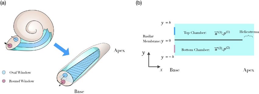
www.nature.com/scientificreports/
down. However, even in that limit the particle paths need to be found numerically, and so we preferred to use
computational simulation from the start, for all parameter values.
We adapted the solver BMicoFoam that was created through the combination of the icoFoam and solidParticle
solvers. The icoFoam solver is a transient incompressible flow solver, which uses an implicit Euler method for time
integration, and a discrete volume Gauss linear scheme for the spatial differentials. The fluid flows were visualized
through paraView. Post-processing was completed separately by analysing the OpenFOAM output in MATLAB.
The timestep, t was determined by the stimulation frequencies. For multi-frequency forcing, in order to
resolve the particle trajectories well, we employed ten timesteps per cycle of the highest frequency used in the
stimulation. For single-frequency forcing a smaller timestep can be used for the same computational cost. The
spatial discretisation used a triangular Delaunay mesh. The mesh-size x was chosen to be small enough to
capture the basilar-membrane motion accurately, and also to resolve the thickness of the Stokes boundary layer
at higher Womersley numbers. The Courant number, C0 = (umax �t)/�x , in which umax is the largest velocity
present in the flow was kept much less than 1 to obtain stable particle trajectories. Accordingly, the mesh size
varied from 22.5 µm near the cochlear boundaries to 11.2 µm at the basilar membrane to provide increased
spatial resolution near the membrane.
The cochlea is filled with perilymph and endolymph, specialized ionic solutions whose density and viscosity
are similar to that of water. We therefore considered a density of ρ0 = 1000 kg m−3 and a kinematic viscosity of
the fluid of ν = 10−6 m2 s−1.
For particle tracking, 5000 neutrally buoyant particles with an almost elastic collision model (coefficient of
restitution of 0.95), a diameter of 200 nm17 and a friction coefficient of 10−3 were injected at zero velocity into
the basal region of the two chambers. Note that these particles have an associated mass and are not passive point
particles as have been considered by other authors27,42. In this way, the work accounts not only for passive particles
in solution, but also for low-solubility particles in suspension as used in certain therapeutical a pproaches26,43.
Particles were evenly spaced on a grid covering most of the basal region of each chamber. Their trajectories
were calculated throughout the simulation. The components of the steady streaming velocity, (uss , vss ) were then
extracted from the particle trajectories through computing the temporal averages of the velocities.
Steady streaming is a nonlinear phenomenon, and its amplitude is proportional to the square of the fluid
velocity27,30,44,45. Small stimulation intensities therefore lead to only tiny steady streaming velocities that require
long computational times to measure. However, while the oscillatory motion must be resolved on the oscillatory
time-scale, the steady component does not. For computational efficacy, and for the multiple-frequency stimula-
tion presented below, we therefore scaled both the basilar membrane motion, as well as the fluid velocities at
80 dB SPL, by a factor of 300. We then computed the resulting fluid flows and steady streaming, and scaled them
back by the factor of 300 as well as by its square, respectively. This method resulted in a significant reduction of
the required computation time without affecting the obtained results.
Results
Pure‑tone stimulation. We first investigated the steady streaming caused by stimulation with a pure tone.
In agreement with analytical descriptions as well as computational results, we found that pure-tone stimulation
elicits oscillatory fluid flow in both cochlear chambers, with an amplitude that is largest near the peak of the
basilar-membrane wave (Fig. 3).
The particle tracking that we employed showed the accompanying steady streaming, in particular the Lagran-
gian streaming field (Fig. 3). We observed the formation of a pair of counter-rotating vortices above and below
the basilar membrane, at the location of the wave’s peak. We then investigated the influence of the stimulation
intensity on the shape of these vortices. We found only a minor change, with the vortices induced by a higher
intensity being slightly wider in the vertical direction (Fig. 4). The steady streaming velocity depends on the
sound pressure level p approximately through a power law with a power of 1.3: uss ∼ p1.3 and vss ∼ p1.3. This
is less than the quadratic increase with pressure expected from a passive cochlea with a linear response, and
reflects the active’s cochlear compressive nonlinearity that we have modelled through a level-dependent damp-
ing parameter (Fig. 2).
Multi‑frequency stimulation. Because pure-tone stimulation elicited significant steady streaming only
near the peak of the basilar-membrane wave, we investigated whether a complex tone with several frequen-
cies could lead to steady streaming within a larger longitudinal range within the cochlea (Fig. 5). We therefore
considered frequencies f1, f2, ..., fn that differed by a constant factor R > 1: fi+1 = Rfi for i = 1, 2, ..., n − 1.
Due to the cochlea’s logarithmic tonotopic map, the locations x1, x2, ..., xn at which the basilar-membrane waves
elicited by stimulation at frequency f1, f2, ..., fn peaked decreased linearly with the logarithm of the stimulation
frequency: xi = −m ln(fi ). The spatial distance δ between two neighbouring peak locations xi and xi+1 was there-
fore constant: xi − xi+1 = δ for i = 1, 2, ..., n − 1. The spatial constant δ was proportional to the logarithm of the
frequency ratio R, δ = m ln(R), with a proportionality constant m = 3.3 mm. In our simulations we explored
how the spatial spacing δ affected the resulting steady streaming.
Two factors limited the spacings that we could meaningfully consider. First, our simulations of pure-tone
stimulation revealed that the spatial extent of the induced steady streaming was on the order of 1 mm. Spacings
of subsequent wave peaks of more than 1 mm will therefore not lead to overlapping vortices, and thus not to a
combined steady streaming. Second, the region around the peak of the wave induced by a pure tone in which
the basilar-membrane response is nonlinear is at least 100 µm long46. Although we accounted for the nonlin-
ear response through a level-dependent drag coefficient in the basilar-membrane impedance, our analytical
approximation did not capture further nonlinear effects. In particular, the approximation we employed did not
account for the two-tone suppression that results when the basilar-membrane waves induced by two nearby
Scientific Reports | (2021) 11:57 | https://doi.org/10.1038/s41598-020-79946-z 5
Vol.:(0123456789)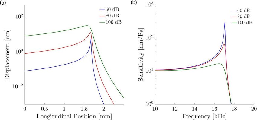
www.nature.com/scientificreports/

                                           Figure 3.  Steady streaming induced by a pure tone of 20 kHz at 80 dB SPL. (a) The wave on the basilar
                                           membrane (blue line) has a wavelength that shortens upon approaching the resonant position. The wave
                                           amplitude (black dashed line) peaks at the resonant position. OpenFOAM results for the instantaneous
                                           longitudinal (b) and vertical (c) fluid field velocity components, (u, v) which are both largest near the resonant
                                           position. (d) The longitudinal component of the steady streaming, uss, is largest near the peak of the basilar-
                                           membrane wave as well. Near the membrane, the velocity is oriented towards the apex, and further away from
                                           the membrane particles move towards the base. (e) The vertical streaming velocity, vss points towards the
                                           membrane basal to the wave’s peak, and away from the membrane apical to the peak. (f) The steady streaming
                                           flow field shows two counter-rotating vortices, one in the upper and one in the lower chamber.

          Scientific Reports |   (2021) 11:57 |                 https://doi.org/10.1038/s41598-020-79946-z                                                      6

Vol:.(1234567890)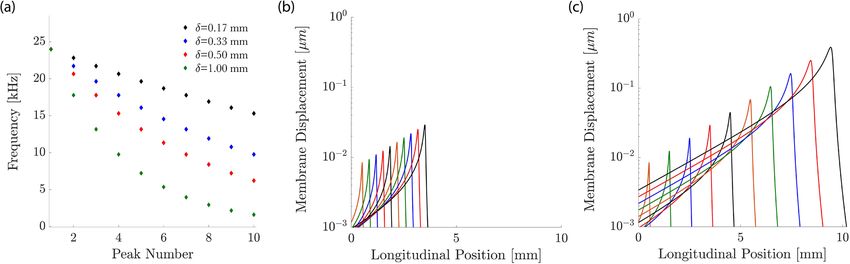
www.nature.com/scientificreports/

                                 Figure 4.  Steady-streaming velocity profiles at different sound intensities for 20 kHz stimulation taken at
                                 x = 1.2 mm, the 20 kHz resonance location. The velocity profiles have been normalized by the maximal velocity.
                                 (a) The longitudinal streaming velocity is largest close to the basilar membrane. The vertical extent around the
                                 basilar membrane over which fluid flow occurs towards the apex is slightly larger at higher sound intensities.
                                 (b) The vertical streaming velocity peaks at a similar distance from the basilar membrane as the longitudinal
                                 component. The kinks seen in these profiles are an artefact of the coarse resolution of the streaming field but
                                 do not affect the overall accuracy of the calculation. (c) The longitudinal streaming velocity close to the basilar
                                 membrane. The inset shows that the maximal value of uss increases approximately as a power law with the
                                 the sound pressure p: uss ∼ p1.3. (d) The vertical streaming velocity shows that, for high frequencies at higher
                                 sound-pressure levels (eg. 100 dB SPL), the boundary at the base of the cochlea influences the flow. The inset
                                 shows the power law dependence of the maximal value of vss on the sound pressure level p which follows
                                 approximately a power law, vss ∼ p1.3.

                                 Figure 5.  Multi-frequency stimulation. (a) We choose discrete frequencies so that successive frequencies differ
                                 by a constant ratio, leading to a constant spacing δ of the peaks of the different induced basilar-membrane waves.
                                 (b) When the frequencies are close such that the spacing between the peaks of the induced waves is small,
                                 δ = 330 µm, we obtain significant overlap between the amplitudes of the individual waves. (c) A larger spacing
                                 between the successive peaks, δ = 1 mm, leads to less overlap.

Scientific Reports |   (2021) 11:57 |                 https://doi.org/10.1038/s41598-020-79946-z                                                  7

                                                                                                                                             Vol.:(0123456789)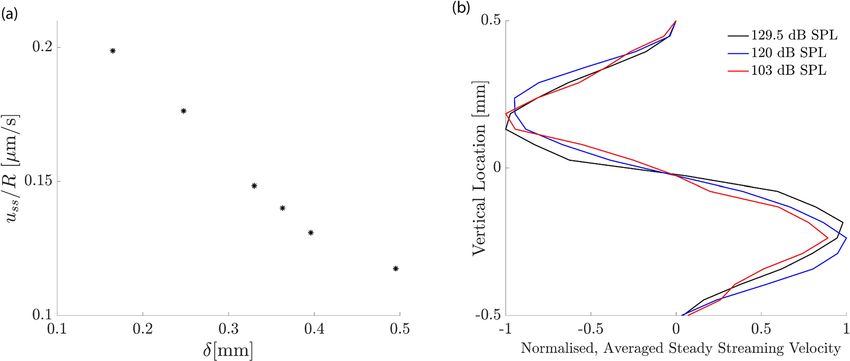
www.nature.com/scientificreports/
Figure 6. Steady streaming induced by multi-frequency stimulation at 80 dB SPL. (a–c), The velocities of
steady streaming that results from stimulation at frequencies whose individual basilar-membrane waves peak
at a spatial distance of δ = 170 µm apart from one another. (a) The longitudinal fluid flow has a significant
amplitude in the extended region from 0 to 2.5 mm, where the waves induced by the individual waves peak. (b)
The vertical velocity is strongest at the beginning and at the end of that region, that is, near the cochlear base as
well as about 2.5 mm apical to it. (c) The vector fields show how the steady-streaming vortices associated with
each individual frequency combine to a large circular flow that extends significantly along the cochlea. (d–f)
The steady streaming velocity induced by a multi-frequency stimulation in which the individual peaks of the
induced basilar-membrane waves are further apart, δ = 330 µm. Significant steady streaming emerges along a
longer longitudinal range of the cochlea, because the multi-frequency stimulation extends to lower frequencies.
Because the wave peaks are further apart, the steady-streaming vortices associated to the individual frequencies
emerge stronger than for the case of closer frequencies.
frequencies overlap in their region of nonlinear response47–49. We could therefore not reliably simulate spacings
of 100 µm or less.
Our numerical results showed that such multi-frequency stimulation leads indeed to significant steady stream-
ing along a considerable extent of the inner ear (Fig. 6). This effect emerged because the steady-streaming vortices
induced by the individual frequencies combined to create an effective “streaming channel” along which the
particles are transported longitudinally. Figure 7 details the positions of the injected particles at different points
in time throughout the simulation in the bottom half of the channel. It shows how the particles are pulled into
a streaming channel near the basilar membrane that is formed by the combined eddy structure. Our results also
evidenced that a smaller separation between frequencies led to stronger longitudinal steady streaming (Fig. 8).
Moreover, our CFD simulations show significant vertical velocity of the steady streaming near the base of the
cochlea. This vertical velocity was, in either chamber, directed towards the basilar membrane, and implied that
particles near the base were transported near the membrane and then propelled longitudinally along it.
We also considered whether particles injected at the base but far from the basilar membrane would be
transported to the membrane as a result of the steady streaming. We found that this was indeed the case: the
steady streaming near the base has a significant vertical component that is directed towards the basilar mem-
brane (Fig. 6). Due to the nonlinear basilar-membrane response to sound intensity, which we modelled through
an intensity-dependent drag coefficient of the basilar membrane, the spatial profiles of the steady streaming
could, in principle, depend on the sound intensity. We confirmed, however, that the vertical velocity profile
depended only little on the intensity level of the sound, up to an overall scaling (Fig. 8b). This result agrees with
our finding that the intensity of a pure tone has little influence on the spatial velocity profile as well (Fig. 4), and
confirms that particles injected at the base are transported effectively towards the membrane, independent of
the sound intensity.
Discussion
This work investigated the effect of different sound stimuli on the fluid mechanics of the inner ear. Although com-
putational simulation of both the fluid m echanics50,51 as well as the steady s treaming27,52 and drug d
istribution25
have been reported, tracking of finite mass particles (i.e. not passive) individual particles in the flow has not.
Here we validated our model against Lighthill’s analytical work on the subject and subsequently used the model
Scientific Reports | (2021) 11:57 | https://doi.org/10.1038/s41598-020-79946-z 8
Vol:.(1234567890)www.nature.com/scientificreports/
Figure 7. Individual particles near the cochlear base (coloured dots) are displaced throughout the cochlea due
to steady streaming. We show the particle displacement in the bottom channel as induced by multi-frequency
stimulation at 80 dB SPL with a frequency separation of δ = 330 µm. Particles near the membrane (purple) are
quickly displaced towards the base, while particles away from the membrane (black) are first moved towards the
membrane and then towards the apex.
Figure 8. Influence of sound intensity. (a) The longitudinal component of the average steady streaming, uss at
the base of the cochlea, normalised by the ratio of the successive frequencies in the multi-frequency stimulation
R. If the steady streaming were proportional to the energy dissipated along a constant cochlear extent, then this
normalized velocity would be constant. However, the normalized velocity increases strongly for smaller spatial
separations, evidencing that the steady streaming becomes stronger when the multi-frequency stimulation
uses many pure tones that successively differ only by a small amount. (b) Vertical profile of the vertical average
steady-streaming velocity 0.1 mm from the base of the cochlea, the basal edge of the circulation region for
δ = 170 µm, as shown in Fig. 6 and its dependence on the sound intensity. The stimulation intensity has only
little influence on the velocity profile. The velocity profiles have been normalized by the maximal value at each
sound pressure level.
to investigate the most effective way to harness the streaming. The aim of the work was to determine whether
there is a combination of tones which could deliver (or at least better redistribute) a drug which has either dif-
fused across the round window or been injected through it directly.
Each pure tone resulted in an eddy in each cochlear chamber whose apical edge caused particle translation
back towards the base of the cochlea. Playing a single tone therefore serves to rotate the particles locally in the
Scientific Reports | (2021) 11:57 | https://doi.org/10.1038/s41598-020-79946-z 9
Vol.:(0123456789)www.nature.com/scientificreports/
region of the characteristic place. By inducing another eddy apical to the first, the backward drift of particles
can be avoided: instead they are “pulled” into the flow of the proceeding eddy. Because the eddy is at a lower
frequency at the same sound-pressure level, the amplitude is higher and so therefore is the velocity, leading to an
acceleration along the channel. By continuing to increase the number of eddies, this “streaming channel” along
which particles are accelerated is extended and hence the region over which the drug is distributed increases. By
choosing the correct frequency combination, the effect can be optimised by reducing the vertical component of
particle velocity. This can be seen in Fig. 6 where panels (a)–(c) show that the closer eddy combination results
in a diminished vertical steady streaming velocity inside the global eddy compared to the edge. Importantly, as
presented in Fig. 8, although the eddy strength is reduced with sound-pressure level, the penetration depth and
hence the region of influence of the eddies does not change. These results therefore show that a multiple fre-
quency stimulus not only generates larger velocities in the cochlea by creating a streaming channel along which
particles are accelerated, but also does not require a specific injection location at the base in order to be effective.
Starting from the proof-of-concept undertaken here, further work will be required to develop the method
towards clinical application. In particular, it will be important to use experimental data for the human coch-
lea rather than for the rodent cochlea that we investigated here. It will also be beneficial to investigate a more
complex, three-dimensional and realistic geometry of the cochlea, as well as to include the interaction of the
fluid with the different membranes, to determine the dependence of the streaming on factors such as boundary
effects. In particular, including the motion of the basilar membrane in a fluid-structure interaction simulation
will allow one to model the longitudinal motion of the basilar membrane, which has been proposed by Edom
et al.27 to increase the streaming velocity. Finally, including the active process will allow to model the width of
the basilar-membrane waves induced by a pure tone and their dependence on the sound-pressure level. This
will be necessary to optimise the sound stimuli for clinical application, in particular regarding the choice of the
frequency separation parameter δ that one can use in order to create the streaming channel.
Sensorineural hearing loss therapies are lacking an effective mode of delivery of the pharmaceuticals to the
damage site. The purpose of this work was to investigate whether or not the phenomenon of steady streaming,
which is already present in the inner ear, could be a viable means of overcoming this hurdle. From the results
presented here, we believe that it shows promise. Specifically, we find that a superposition of optimally separated
frequencies results in the formation of a “streaming channel”, capable of transporting a drug from a broad region
near the base of the cochlea a predetermined distance along it. In all simulations presented here, 10 frequencies
were chosen to be superposed. By increasing or decreasing this value, for a constant δ, the longitudinal reach of
the streaming channel will either increase or decrease. In this way, we can achieve targeted delivery relatively
simply.
We also show that the timescales over which these stimuli must be played to achieve this transport are reason-
able for therapies. From Fig. 6d, considering a particle initially injected at 0.25 mm, the time taken to reach 3
mm (approx 1/3 of the length of the cochlea) by accelerating from 1 µm/s to 3 µm/s is around 5 h. This timescale
is well within the recommended exposure limit suggested by both the OSHA (16 h at 80 dB SPL) as well as the
NIOSH (25 h at 80 dB SPL) in the CDC guidelines53,54.
Previous work by Edom et al.27 has estimated the steady streaming velocity for a pure tone at 1 kHz and
94 dB SPL to be about 300 µm/s. This result is in approximate agreement with the steady streaming velocities
that we reported here, such as our finding that a 20 kHz tone presented at 80 dB SPL leads to a steady stream-
ing velocity of about 300 nm/s. The difference in stimulation intensity of 14 dB SPL between our computation
and the previous one indeed leads to a factor of 5 in the stimulation amplitude, and hence to a factor of 25 in
the resulting steady streaming. This leaves a difference of a factor of 40 between the steady streaming velocity
reported by Edom et al.27 and the one that we have computed. This factor presumably results from the different
frequencies that were considered: lower frequencies are indeed known to lead to larger basilar-membrane dis-
placement than higher frequencies, at the peak of the traveling wave. We note that, since the steady-streaming
velocity increases in proportion to the square of the basilar-membrane velocity, a factor of about 7 in the basilar-
membrane displacement at the different frequencies and characteristic locations already suffices to explain the
additional factor of 40 between the obtained steady-streaming velocities.
Although the streaming velocities are admittedly small, over long enough therapy times and with optimised
sound stimuli, there is potential for steady streaming to become a way to non-invasively deliver a drug once
administered to the cochlea. Now that we have demonstrated a proof of concept for non-invasive drug delivery
in the passive cochlea, there is a rich landscape of sound stimuli to explore.
Received: 3 August 2020; Accepted: 15 December 2020
References
1. Dai, C. et al. Rhesus cochlear and vestibular functions are preserved after inner ear injection of saline volume sufficient for gene
therapy delivery. J. Assoc. Res. Otolaryngol. 18, 601–617. https://doi.org/10.1007/s10162-017-0628-6 (2017).
2. Wise, A. & Gillespie, L. Drug delivery to the inner ear. J. Neural Eng. 9, 065002 (2012).
3. Richardson, R. & Atkinson, P. Atoh1 gene therapy in the cochlea for hair cell regeneration. Expert Opin. Biol. Ther. 15, 417–430
(2015).
4. Chien, W. et al. Gene therapy restores hair cell stereocilia morphology in inner ears of deaf whirler mice. Mol. Ther. 24, 17–25
(2016).
5. Suzuki, J., Hashimoto, K., Xiao, R., Vandenberghe, L. & Liberman, M. Cochlear gene therapy with ancestral aav in adult mice:
complete transduction of inner hair cells without cochlear dysfunction. Sci. Rep. 7, 45524 (2017).
6. Pickles, J. An Introduction to the Physiology of Hearing (Brill, Leiden, 2013).
7. Wangemann, P. Supporting sensory transduction: Cochlear fluid homeostasis and the endocochlear potential. J. Physiol. 576,
11–21 (2006).
Scientific Reports | (2021) 11:57 | https://doi.org/10.1038/s41598-020-79946-z 10
Vol:.(1234567890)www.nature.com/scientificreports/
8. Jahnke, K. The blood-perilymph barrier. Arch. Rhino-Laryngol 228, 29–34 (1980).
9. Juhn, S. K., Rybak, L. P. & Fowlks, W. L. Transport characteristics of the blood–perilymph barrier. Am. J. Otolaryngol. 3, 392–396
(1982).
10. Engleder, E. et al. Preclinical evaluation of thermoreversible triamcinolone acetonide hydrogels for drug delivery to the inner ear.
Int. J. Pharm. 471, 297–302 (2014).
11. Wang, X. et al. Dose-dependent sustained release of dexamethasone in inner ear cochlear fluids using a novel local delivery
approach. Audiol. Neurotol. 14, 393–401 (2009).
12. Piu, F. et al. Oto-104: a sustained-release dexamethasone hydrogel for the treatment of otic disorders. Otol. Neurotol. 32, 171–179
(2011).
13. Silverstein, H., Arruda, J., Rosenberg, S. I., Deems, D. & Hester, T. O. Direct round window membrane application of gentamicin
in the treatment of meniere’s disease. Otolaryngol. Head Neck Surg. 120, 649–655 (1999).
14. Havenith, S. et al. Spiral ganglion cell survival after round window membrane application of brain-derived neurotrophic factor
using gelfoam as carrier. Hear. Res. 272, 168–177 (2011).
15. Yoon, J. Y. et al. Intratympanic delivery of oligoarginine-conjugated nanoparticles as a gene (or drug) carrier to the inner ear.
Biomaterials 73, 243–253 (2015).
16. Takeda, H. et al. Protein transduction therapy into cochleae via the round window niche in guinea pigs. Mol. Ther. Methods Clin.
Dev. 3, 16055 (2016).
17. Lajud, S. A. et al. A novel chitosan-hydrogel-based nanoparticle delivery system for local inner ear application. Otol. Neurotol. 36,
341 (2015).
18. Xu, L. et al. A controlled and sustained local gentamicin delivery system for inner ear applications. Otol. Neurotol. 31, 1115–1121
(2010).
19. Thaler, M. et al. Visualization and analysis of superparamagnetic iron oxide nanoparticles in the inner ear by light microscopy and
energy filtered tem. Nanomed. Nanotechnol. Biol. Med. 7, 360–369 (2011).
20. Mäder, K., Lehner, E., Liebau, A. & Plontke, S. K. Controlled drug release to the inner ear: concepts, materials, mechanisms, and
performance. Hear. Res. 368, 49–66 (2018).
21. Jiam, N. T. & Limb, C. J. The impact of round window vs cochleostomy surgical approaches on interscalar excursions in the cochlea:
Preliminary results from a flat-panel computed tomography study. World J. Otorhinolaryngol. Head Neck Surg. 2, 142–147 (2016).
22. Robles, L. & Ruggero, M. A. Mechanics of the mammalian cochlea. Physiol. Rev. 81, 1305–1352 (2001).
23. Peppi, M., Marie, A., Belline, C. & Borenstein, J. Intracochlear drug delivery systems: a novel approach whose time has come
(2018).
24. McCall, A. A. et al. Drug delivery for treatment of inner ear disease: current state of knowledge. Ear Hear. 31, 156 (2010).
25. Salt, A. N. & Plontke, S. K. Principles of local drug delivery to the inner ear. Audiol. Neurotol. 14, 350–360 (2009).
26. Salt, A. N. & Plontke, S. K. Pharmacokinetic principles in the inner ear: influence of drug properties on intratympanic applications.
Hear. Res. 368, 28–40 (2018).
27. Edom, E., Obrist, D. & Kleiser, L. Steady streaming in a two-dimensional box model of a passive cochlea. J. Fluid Mech. 753,
254–278 (2014).
28. Obrist, D. Flow phenomena in the inner ear. Annu. Rev. Fluid Mech. 51, 487–510 (2019).
29. Riley, N. Steady streaming. Annu. Rev. Fluid Mech. 33, 43–65 (2001).
30. Lighthill, J. Acoustic streaming in the ear itself. J. Fluid Mech. 239, 551–606 (1992).
31. Reichenbach, T. & Hudspeth, A. The physics of hearing: fluid mechanics and the active process of the inner ear. Rep. Prog. Phys.
77, 076601 (2014).
32. Lighthill, J. Energy flow in the cochlea. J. Fluid Mech. 106, 149–213 (1981).
33. MacKay, R. Mode conversion in the cochlea?. Trans. Math. Appl. 1, 1–37 (2017).
34. Emadi, G., Richter, C. & Dallos, P. Stiffness of the gerbil basilar membrane: Radial and longitudinal variations. J. Neurophysiol. 91,
474–488 (2004).
35. Richter, C.-P., Edge, R., He, D. Z. & Dallos, P. Development of the gerbil inner ear observed in the hemicochlea. J. Assoc. Res.
Otolaryngol. 1, 195–210 (2000).
36. Dallos, P. The active cochlea. J. Neurosci. 12, 4575–4585 (1992).
37. Hudspeth, A. Integrating the active process of hair cells with cochlear function. Nat. Rev. Neurosci. 15, 600 (2014).
38. Magnasco, M. O. A wave traveling over a hopf instability shapes the cochlear tuning curve. Phys. Rev. Lett. 90, 058101 (2003).
39. Kern, A. & Stoop, R. Essential role of couplings between hearing nonlinearities. Phys. Rev. Lett. 91, 128101 (2003).
40. Duke, T. & Jülicher, F. Active traveling wave in the cochlea. Phys. Rev. Lett. 90, 158101 (2003).
41. Cooper, N. & Rhode, W. Mechanical responses to two-tone distortion products in the apical and basal turns of the mammalian
cochlea. J. Neurophysiol. 78, 261–270 (1997).
42. Lesser, M. B. & Berkley, D. A. Fluid mechanics of the cochlea. Part 1. J. Fluid Mech. 51, 497–512. https://doi.org/10.1017/S0022
112072002320 (1972).
43. Honeder, C., Engleder, E. & Schöpper, H. E. A. Sustained release of triamcinolone acetonide from an intratympanically applied
hydrogel designed for the delivery of high glucocorticoid doses. Audiol. Neurotol. 19, 193–202 (2014).
44. Lighthill, J. Acoustic streaming. J. Sound Vib. 61, 391–418 (1978).
45. Amit, R., Abadi, A. & Kosa, G. Characterization of steady streaming for a particle manipulation system. Biomed. Microdevices 18,
39 (2016).
46. Fisher, J., Nin, F., Reichenbach, T., Uthaiah, R. & Hudspeth, A. The spatial pattern of cochlear amplification. Neuron 76, 989–997
(2012).
47. Ruggero, M. A. et al. Two-tone suppression in the basilar membrane of the cochlea: Mechanical basis of auditory-nerve rate sup-
pression. J. Neurophysiol. 68, 1087–1087 (1992).
48. Cooper, N. P. Two-tone suppression in cochlear mechanics. J. Acoust. Soc. Am. 99, 3087–3098 (1996).
49. Jülicher, F., Andor, D. & Duke, T. Physical basis of two-tone interference in hearing. Proc. Natl. Acad. Sci. 98, 9080–9085 (2001).
50. Beyer, R. P. Jr. A computational model of the cochlea using the immersed boundary method. J. Comput. Phys. 98, 145–162 (1992).
51. Givelberg, E. & Bunn, J. A comprehensive three-dimensional model of the cochlea. J. Comput. Phys. 191, 377–391 (2003).
52. Gerstenberger, C. & Wolter, F.-E. Numerical simulation of acoustic streaming within the cochlea. J. Comput. Acoust.https://doi.
org/10.1142/S0218396X13500197 (2013).
53. Noise exposure computation. In Occupational Safety and Health Standards, 1910 Subpart G, Occupational Health and Environmental
Control, 1910.95 App A (Occupational Safety and Health Administration, United States Department of Labor).
54. Occupational noise exposure (U.S. Department of Health and Human Services, Public Health Services, Centers for Disease Control
and Prevention, National Institute for Occupational Safety and Health, 1998).
Acknowledgements
The work was funded by the EPSRC Centre for Doctoral Training in Fluid Dynamics Across Scales. The authors
would also like to thank Dr Nikola Ciganovic for supplying code in the early stages of this project.
Scientific Reports | (2021) 11:57 | https://doi.org/10.1038/s41598-020-79946-z 11
Vol.:(0123456789)www.nature.com/scientificreports/
Author contributions
T.R. conceived the project and provided guidance. L.S. undertook the numerical work and completed the data
analysis. J.M. and T.R. oversaw the project. J.M, T.R. and L.S. wrote the manuscript.
Competing interests
The authors declare no competing interests.
Additional information
Correspondence and requests for materials should be addressed to T.R.
Reprints and permissions information is available at www.nature.com/reprints.
Publisher’s note Springer Nature remains neutral with regard to jurisdictional claims in published maps and
institutional affiliations.
Open Access This article is licensed under a Creative Commons Attribution 4.0 International
License, which permits use, sharing, adaptation, distribution and reproduction in any medium or
format, as long as you give appropriate credit to the original author(s) and the source, provide a link to the
Creative Commons licence, and indicate if changes were made. The images or other third party material in this
article are included in the article’s Creative Commons licence, unless indicated otherwise in a credit line to the
material. If material is not included in the article’s Creative Commons licence and your intended use is not
permitted by statutory regulation or exceeds the permitted use, you will need to obtain permission directly from
the copyright holder. To view a copy of this licence, visit http://creativecommons.org/licenses/by/4.0/.
© The Author(s) 2021
Scientific Reports | (2021) 11:57 | https://doi.org/10.1038/s41598-020-79946-z 12
Vol:.(1234567890)You can also read