Stingray envenomation and injury in a dog - De Gruyter
←
→
Page content transcription
If your browser does not render page correctly, please read the page content below

Open Vets. 2021; 2; 1–5

Rapid Communication

Olutoye Adegboye*, Olusegun Adegboye

Stingray envenomation and injury in a dog
https://10.1515/ovs-2020-0103
                                                                            In some coastal areas in Gambia, these fishes
Received Oct 23, 2020; accepted Jan 20, 2021
                                                                       sometimes wash ashore or are caught in fishing nets. It
Abstract: The stingray, a seemingly harmless cartilaginous             is a common practice amongst dog owners living near the
fish, is capable of causing painful injuries and envenomation          coast to walk their dogs along the beaches. These dogs
in humans. There is no known peer reviewed case report                 encounter various marine animals while nosing around
involving dogs in a veterinary journal at the time of writing          in the beach or swimming in the open sea. During this
this case report. Poor management of the condition or                  period, they sometimes encounter the stingrays and could
overzealous attempts to remove embedded barbs has                      get stung.
resulted in complications in humans. This case report                       As reported in all published case reports, human
presents an effective approach to the treatment of stingray            stingray injuries and envenomation are painful, they
envenomation in dogs which is likely to be reproducible in             often lead to tissue necrosis and wound sepsis. They are
other domestic animals. Clearly elucidated are the principles          usually accidental during swimming or fishing, most of
behind antibiotic therapy in the treatment of stingray injuries        the injuries are sustained on the lower extremities [3,6-11].
and the benefit of lignocaine injection in cases of embedded           Based on the authors’ clinical experience, among the few
stingers. There is a need for case reports to enhance clinical         cases encountered, the common site of envenomation in
knowledge of stingray management in domestic animals.                  dogs in Gambia is the submandibular area. This is a logical
This case report, thus, serves as an impetus for future                observation, as most of these unpleasant encounters
research in this area of veterinary medicine.                          occur when they are nosing around on the beach. It must
                                                                       be noted that stingray envenomation in dogs is a rare
Keywords: stingray, envenomation, stinger, barb, venom                 clinical occurrence in Gambia likely due to poor reporting
                                                                       or misdiagnosis.
                                                                            Almost the entire report of stingray injuries and
                                                                       envenomation in scientific journals are human cases,
1 Introduction                                                         only a few reports are related to animals. These few
                                                                       reports are of laboratory animals in research scenarios,
The Stingray is an aquatic animal found worldwide;                     except for a case report involving a loggerhead turtle
they are dorsoventrally flattened cartilaginous fishes                 [5,12-14]. We present to you the first case report of stingray
belonging to the suborder myliobatoidei and the order                  envenomation with thoracic injury in a dog.
myliobatiformes. There are 7 families, among which is
the Dasyatidae (the whip stingrays). Stingrays are found
along the west coast of Africa, the Dasyatis margarita and
                                                                       2 Case
Dasyatis chrysonota can be found in the coast of Senegal
                                                                       An owner presented a 6 months old mongrel male dog
and Gambia. They possess a distinctive tail with a barb-
                                                                       weighing 15.7 kg, after noticing a barb subcutaneously
like stinger projecting dorso-caudally. This is used as a
                                                                       buried in the mid left lateral aspect of the thorax with a
defensive weapon rather than an offensive one, they are
                                                                       significant part sticking out. The point of entrance was
usually not aggressive in nature [1-5].
                                                                       about 6.5 cm caudal to the mid-scapular spine. The dog
                                                                       and its owner regularly visited the beach. According to
                                                                       the owner, the dog had encountered the stingray on the
*Corresponding author: Olutoye Adegboye, AHS Veterinary clinic,
                                                                       beach less than 8 hours before presentation. The dog
1.95 Brufut AUV, Brufut, BAC, Gambia. email:
toyeanimalhealthservices@hotmail.com                                   showed mild discomfort when the affected area was
Olusegun Adegboye, AHS Veterinary Clinic (ANIHERO) 1.95 Brufut AUV,    palpated, rectal temperature was 39.4oC, respiratory
Brufut , BAC, Gambia. email: animalhealthservices@protonmail.com       rate; 28 breaths per minute and heart rate was 105 beats

 Open Access. © 2020 Olutoye Adegboye and Olusegun Adegboye, published by De Gruyter.       This work is licensed under the Creative Com-
mons Attribution 4.0 Public License.
2       Olutoye Adegboye and Olusegun Adegboye

per minute, the skin or the site of insertion of the barb     is covered with venom secreting epithelial secretory
showed no obvious sign of infection. There were no other      cells. The venom contains certain enzymes and the
significant clinical findings.                                neurotransmitter, serotonin. While some of these enzymes
    The non-visible part of the barb lay parallel to the      induce apoptosis, the serotonin component of the venom
thoracic wall. It could be palpated as a subcutaneously       is likely responsible for marked muscle contraction
embedded, stiff, pointed material with bilaterally            generating the infamous pain associated with stingray
serrated edges and a pointed end like a hypodermic            envenomation [14,15]. The venom and the induced trauma
needle in the hypodermis. The visible part of the barb        set off a cascade of inflammatory responses, which need
(>3cm in length) tapered as it intruded cranially into the    to be controlled to avoid serious tissue damage. Non-
thoracic wall cephalad. The placement (as discerned           steroidal anti-inflammatories and opioids have been used
through palpation) of the barb indicated there was no         successfully in the management of this inflammatory
deeper penetration of the thorax beneath the hypodermis.      response in stingray injury [16]. Heat immersion has also
A ring block was done with 4 ml of 2% lignocaine (LIGNO       been used in human cases but the mechanism of action is
2%, Kopran, Mumbai, India) injected subcutaneously            still debatable [10]. A single injection of tolfenamic acid,
around the barb, blocking all the cutaneous spinal nerve      a non-steroidal anti-inflammatory drug with good safety
branches in the affected area. Blunt-blunt scissors were      margin (LD50 200 mg – 1000 mg/Kg b.w) combined with
used to widen and loosen the skin at the entry point          local anaesthetic, was effective in this case. Generally,
slightly in a dorsoventral direction. Using haemostatic       tolfenamic acid at a dose rate of 4mg/kg administered
forceps, the protruding end of the barb was grasped           parenterally followed by a repeat treatment after
firmly, with caudally directed traction parallel to the       24-48 hours is recommended. A single injection might
thorax, the intact barb was carefully pulled out. The         suffice depending on the clinician’s assessment [17,18].
barb (7.3 cm in length, about 0.45 cm in width) was           Lignocaine has antiarrhythmic and antinociceptive
examined under a microscope at x10 magnification              properties. Its ability to induce analgesia via alteration of
for microfractures, to ascertain if any fragment of the       sodium channels, reversibly blocking nerve fibre impulse
stinger was left embedded in the wound. The barb had          propagation, and its quick action makes it a good choice
no significant damage and no blood stains. The resultant      of analgesic in the treatment of stingray envenomation
wound was carefully flushed using sterile normal saline       as seen in this case. Lignocaine has anti-inflammatory
in 20G syringe. Penicillin 600000IU-Streptomycin              and bacterial inhibitory properties [19], which makes
600mg injection (Penstrep-400, Interchemie, Holland,          it desirable in the symptomatic treatment of stingray
recommended route of administration; IM and SQ) was           envenomation. Stingray envenomation should be taken
injected subcutaneously around and into the wound.            seriously no matter how insignificant the injury appears
64 mg of Tolfenamic acid (4% Tolfedine, Vetoquinol            [7,20,21].. Severe wounds to the thorax have been reported
S.A) was administered via subcutaneous injection. The         in human cases [8,9].
wound was left to heal by secondary intention.                      Complete removal of a stingray barb from a wound
    From day 3 to day 8 of treatment, 200 mg Amoxicillin-50   requires much care, as the serrated edges can easily
mg clavulanate (Noroclav 250 mg, Norbrook UK) tablet          break off or create more injury when extracted [3,16].
was administered twice daily. The dog was reassessed          In this case report, since a considerable portion of the
one week after presentation followed by 4 weeks after         tapered barb was not buried in the skin, it was possible
presentation. Recovery was uneventful and the wound           to remove the barb by expanding the entry point dorsally
healed without a major scar.                                  and ventrally with blunt-blunt scissors. Alternatively,
                                                              the barb can be removed after an incision over the
                                                              embedded part. The incised skin can be closed after
3 Discussion                                                  cleaning with intradermal suture pattern using 2-0 or
                                                              3-0 absorbable sutures. Because the wound size is larger,
As shown in figure 1 below, the stingray barb has sharp,      management and healing may take a relatively longer
pointed and caudally directed spines on both edges.           time. The method of removal used in this case did not
The anatomy of the stinger makes it a mechanically            result in any complication, recovery was good, cost and
efficient piercing device. An overzealous attempt to          time of treatment was minimal. Where the entire barb
remove an embedded stinger will result in severe injury.      can be retrieved, the embedded part should be carefully
Control of pain and infection is key in the treatment of      examined for the presence of macro- and microfractures
stingray injuries. The stinger possesses a groove that        with a microscope or magnifying glass of x10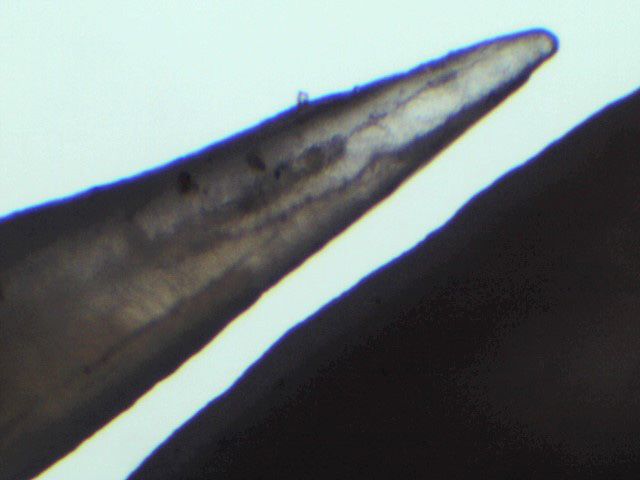
                                                                              Stingray envenomation and injury in a dog          3

     a                                                                  b

     c                                                                  d

Figure 1 x10 magnification of the serrated edges of a stingray stinger removed from a dog. a. sharp pointed cranial spine with a broken tip,
b. blunt caudal spines, c. sharp pointed mid spines, d. sharp pointed caudal spines.

magnification. This also allows the clinician to determine              antibiotics. These injuries could lead to septicaemia
whether there is a real need for diagnostic imaging. As                 and osteomyelitis. Many marine infectious bacteria are
seen in this report, the barb had no significant fractures              sensitive to aminoglycosides and fluoroquinolones, a
indicating there was no remnant fragment of clinical                    few, such as Erysipelothrix rusiopathiae are sensitive to
importance in the wound. The knowledge of the anatomy                   the penicillin. [3,16,25-28]. Flushing with normal saline
of the stingray barb was useful in this method. Diagnostic              solution helps to remove contaminants, debris and also
imaging, such as MRI, radiograph, and ultrasound have                   impact on the venom. Since the point of introduction of
been useful in detecting embedded remains of stingray                   contaminant was the location of the barb, it is logical to
barbs, although diagnostic value of radiographs in some                 conclude that microbial multiplication will commence
cases is still uncertain. MRI has been recommended but                  from that point. Subcutaneous injection of a broad-
it is mostly expensive and less accessible for veterinary               spectrum antibiotic around and within the puncture
use [3,16,23,24].                                                       wound site, increases initial antibiotic concentration
     A puncture wound sustained from a marine stingray                  in the wound site. This also decreases the possibility of
is a contaminated wound with high possibility of                        rapid multiplication and assimilation of bacteria into the
infection, it could easily become a recalcitrant wound if               blood stream [29-37]. In this case, a successful treatment
not treated properly. It is pragmatic to consider broad-                was achieved with the use of local subcutaneous injection
spectrum antibiotic prophylaxis in the management of                    of Penicillin-streptomycin and oral administration
such injury [16,23]. Bacteria such as Aeromonas, vibrios                amoxicillin-clavulanate [38,39]. There is currently no
and clostridia are possible microbial contaminants of                   research in canine stingray envenomation to support this
such wounds and some are resistant to routinely used                    approach.
4          Olutoye Adegboye and Olusegun Adegboye

                                                                            [14] Russel FE, Fairchild MD, Michaelson J. Some properties of the
4 Conclusion                                                                     venom of the stingray. Med Arts Sci. 1958;12(2):78–86.
                                                                            [15] Acott C, Meier J. Clinical toxicology of venomous stingray
Stingray envenomation in Africa has not been extensively                         injuries. In: Meier J, White T, editors. Handbook of clinical
                                                                                 toxicology of animal venoms and poisons. Boca Raton: Press;
studied, and there is a dearth of knowledge of the pathologies
                                                                                 1995. pp. 135–40.
of this condition in veterinary science. Although research                  [16] Clark RF, Girard RH, Rao D, Ly BT, Davis DP. Stingray
in this field in veterinary science is virtually non-existent,                   envenomation: a retrospective review of clinical presentation
case reports such as this will provide impetus for research                      and treatment in 119 cases. J Emerg Med. 2007 Jul;33(1):33–7.
and a knowledge base for practicing veterinarians, vets in                  [17] The European Agency for the Evaluation of Medicinal
training and human clinicians.                                                   Products. Veterinary Medicines Evaluation Unit. EMEA/
                                                                                 MRL/183/97-FINAL. (1997). Available from: https://
                                                                                 www.ema.europa.eu/en/documents/mrl-report/
Conflict of interest: Authors state no conflict of interest                      tolfenamic-acid-summary-report-committee-veterinary-
                                                                                 medicinal-products_en.pdf
Data availability: Data sharing is not applicable to this                   [18] Veterinaria SP. Tolfedin 40mg/ml Quick and Convincing.
article as no datasets were generated or analysed during                         Available from: https://www.spveterinaria.eu/download/
                                                                                 m46IPCaRMP8jwDd/778/TOLFEDOL.pdf [Accessed on February
the current study.
                                                                                 10, 2020]
                                                                            [19] Weinberg L, Peake B, Tan C, Nikfarjan M. Pharmacokinetics
                                                                                 and Pharmacodynamics of Lignocaine : A review. World Journal
References                                                                       Anesthiology. 2015;4(2):17–29.
                                                                            [20] Germain M, Smith KJ, Skelton H. The cutaneous cellular
[1]    Compagno, LJV and LJ Marshall. ‘Fontityrygon margarita’ IUCN              infiltrate to stingray envenomization contains increased TIA+
       Red List of Threatened species (2016)                                     cells. British Journal of Dermatology. 2000; 143(5):1074-7. doi:
[2]    Ratton B. Mebs D. Venomous and poisonous animals.                         https://doi.org/10.1046/j.1365-2133.2000.03848.. x. PMID:
       A handbook for biologist, toxicologists, physicians and                   11069525.
       pharmacist. CRC press; 2002.                                         [21] Rodriguez HG, Sanchez EC, Mendez JD. Stingray poisoning, a
[3]    Diaz JH. The evaluation, management, and prevention                       careless aspect in Mexico. Adv Environ Biol. 2008;2(2):54–62.
       of stingray injuries in travelers. J Travel Med. 2008                [22] Enad JG, Espiritu JM, Fisher D. Stingray injury of the hand:
       Mar-Apr;15(2):102–9.                                                      review of management. Trop Doct. 2001 Jul;31(3):174–5.
[4]    Snyderman M, Wiseman C. Guide to marine life. Carribbean,            [23] O’Malley GF, O’Malley RN, Pham O, Randolph F. Retained
       Bahamas. New york (Florida): Aqua Quest Publication; 1996.                Stingray Barb and the Importance of Imaging. Wilderness
[5]    Isbister GK. Marine envenomation from coral, sea urchins, or              Environ Med. 2015 Sep;26(3):375–9.
       stingrays. 2020. Available from: http://www.uptodate.com/            [24] Weinberg L, Peake B, Tan C, Nikfarjan M. Pharmacokinetics
       contents/ marine-envenomations-from-coral-sea-urchins-or-                 and Pharmacodynamics of Lignocaine : A review. World Journal
       stingrays [Accessed on October 10, 2020].                                 Anesthiology. 2015;4(2):17–29.
[6]    Perkins RA, Morgan SS. Poisoning, envenomation, and                  [25] Finkelstein R, Oren I. Soft tissue infections caused by
       trauma from marine creatures. Am Fam Physician. 2004                      marine bacterial pathogens: epidemiology, diagnosis, and
       Feb;69(4):885–90.                                                         management. Curr Infect Dis Rep. 2011 Oct;13(5):470–7.
[7]    Masson AA. Ormonde do carno PHA, Carvalho JLV.                       [26] Midani S, Rathore M. Chromobacterium violaceum infection.
       Rhabdomylysis secondary to an accident with marine                        South Med J. 1998 May;91(5):464–6.
       stingray (Dasyatis family). J Venom Anim Toxins Incl Trop Dis.       [27] Yang CH. Nonpigmented Chromobacterium violaceum
       2012;18(3):344–8.                                                         bacteremic cellulitis after fish bite. J Microbiol Immunol Infect.
[8]    Fenner PJ, Williamson JA, Skinner RA. Fatal and non-fatal                 2011 Oct;44(5):401–5.
       stingray envenomation. Med J Aust. 1989 Dec;151(11-12):621–5.        [28] Lee J, Kim JS, Nahm CH, Choi JW, Kim J, Pai SH, et al. Two cases
[9]    Weiss BF, Wolfenden HD. Survivor of a stingray injury to the              of Chromobacterium violaceum infection after injury in a
       heart. Med J Aust. 2001 Jul;175(1):33–4.                                  subtropical region. J Clin Microbiol. 1999 Jun;37(6):2068–70.
[10]   Cook MD, Matteucci MJ, Lall R, Ly BT. Stingray envenomation. J       [29] Fleischman AN, Austin MS. Local Intra-wound Administration of
       Emerg Med. 2006 Apr;30(3):345–7.                                          Powdered Antibiotics in Orthopaedic Surgery. J Bone Jt Infect.
[11]   Smarrito S, Smarrito F, Leclair O, Labbe JL. [Surgical                    2017 Jan;2(1):23–8.
       management of stingray injuries. About two clinical cases]. Ann      [30] van der Horst AS, Medda S, Ledbetter E, Liu A, Weinhold P,
       Chir Plast Esthet. 2004 Aug;49(4):383–6.                                  Del Gaizo DJ, et al. Combined local and systemic antibiotic
[12]   Russell FE, Van Harreveld A. Cardiovascular effects of the                treatment is effective against experimental Staphylococcus
       venom of the round stingray, Urobatis halleri. Arch Int Physiol           aureus peri-implant biofilm infection. J Orthop Res. 2015
       Biochim. 1954 Sep;62(3):322–33.                                           Sep;33(9):1320–6.
[13]   Rusell FF, Baririt WC, Fairchild MO. Electrocardiographic patterns   [31] Venkatesh P, Temkar S, Tripathy K, Chawla R. Intralesional
       evoked by venom of the stingray. Proceedings of the Society for           antibiotic injection using 41G needle for the management of
       Experimental Biology and Medicine. Society for Experimental               subretinal abscess in endogenous endophthalmitis. Int J Retina
       Biology and Medicine (New York, N.Y.). 1957; 96: 634-635.                 Vitreous. 2016 Aug;2(1):17.
Stingray envenomation and injury in a dog 5
[32] Silberg BN. Direct Antibiotic Delivery into Soft Tissue environmental bacteria in freshwater stingray wound-healing.
Infections Using Ultrasonic Dispersion. Plast Reconstr Surg. Toxicon. 2011 Aug;58(2):147–53.
2013;132(4):51–2. [37] Nelson Jr., Kalley F. Raimundo P., Steven A. A Severe Accident
[33] Cavanaugh DL, Berry J, Yarboro SR, Dahners LE. Better Caused by an Ocellate River Stingray (Potamotrygon motoro)
prophylaxis against surgical site infection with local as well in Central Brazil: How Well Do We Really Understand Stingray
as systemic antibiotics. An in vivo study. J Bone Joint Surg Am. Venom Chemistry, Envenomation, and Therapeutics?
2009 Aug;91(8):1907–12. Toxin. 2015; 7:2272-2288. DO - https://doi.org/10.3390/
[34] Koulakis JP, Rouch J, Huynh N, Wu HH, Dunn JC, Putterman S. toxins7062272.
Tumescent Injections in Subcutaneous Pig Tissue Disperse Fluids [38] Yarboro S.R., Baum E. J., Dahners L. E., Locally
Volumetrically and Maintain Elevated Local Concentrations of Administered Antibiotics for Prophylaxis Against Surgical
Additives for Several Hours, Suggesting a Treatment for Drug Wound Infection - An in Vivo Study. Journal of Bone and Joint
Resistant Wounds. Pharm Res. 2020 Feb;37(3):51. Surgery 2007:89:929-33 • doi:https://doi.org/10.2106/
[35] U.S. National Library Of Medicine Tumescent Anesthesia J3JS.F.00919.
Antibiotic Delivery (TAAD) Available from. https://clinicaltrials. [39] Huether MJ, Griego RD, Brodland DG, Zitelli JA.
gov/ct2/show/NCT03226626 [Accessed September 3, 2020] Clindamycin for intraincisional antibiotic prophylaxis in
[36] Domingos MO, Franzolin MR, dos Anjos MT, Franzolin TM, dermatologic surgery. Arch Dermatol. 2002 Sep;138(9):
Barbosa Albes RC, de Andrade GR, et al. The influence of 1145–8.You can also read