Synergistic Effect and Antibiofilm Activity of a Skin and Wound Cleanser
←
→
Page content transcription
If your browser does not render page correctly, please read the page content below
ORIGINAL RESEARCH

Synergistic Effect and Antibiofilm Activity of
a Skin and Wound Cleanser
Ann Beal Salamone, MA1; Joseph C. Salamone, PhD1; Rebecca Erin McMahon, PhD1; Suprena Poleon, BS2; Nina Bionda, PhD3; and Peter D’Arpa, PhD4

ABSTRACT
Introduction. Biofilm in chronic wounds impedes the wound healing process. Each biofilm has differing characteristics requiring a
multifaceted approach for removal while maintaining a surrounding environment conducive to wound healing. Objective. In this study, 3
of the components in a wound cleanser are tested to determine synergy in eradicating biofilms of methicillin-resistant Staphylococcus
aureus (MRSA) and Pseudomonas aeruginosa in vitro. Materials and Methods. The 3 components assessed for synergy were
ethylenediamine tetraacetic acid sodium salts (EDTA), vicinal diols (VD; ethylhexylglycerin and octane-1,2-diol), and polyhexamethylene
biguanide (PHMB). Each component was assessed individually and in combination while dissolved in a base solution. The Calgary assay
method was used for biofilm growth and treatment. Kull Equation analysis for synergy was conducted using viable count results. Results.
Synergy is defined as the interaction of components to produce a combined effect greater than the sum of their separate effects. The
base solution containing all 3 components (EDTA, VD, and PHMB) reduced biofilm viability by more than 5 logs, demonstrating statistically
significant synergy. The 3 components tested individually in the base solution resulted in the following: EDTA did not reduce bacteria
viability; VD reduced viability by about 1 log; and PHMB reduced P aeruginosa viability by about 2.5 logs and MRSA viability by about 4 logs.
Of importance, the MRSA biofilm failed to regrow in the recovery plates after combined treatment, indicating complete elimination of the
biofilm bacteria. Conclusions. The experimental and calculated results indicate the 3 components (VD, EDTA, and PHMB) when used
together act synergistically to eradicate MRSA and P aeruginosa biofilms in vitro.

KEY WORDS
wound cleanser, biofilm, MRSA, Staphylococcus aureus, Pseudomonas aeruginosa, synergy, EDTA, vicinal diols, polyhexamethylene biguanide

INDEX
Wounds 2020;32(8):208–216. Epub 2020 May 7

In most natural environments, free-float-       and London dispersion forces.8,9 The EPS              Components of the wound cleanser prod-
ing bacteria exist transiently and only as a    comprises about 50% to 90% of the total            uct studied herein were selected based on
minor population, while the predominant         biofilm organic matter and varies depend-          the authors’ experiences in eye care and wa-
form is biofilm,1 aggregates of microorgan-     ing on the microorganisms, environment,            ter treatment as well as taking into consid-
isms within a self-created polymeric matrix     and biofilm age.8,10,11 The EPS of biofilm in      eration the wound milieu to target a breadth
wherein they are resistant to host defenses     wounds is comprised of dead host tissues,          of biofilms. These components comprise:
and antimicrobial agents. Biofilm has been      in addition to the substances the microor-         (1) polyhexamethylene biguanide (PHMB),
reported to be involved in 78% to 90% of        ganisms secrete, as well as proteins, nucleic      a broad-spectrum polycationic wound
human chronic wounds2,3 and is associated       acids, lipids, polysaccharides, and humic          care antimicrobial14-16 that also is used in
with delayed wound healing4 and other           substances.8,10,11 These substances and their      multipurpose contact lens solutions,17 water
negative wound healing outcomes.5-7             interactions are targets for biofilm elimina-      treatment, and numerous consumer prod-
   Solutions for the elimination of biofilm     tion from wounds.                                  ucts; (2) ethylenediamine tetraacetic acid
have demonstrated significant challenges,          Control of the wound bioburden and              sodium salts (EDTA), a chelator of divalent
in part due to the complexity of the biofilm    biofilm involves multiple treatment                metal ions used in wound care, contact
structure. Biofilm is the construct (mi-        modalities and components that impact              lens cleaners, and numerous personal care
crobial community) created through the          microbial activity and the integrity and           products18; and (3) vicinal diols (VD), eth-
attachment of microorganisms to substrata       attachment of EPS.12,13 Optimally, com-            ylhexylglycerin and octane-1,2-diol, which
within an extracellular polymeric substance     ponents are synergistic not just additive.         are amphiphilic surfactants with moistur-
(EPS).8,9 This construct is stabilized by       In other words, with synergy, 1 plus 1 is          izing, antimicrobial, and odor-reducing
electrostatic interactions, hydrogen bonds,     greater than 2 (1 + 1 > 2).                        functions used in underarm deodorants.19,20

                            Licensed and used with permission from HMP from August 21, 2020 through August 21, 2021.

208                                                                                                                    August 2020 | vol. 32, no. 8
Salamone et al
Table 1. Wound cleanser components and their properties
INGREDIENT PROPERTIES
Antibiofilm components EPS disruption Microbial kill
PHMB In vitro: Cationic polymer that forms Clinical: Antimicrobial44,50
polyelectrolyte complexes with polyanions46 In vitro: Disrupts microbe cell wall47,48
Flocculant45 In vitro: Enters bacterial cells49
EDTA (di- and tri-Na) Clinical/in vitro: Broad pH range chelator18 Clinical: Antimicrobial18
In vitro: Chelates Ca+2 57 In vitro: Disrupts microbe cell wall56
In vitro: Destabilizes matrix integrity18
VD Clinical: Reduces odor62,63 Clinical: Antimicrobial61,62
In vitro: Dissolves and swells lipids63,64 In vitro: Antimicrobial61,62
Amphiphilic surfactants63,64 In vitro: Disrupts microbe cell wall19
HLB 7–7.563,64 In vitro: Enters microbe cells19
BASE SOLUTION IN WATER PROPERTIES
P-407 Clinical: Detergent70
Triblock copolymer surfactant69
Mw 9,840-14,600 Daltons69
HLB 18–21.569
HPMC Clinical: Mucoadhesive73
Neutral charge73
NaCl Clinical: Osmolality balance74
EPS: extracellular polymeric substance; PHMB: polyhexamethylene biguanide; EDTA: ethylenediamine tetraacetic acid sodium salts;
VD: vicinal diols; HLB: hydrophilic-lipophilic balance; P-407: Poloxamer 407; HPMC: hydroxypropylmethylcellulose; NaCl: sodium chloride
The 3 components (PHMB, EDTA, and VD) di- and tri-sodium salts, ethylhexyl- under specified conditions with the test
were theorized to synergistically disrupt glycerin and octane-1,2-diol (ie, VD, solutions. The log survival of the biofilm
EPS, providing access to the microbes, and monoalkyl glycerol, and monoalkyl glycol, microbes (the data) was mathematically as-
then to synergistically permeabilize cell respectively) (Sensiva SC 50 and Sensiva sessed for synergy using the Kull equation.
membranes and impair processes needed SC 10; Schülke & Mayr GmbH), P-407,
for viability. hydroxypropylmethylcellulose (HPMC), Calgary assay for survival of S aureus or
Each component was evaluated individ- and sodium chloride (NaCl) in water at P aeruginosa gown in biofilm
ually and compared with the 3 components physiological osmolality and pH 5.5. The minimum biofilm eradication (MBEC)
combined in the cleanser product for assay with the biofilm device was utilized.23
synergistic activity in disrupting and killing Bacterial strains An MBEC 96-peg lid (Innovotech Inc) was
Pseudomonas aeruginosa and Staphylococcus Methicillin-resistant S aureus strain placed into a 96-well plate filled with 150 µL
aureus biofilms. Each test solution was (MRSA) USA-300 is a predominant com- of an overnight culture of S aureus or P aeru-
dissolved in a base solution containing a munity-associated methicillin-resistant ginosa diluted to 0.1 OD₆₀₀. During method
non-ionic surfactant poloxamer 407 (P- strain that causes significant morbidity and development, 2 inoculation volumes were
407) , a mucoadhesive hydroxypropylmeth- mortality.21 Pseudomonas aeruginosa (Schro- investigated (100 µL and150 µL). There was
ylcellulose (HPMC), and sodium chloride eter) Migula (ATCC 27312) was originally no significant difference in the overall num-
(NaCl). A statistically significant synergy isolated from an infected wound.22 ber of biofilm-associated bacteria for either
index of less than 1 was determined, which species with respect to different inoculation
proved synergy. Experimental design volumes. The higher inoculation volume of
Correlational research was conducted to 150 µL was selected for this synergy study.
MATERIALS AND METHODS determine the synergy between 3 com- The MBEC plate was covered with
Wound cleanser materials and ponents of a wound cleanser based on parafilm to prevent evaporation and was in-
compositions anti-biofilm effectiveness. Biofilm was cubated with shaking (75 rpm) for 48 hours.
Aqueous wound cleanser compositions grown using the Calgary Biofilm Device The biofilm-coated pegs were removed
were created by solvating PHMB, EDTA (Innovotech Inc). The biofilm was treated and rinsed with 200 µL/well of PBS twice
woundsresearch.com 209Antibiofilm Activity of a Wound Cleanser
Figure 1. Wound cleanser components tested for synergism in eradicating biofilms of Pseudomonas aeruginosa and Staphylococcus aureus.
Structures obtained from PubChem.30
EDTA: ethylenediamine tetraacetic acid sodium salts; PHMB: polyhexamethylene biguanide
to remove loosely adhered bacteria. The sd = FALSE and Benjamini-Hochberg cor- RESULTS
volume of 200 µL/well of PBS, test solution, rection for multiple comparisons).24 This Polymeric biguanides (PHMB), VD
or neutralizer was selected to provide for test performs better than the Mann-Whit- (ethylhexyl glycerin and octane-1,2-diol),
complete submersion of the biofilm-coat- ney U test in controlling Type I errors when and EDTA have demonstrated qualitative
ed pegs. Submersion of the pegs in the variances are unequal.25-27 synergistic biocidal activity in antibiofilm
same volume of each solution eliminates Synergy among the test solutions was research studies.29 Here, quantitative stud-
potential data variations due to a portion determined using the Kull equation,28 which ies with statistical analysis are presented,
of the biofilm surface not being exposed to calculates a Synergy Index (SI), equaling which verify statistically significant synergy.
the solutions. In clinical practice, washing/ 1 for simple additivity, greater than 1 for Together, the 3 components with the
soaking of a wound’s complete biofilm antagonism, and less than 1 for synergy. The base components of the complete wound
surface with a cleanser is desired. The pegs SI is the sum of the terms that are the ratios cleanser are listed in Table 1, and the
then were transferred into a 96-well plate of the viable counts (vc) resulting from structures of the 3 components under
containing the test or control solutions treatment with the complete cleanser (CC) study for synergistic antibiofilm activity
(200 µL/well) for a 15-minute incubation at — multiplied by the weight fraction (wf) of are shown in Figure 1.30
room temperature with shaking (75 rpm). the ingredient evaluated in that term — to For assessing the synergistic activity
After treatment, the pegs were transferred the vc resulting from the single ingredient of EDTA, VD, and PHMB in eliminating
into the neutralization plate containing (either EDTA, VD, or PHMB) (Formula). single-species biofilms of P aeruginosa
200 µL/well of Dey-Engley broth (Becton
Dickinson) for 5 minutes before being trans-
ferred into the recovery plate (200 µL/well FORMULA
of tryptic soy broth; Becton, Dickinson and
(CCvc * EDTAwf ) (CCvc * VDwf ) CCvc * PHMBwf
Company), which then was sonicated for 20 SI = + +
minutes to disintegrate the biofilms. From EDTAvc VDvc PHMBvc
each well of the recovery plate, 100 µL was Where:
removed, serially diluted, and plated to enu- SI: Synergy Index (Synergy: SI1); EDTAvc: vc after EDTA treatment; VDvc: vc after VD
merate colony forming units (CFUs). The treatment; PHMBvc: vc after PHMB treatment; CCvc: vc after treatment with CC (ie, EDTA, VD, and PHMB); EDTAwf: wf of
remaining volume of the recovery cultures EDTA=0.115; VDwf: wf of VD=0.708; PHMBwf: wf of PHMB=0.177
was incubated overnight to assess regrowth.
The confidence interval of the SI was and S aureus, the solutions listed in
Data analysis constructed using bootstrap resampling Table 2 were prepared. These solutions
For this study, t tests with unequal varianc- (R package boot, bootstrap replicates = 10 all contain the base solution compo-
es were performed on the ranked data using 000, set.seed = 1) of the vc data (replacing nents: the neutral-charged, amphiphilic
the R function pairwise t test (with pooled. 0 CFU with 0.1 CFU). surfactant P-407 and the neutral-charged,
210 August 2020 | vol. 32, no. 8Salamone et al
Table 2. Test solutions used to assess synergistic antibiofilm activity of EDTA, VD, and PHMB
SOLUTION EDTA-2Na EDTA-3Na VDa1 VDb2 PHMB P-407 HPMC OSMOLALITY pH
(wt %) (wt %) (wt %) (wt %) (ppm/wt %) (wt %) (wt %) (mOsm/kg)
A 0.05 0.015 – – – 2 0.2 334 5.5
B – – 0.3 0.1 – 2 0.2 347 6.2
C – – – – 1,000/0.1 2 0.2 335 6.3
Dc 0.05 0.015 0.3 0.1 1,000/0.1 2 0.2 352 5.7
a
VD-1: Sensiva SC-50
b
VD-2: Sensiva SC-10
c
Composition of BIAKŌS™ Antimicrobial Skin and Wound Cleanser (Sanara MedTech Inc)
EDTA: ethylenediamine tetraacetic acid sodium salts; VD: vicinal diol; PHMB: polyhexamethylene biguanide; P-407: Poloxamer 407; HPMC:
hydroxypropylmethylcellulose
mucoadhesive HPMC. In addition,
solutions A through C contain 1 of the
biofilm-disrupting components: EDTA,
VD, or PHMB, respectively. Solution D is
the CC comprised of all 3 biofilm-disrupt-
ing components in the base solution.
Biocidal activity of the test solutions
was evaluated on biofilms grown using
the biofilm device (ie, on pegs attached to
the lids of 96-well culture plates).23,31 The
biofilms were treated in blinded fashion
with test solutions A–D (Table 2). After the
treatments, the viable bacteria remaining
adherent to the pegs were enumerated to
determine viable bacterial counts (CFUs).
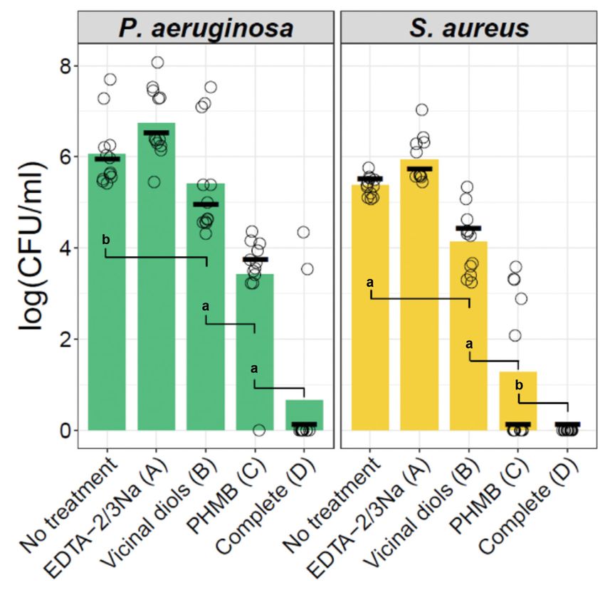
As shown in Figure 2, solution A, contain-
ing EDTA and without VD or PHMB, was
not effective at reducing the viability of
either species’ biofilm. Slightly effective was
solution B, containing VD, which reduced
the viability of both species by about 1 log.
Solution C, containing PHMB, was the most
effective of the single components, reducing
viability by about 2.5 logs or about 4 logs.
However, the complete wound cleanser,
solution D, reduced the viability of the S au-
reus and P aeruginosa monospecies biofilms
by more than 5 logs (Figure 2 and Tables
Figure 2. Viable cell counts after treatment of Staphylococcus aureus and Pseudomonas aeruginosa
3, 4). Of importance, S aureus biofilms that
biofilms with the base solution containing either EDTA, VD, or PHMB; or the complete wound cleanser
yielded 0 CFU after treatment also failed to solution containing EDTA, VD, and PHMB.
regrow after incubating the recovery plates Individual viable cell count determinations (CFU) are shown as open circles (n=12). Letters in
overnight, demonstrating complete biofilm parentheses correspond to the solutions listed in Table 2. The height of the green and yellow bars
elimination from the pegs. represents mean CFUs. The median is marked by thick horizontal lines.
Significant differences between the groups are shown for a one-sided t test for unequal variances on
To assess synergy among EDTA, VD,
ranked data: aPAntibiofilm Activity of a Wound Cleanser
components in eradicating P aeruginosa combination of PHMB, EDTA salts, and Biofilm infections persist in wounds
and S aureus biofilms was 0.148 (95% CI, VD had been found to provide synergistic, as a result of biofilm EPS blocking access
0.019–0.340) and 0.032 (95% CI, 0.027– not simply additive, antibiofilm effective- of antimicrobial agents to their sites of
0.050), respectively. These SI values ness in this report. The Kull equation was action as well as microbes in biofilm having
demonstrate evidence of highly synergis- created to determine synergy of anti- depressed metabolism and activated
tic biocidal activity by the components. fungal mixtures that were tested against protective stress responses.33-36 Thus,
Similar values of SI were calculated using planktonic fungi. Subsequently, Schmaus the removal of biofilm from wounds is
median instead of mean log (CFUs) or et al32 used the Kull equation for synergy important to promote wound healing,37
when only VD and PHMB solutions were determination of mixtures of 1,2 alkane and an aggressive multimodal therapy that
included in the analysis (ie, omitting diols as antimicrobial agents when tested includes debridement, frequent lavage,
solution A from the equation). on planktonic microorganisms. To the and antimicrobial treatment is supported
authors’ knowledge, the use of the Kull by clinical evidence.35,37 Such repeated
DISCUSSION equation to prove antibiofilm synergy for attacks on biofilm forces it to reattach
Using the Kull equation to analyze the any application, including wound cleans- and reform, temporarily driving it into an
biofilm viability experimental results, the ers, has not previously been published. immature state more susceptible to host
defenses and antimicrobials.12,38 However
within hours posttreatment, biofilm can
reform39 and spread into tissues where it
Table 3. Mean CFU differences between treatments adheres firmly,2 thus repeatedly applied
debridement40 and topical biocides have
STRAIN TREATMENT MEAN LOG10 CFU (% OF CONTROL) SD been used to deter biofilm from reforming
Pseudomonas No treatment 6.06 (100) 0.73 and entrenching into tissue.2,35 Of note,
aeruginosa EDTA-2/3Na (A) 6.75 (111) 0.76 microbes do not proliferate unchecked
in healthy tissue.41 This suggests that a
VD (B) 5.41 (89) 1.17
multifaceted approach is required in vivo
PHMB (C) 3.41 (56) 1.13 to remove all causes of tissue damage, such
Complete (D) 0.66 (11) 1.55 as compromised circulation, edema, or
Staphylococcus No treatment 5.37 (100) 0.21 repeated trauma, while protecting injured
aureus tissue from microbial invasion and/or
EDTA-2/3Na (A) 5.93 (110) 0.5
biofilm formation.42
VD (B) 4.14 (77) 0.69 As listed in Table 1 and, discussed
PHMB (C) 1.27 (24) 1.61 further on, PHMB, EDTA, and the VD each
Complete (D) 0 (0) 0 individually are known to disrupt EPS and
permeabilize microbial cell membranes
CFU: colony forming unit; SD: standard deviation; EDTA: ethylenediamine tetraacetic acid
and impair processes needed for viability.
sodium salts; VD: vicinal diol; PHMB: polyhexamethylene biguanide
Polyhexamethylene biguanide is a
Table 4. P values for differences between treatments
NO TX EDTA-2/3Na (A) VD (B) PHMB (C)
Pseudomonas aeruginosa EDTA-2/3Na (A) 0.99 - - -
VD (B) 0.39 7.2e-04 - -
PHMB (C) 6.2e-09 5.8e-11 1.3e-04 -
Complete (D) 5.8e-11 1.7e-12 2.1e-06 6.7e-04
Staphylococcus aureus EDTA-2/3Na (A) 0.99 - - -
VD (B) 3.1e-05 4.6e-07 - -
PHMB (C) 1.8e-06 3.4e-07 3.1e-05 -
Complete (D) 2.3e-16 5.1e-13 5.9e-10 0.011
Tx: treatment; EDTA: ethylenediamine tetraacetic acid sodium salts; VD: vicinal diol; PHMB: polyhexamethylene biguanide
212 August 2020 | vol. 32, no. 8Salamone et al broad-spectrum antimicrobial, biocom- um) provides a slightly acidic pH, which, biofilm from the wound. Very hydrophilic patible43,44 cationic polymer that has a low together with physiologic osmolarity to surfactants, such as poloxamer 188 with an average molecular weight range of 2000 prevent cell dehydration from high osmo- HLB of 29, have been used to aid in the re- to 4500 Daltons. Polyhexamethylene larity or cell swelling from low osmolarity, moval of biofilm from wound surfaces.12,67,68 biguanide can penetrate EPS and form contribute to mitigating pain while being However, the nonionic surfactant, P-407 polyelectrolyte complexes with polyanions, non-cytotoxic to human tissue.59,60 in the base solution, not only lowers inter- such as DNA and polysaccharides, causing The VD, ethylhexylglycerin and facial tension to aid in removal of debris flocculation and large aggregates that may octane-1,2-diol, are multifunctional but also is a detergent (HLB 18–21.5)69 that be removed more easily.45 When PHMB personal care ingredients commonly used incorporates (ie, emulsifies) hydrophobic is present, aggregation of EPS polyan- in underarm deodorants, with moisturiz- materials into water.70 These hydropho- ions (alginates) in P aeruginosa biofilm is ing and antimicrobial activities resulting bic materials may be present as lipids, visually observable as “clumps.” Bueno and in odor reduction due to inhibition of proteins, and polysaccharides, for instance. Moraes46 used this effect to bind PHMB to odor-causing Gram-positive bacteria (eg, By incorporating these organic substanc- chitosan-alginate wound dressings for sus- Corynebacterium spp, Leifsonia aquaticum, es into water, they are easier to remove tained release of PHMB. Once in contact Ochrobactrum anthropi, Kocuria rhizophi- by irrigation. In addition to serving as a with microbes, the polycation PHMB inter- la).61-64 They are known to disrupt micro- detergent and surfactant, P-407 aids in the acts with microbial membrane anions to bial membranes and synergistically boost solubility of the VD due to its amphiphilic disrupt the cell membrane with resultant the effectiveness of preservatives such as competency.71 Poloxamer 407 (Pluronic leakage of cytoplasmic components and in- parabens or phenoxyethanol19 — eg, eth- 127; Sigma-Aldrich) is a triblock copolymer hibition of membrane-bound enzymes17,47,48; ylhexylglycerin potentiated the lethality consisting of a central hydrophobic block additionally, PHMB enters bacterial cells, of phenoxyethanol against P aeruginosa of about 101 repeats of polypropylene condenses chromosomal DNA, and arrests and Aspergillus niger.65 With 8 carbon glycol flanked by 2 hydrophilic blocks of cell division.49 Also, PHMB has been rec- atoms and 2 hydroxyl groups on adjacent about 56 repeats of polyethylene glycol. Of ommended as an antimicrobial compound carbons, these amphiphilic surfactants, note, P-407 helps to maintain the activity of choice for chronic wounds and burns.50 with an hydrophilic-lipophilic balance of PHMB17 and VD. Ethylenediamine tetraacetic acid (HLB) of 7 to 7.5, provide humectance Mucoadhesives are used to increase sodium salts (di- and tri-) functions (hydration) and emollience (occlusivity, residence time of a composition on a over a wide pH range (2–12), which is a softening, lubrication, spreading, and mucosal membrane such as found in the requirement as both the wound milieu pH delivery of actives), solvate out lipids and gastrointestinal tract, lungs, and eyes. range can be from acidic to basic depend- humic components (ie, from biofilm), Mucosal membranes contain up to 95% ing on native biochemical processes for and have antimicrobial activities differ- water, with the remaining components wound healing51 and multispecies biofilms ing from PHMB.63,64 While PHMB has comprising glycoproteins, lipids, and oth- (aerobic, facultative, and anaerobic) can broad-spectrum activity against bacteria, er hydrophilic organic matter.72 In general, have pH gradient ranges from acidic to fungi, protozoa, and viruses, the VD are a wound bed has similarities to mucosal basic.52 The EDTA is known to chelate particularly effective against Gram-posi- tissue, such as higher water content com- divalent metal cations essential for bac- tive bacteria and yeasts.65,66 bined with the presence of hydrophilic or- terial growth53 and destabilize bacterial By incorporating the combination of ganic matter, as found in wound exudate membranes and matrix integrity.18,53-55 As PHMB, EDTA, and VD, synergistic antibio- as well as biofilm. Therefore, the muco- an example, EDTA is reported to potenti- film effectiveness was found, not just addi- adhesive, water-soluble, neutral-charged ate the antimicrobial effects of quaternary tive effectiveness. The SI values determined HPMC73 forms a hydrated film on the ammonium compounds by extraction of were 0.148 with P aeruginosa and 0.032 with wound surface and, hence, increases the lipopolysaccharide from P aeruginosa cell MRSA. The SI values are notably lower than cleanser’s residence time. walls.56 Cationic quaternary ammonium 1, thus indicating high synergy. In order to adjust osmolality to a compounds absorb on negatively charged The base solution for these studies physiologically normal range (290–320 cell walls concurrently with EDTA che- and used in the wound cleanser was mOsm/kg), sodium chloride is added as lation of cell wall metal cations with a developed to complement the synergistic a component of the base solution. When resulting loss of lipopolysaccharides and antibiofilm efficacy of the 3 components products used on wounds are hyperton- increased cell permeability.56 Also, EDTA (PHMB, EDTA, and VD). The base solu- ic, water is pulled out of surrounding is reported to inhibit excess matrix metal- tion comprises water, a salt, a mucoadhe- tissue through osmosis, which causes loproteases by chelating zinc and calci- sive, and a surfactant. dehydration and cell size shrinkage.74 um,57 thereby facilitate wound healing.58 Surfactants lower the interfacial tension The opposite effect occurs when os- Consistent with healthy human tissue, the between substances and can loosen and molality is hypotonic; the surrounding wound cleanser’s EDTA (di- and tri-sodi- remove dirt, debris, slough and loosen tissue pulls in water causing cell size woundsresearch.com 213
Antibiofilm Activity of a Wound Cleanser

enlargement.74 Pain is created with either    tion and density distribution (both EPS                         prevalence of biofilms in chronic wounds:

too high or too low osmolality, and neu-      and microbial cells) will be influenced by                      a systematic review and meta-analysis of

rological damage is suffered.74 Therefore,    these factors in each biofilm-containing                        published data. J Wound Care. 2017;26(1):20–25.

a physiologically balanced osmolality is      wound. The synergistic biocidal activ-                          doi:10.12968/jowc.2017.26.1.20

preferred for protection of healthy tissue    ity likely extends to biofilms of other                   4.    Metcalf DG, Bowler PG. Biofilm delays wound

in and surrounding the wound.                 microbial species because of the gross                          healing: a review of the evidence. Burns Trauma.

   In summary, the targeted points of         similarity (proteins, polysaccharides,                          2013;1(1):5–12. doi:10.4103/2321-3868.113329

the wound cleanser are (1) synergistic        lipids, humic substances) of EPS across                   5.    Bloemsma GC, Dokter J, Boxma H, Oen IM.

antibiofilm components (PHMB, EDTA,           species; however, the current findings                          Mortality and causes of death in a burn centre.

and VD) complemented by (2) P-407 to          are limited to monospecies biofilms                             Burns. 2008;34(8):1103–1107. doi:10.1016/j.

remove loose debris from the wound            of P aeruginosa and S aureus, common                            burns.2008.02.010

surface through interfacial tension re-       species that infect wounds. Translational                 6.    Omar A, Wright JB, Schultz G, Burrell R, Nad-

duction as well as to incorporate hydro-      research is needed to verify these results                      worny P. Microbial biofilms and chronic wounds.

phobic materials (ie, emulsification) into    in clinical wounds.                                             Microorganisms. 2017;5(1). doi:10.3390/microor-

water and (3) the mucoadhesive HPMC                                                                           ganisms5010009

to increase residence time on wound           CONCLUSIONS                                               7.    Withycombe C, Purdy KJ, Maddocks SE.

surfaces. Additionally, physiologically       Highly synergistic antibiofilm activity                         Micro-management: curbing chronic wound
balanced pH and osmolality are gentle to      was observed for the wound cleanser                             infection. Mol Oral Microbiol. 2017;32(4):263–274.
human tissue.59,60,74                         components — VD, EDTA, and PHMB,                                doi:10.1111/omi.12174
   Clinical validation of the antimicrobi-    in an aqueous base solution of P-407 and                  8.    Flemming HC, Wingender J. Relevance of
al cleanser is in progress with bacterial     HPMC where osmolality and pH were at                            microbial extracellular polymeric substances
fluorescence and DNA sequencing. This         physiological levels — against P aeruginosa                     (EPSs)--part I: Structural and ecological aspects.
also includes targeted data points of         and MRSA monospecies biofilms in vitro.                         Water Sci Technol. 2001;43(6):1–8.
wound healing progression and econom-         Clinical studies to compare these in vitro                9.    Mayer C, Moritz R, Kirschner C, et al. The role
ic evaluation. The algorithm for use of       synergy results with clinical outcomes is                       of intermolecular interactions: studies on model
this wound cleanser is provided under         the next research step.                                         systems for bacterial biofilms. Int J Biol Macro-
the guidelines from the International                                                                         mol. 1999;26(1):3–16.
Wound Infection Institute International                                                                 10.   Vu B, Chen M, Crawford RJ, Ivanova EP. Bacteri-
Consensus Update 2016/Wound Infec-                                                                            al extracellular polysaccharides involved in bio-
                                              acknowledgments
tion in Clinical Practice for “Effective                                                                      film formation. Molecules. 2009;14(7):2535–2554.
                                              Affiliations: 1Rochal Industries LLC, San Antonio, TX;
Wound Infection Management,”75 which                                                                          doi:10.3390/molecules14072535
                                              2
                                               University of North Texas Health Science Center, Fort
recommends regular wound evaluation           Worth, TX; 3iFyber, Ithaca, NY; and 4BioMedWrite,         11.   Donlan RM. Biofilms: microbial life on surfaces.
for signs of infection and to “cleanse the    North Potomac, MD                                               Emerg Infect Dis. 2002;8(9):881–890.
wound with each dressing change.” The                                                                   12.   Das Ghatak P, Mathew-Steiner SS, Pandey P,
                                              Correspondence: Ann Beal Salamone, MA, Chairman of
cleanser effectiveness may be impacted        the Board, Rochal Industries LLC, Research &                    Roy S, Sen CK. A surfactant polymer dressing
by enzymes, ointments, or oils in the         Development, 12000 Network Blvd, B-200, San                     potentiates antimicrobial efficacy in biofilm
                                              Antonio, TX 78249; absalamone@rochalindustries.com
wound bed. Irrigation of the wound bed                                                                        disruption. Sci Rep. 2018;8(1):873. doi:10.1038/
may be performed with the cleanser to         Disclosure: This study was funded by Rochal                     s41598-018-19175-7
thoroughly rinse the wound bed from           Industries LLC (San Antonio, TX). Dr. Joseph C.           13.   Kostakioti M, Hadjifrangiskou M, Hultgren
                                              Salamone, Ann Beal Salamone, Dr. Rebecca E.
these agents.                                                                                                 SJ. Bacterial biofilms: development, dispersal,
                                              McMahon and Suprena Poleon are/were employees of
                                              Rochal Industries LLC. iFyber was paid to design and            and therapeutic strategies in the dawn of the
LIMITATIONS                                   conduct the blinded biofilm studies. BioMedWrite was            postantibiotic era. Cold Spring Harb Perspect
Biofilm in wounds treated in vivo is ex-      paid to co-write this manuscript and to independently           Med. 2013;3(4):a010306. doi:10.1101/cshperspect.
                                              conduct statistical analyses.
pected to be susceptible to the same syn-                                                                     a010306
ergistic biocidal activities that have been                                                             14.   Moore K, Gray D. Using PHMB antimicrobi-
observed for biofilm treated in vitro due     references                                                      al to prevent wound infection. Wounds UK.
to the multitargeted chemical approach.       1.    Wolcott RD, Ehrlich GD. Biofilms and chronic              2007;3(2):96–102.
However, biofilm in vivo is affected by             infections. JAMA. 2008;299(22):2682–2684.           15.   Wiegand C, Abel M, Ruth P, Hipler UC, HaCaT
systemic environmental factors, such                doi:10.1001/jama.299.22.2682                              keratinocytes in co-culture with Staphylococ-
as host immune response, cardiovascu-         2.    Attinger C, Wolcott R. Clinically addressing bio-         cus aureus can be protected from bacterial
lar sufficiency, age, nutrition, and local          film in chronic wounds. Adv Wound Care (New               damage by polihexanide. Wound Repair
environmental factors, such as repeated             Rochelle). 2012;1(3):127–132.                             Regen. 2009;17(5):730–738. doi:10.1111/j.1524-
trauma. Therefore, the biofilm composi-       3.    Malone M, Bjarnsholt T, McBain AJ, et al. The             475X.2009.00536.x

214                                                                                                                            August 2020 | vol. 32, no. 8
Salamone et al
16. Kamaruzzaman NF, Tan LP, Mat Yazid KA, et al. Tests for treatment group equality when data techniques for wound debridement. Int Wound J.
Targeting the bacterial protective armour; chal- are nonnormal and heteroscedastic. J Modern 2013;10(3):247–251. doi:10.1111/iwj.12045
lenges and novel strategies in the treatment of Applied Statistical Methods. 2007;6(1):117–132. 41. Nakatsuji T, Chiang H-I, Jiang SB, Nagarajan H,
microbial biofilm. Materials (Basel). 2018;11(9). doi:10.22237/jmasm/1177992660 Zengler K, Gallo RL, The microbiome extends
doi:10.3390/ma11091705 28. Kull FC, Eisman PC, Sylwestrowicz HD, Mayer to subepidermal compartments of normal skin.
17. Yanai R, Ueda K, Nishida T, Toyohara M, Mori RL. Mixturs of quaternary ammonium com- Nat Commun. 2013;4:1431–1447. doi:10.1038/
O. Effects of ionic and surfactant agents on the pounds and long-chain fatty acids as antifungal ncomms2441
antimicrobial activity of polyhexamethylene agents. Appl Microbiol. 1961;9(6):538–541. 42. Hurlow J, Couch K, Laforet K, Bolton L, Metcalf
biguanide. Eye Contact Lens. 2011;37(2):85–89. 29. Salamone J, Salamone AB, inventors; Rochal In- D, Bowler P. Clinical biofilms: a challenging
doi:10.1097/ICL.0b013e31820cebc3 dustries LLC, assignee. Biocidal compositions and frontier in wound care. Adv Wound Care (New
18. Finnegan S, Percival SL. EDTA: an antimicrobial methods of using the same. US patent 8,829,053 Rochelle). 2015;4(5):295–301.
and antibiofilm agent for use in wound care. Adv B2. September 9, 2014. 43. Müller G, Kramer A. Biocompatibility index
Wound Care (New Rochelle). 2015;4(7):415–421. 30. Kim S, Chen J, Cheng T, et al. PubChem 2019 of antiseptic agents by parallel assessment of
19. Langsrud S, Steinhauer K, Luthje S, Weber K, update: improved access to chemical data. antimicrobial activity and cellular cytotoxicity.
Goroncy-Bermes P, Holck AL. Ethylhexylglyc- Nucleic Acids Res. 2019;47(D1):D1102–D1109. J Antimicrob Chemother. 2008;61(6):1281–1287.
erin impairs membrane integrity and enhances doi:10.1093/nar/gky1033 doi:10.1093/jac/dkn125
the lethal effect of phenoxyethanol. PLoS 31. Harrison JJ, Stremick CA, Turner RJ, Allan 44. Daeschlein G, Assadian O, Bruck JC, Meinl
One. 2016;11(10):e0165228. doi:10.1371/journal. ND, Olson ME, Ceri H. Microtiter susceptibil- C, Kramer A, Koch S. Feasibility and clinical
pone.0165228 ity testing of microbes growing on peg lids: a applicability of polihexanide for treatment of
20. Okukawa M, Watanabe T, Miura M, Konno H, miniaturized biofilm model for high-throughput second-degree burn wounds. Skin Pharmacol
Yano S, Nonomura Y, Antibacterial activity of screening. Nat Protoc. 2010;5(7):1236–1254. Physiol. 2007;20(6):292–296.
1,2-alkanediol against Staphylococcus aureus doi:10.1038/nprot.2010.71 45. Lopez-Maldonado EA, Oropeza-Guzman MT,
and Staphylococcus epidermidis. J Oleo Sci. 32. Schmaus G, Lange S, Joppe H. Synergistic mix- Ochoa-Teran A. Improving the efficiency of
2019;68(8):759–763. doi:10.5650/jos.ess19074 tures of 1,2 -alkane diols. US7582681. 2009. a coagulation-flocculation wastewater treat-
21. McClure JA, Zhang K. Complete genome 33. Costerton J, Stewart PS, Greenberg EP. Bacterial ment of the semiconductor industry through
sequence of a community-associated methicil- biofilms: a common cause of persistent infec- zeta potential measurements. J Chem. 2014.
lin-resistant Staphylococcus aureus hypervirulent tions. Science. 1999;284(5418):1318–22. doi:10.1155/2014/969720
strain, USA300-C2406, isolated from a patient 34. Stewart PS, Mechanisms of antibiotic resistance 46. Bueno CZ, Moraes AM. Influence of the incor-
with a lethal case of necrotizing pneumonia. Ge- in bacterial biofilms. Int J Med Microbiol. poration of the antimicrobial agent polyhexam-
nome Announc. 2017;5(22):e00461–17. doi:10.1128/ 2002;292(2):107–113. ethylene biguanide on the properties of dense
genomeA.00461-17 35. Seth AK, Geringer MR, Gurjala AN, et al. Treat- and porous chitosan-alginate membranes. Mater
22. Schroeter J. Über einige durch Bacterien ment of Pseudomonas aeruginosa biofilm-infected Sci Eng C Mater Biol Appl. 2018;93:671–678.
gebildete Pigmente. In: Cohn F, ed. Beitrage zur wounds with clinical wound care strategies: a doi:10.1016/j.msec.2018.07.076
Biologie der Pflanzen. J. U. Kern’s Verlag. Breslau, quantitative study using an in vivo rabbit ear 47. Ikeda T, Ledwith A, Bamford CH, Hann RA.
1875:109 –126. model. Plast Reconstr Surg. 2012;129(2):262e–274e. Interaction of a polymeric biguanide biocide
23. Ceri H, Olson ME, Stremick C, Read RR, Morck doi:10.1097/PRS.0b013e31823aeb3b with phospholipid membranes. Biochim Biophys
D, Buret A. The Calgary Biofilm Device: new 36. Rani SA, Pitts B, Beyenal H, et al. Spatial pat- Acta. 1984;769(1):57–66.
technology for rapid determination of antibiotic terns of DNA replication, protein synthesis, and 48. Broxton P, Woodcock PM, Gilbert P. Binding of
susceptibilities of bacterial biofilms. J Clin Micro- oxygen concentration within bacterial biofilms some polyhexamethylene biguanides to the cell
biol. 1999;37(6):1771–1776. reveal diverse physiological states. J Bacteriol. envelope of Escherichia coli ATCC 8739. Microbi-
24. R Core Team. R: a language and environment for 2007;189(11):4223–4233. os. 1984;41(163):15–22.
statistical computing. R Foundation for Statistical 37. Snyder RJ, Bohn G, Hanft J, et al. Wound 49. Chindera K, Mahato M, Sharma AK, et al. The
Computing, 2016. biofilm: current perspectives and strategies on antimicrobial polymer PHMB enters cells and
25. Zimmerman DW, Zumbo BD. Rank transfor- biofilm disruption and treatments. Wounds. selectively condenses bacterial chromosomes.
mations and the power of the Student t-test 2017;29(6):S1–S17. Sci Rep. 2016;6:23121. doi:10.1038/srep23121
and Welch t-test for non-normal populations 38. Stewart PS, Grab L, Diemer JA. Analysis of bio- 50. Kramer A, Dissemond J, Kim S, et al. Con-
with unequal variances. Can J Exp Psychol. cide transport limitation in an artificial biofilm sensus on wound antisepsis: update 2018.
1993;47(3):523–539. doi:10.1037/h0078850 system. J Appl Microbiol. 1998;85(3):495–500. Skin Pharmacol Physiol. 2018;31(1):28–58. doi:
26. Ruxton G, The unequal variance t-test is an 39. Wolcott RD, Rumbaugh KP, James G, et al. 10.1159/000481545
underused alternative to Student’s t-test and Biofilm maturity studies indicate sharp debride- 51. Schneider LA, Korber A, Grabbe S, Dissemond
the Mann–Whitney U test. Behavioral Ecology. ment opens a time- dependent therapeutic J. Influence of pH on wound-healing: a new
2006;17(4):688–690. window. J Wound Care. 2010;19(8):320–328. perspective for wound-therapy? Arch Dermatol
27. Cribbie RA, Bewell C, Wilcox RR, Keselman HJ. 40. Madhok BM, Vowden K, Vowden P. New Res. 2007;298(9):413–420.
woundsresearch.com 215Antibiofilm Activity of a Wound Cleanser
52. Vroom JM, De Grauw KJ, Gerritsen HC, to boost the preservative efficacy of phenoxyeth-
et al. Depth penetration and detection of anol. SOFW J. 2005;131(11):2–7.
pH gradients in biofilms by two-photon 66. Beilfuss W. A multifunctional ingredient for
excitation microscopy. Appl Environ Microbiol. deodorants. SOFW J. 1998;124(6):360–366.
1999;65(8):3502–3511. 67. Anglen JO, Gainor BJ, Simpson WA, Christensen
53. Chew BP, Tjoelker LW, Tanaka TS. In vitro G. The use of detergent irrigation for musculo-
growth inhibition of mastitis causing bacteria skeletal wounds. Int Orthop. 2003;27(1):40–46.
by phenolics and metal chelators. J Dairy Sci. 68. Yang Q, Larose C, Della Porta AC, Schultz GS,
1985;68(11):3037–3046. Gibson DJ. A surfactant-based wound dressing
54. Metcalf D, Parsons D, Bowler PG. Development can reduce bacterial biofilms in a porcine skin
of a next-generation antimicrobial wound dress- explant model. Int Wound J. 2017;14(2):408–413.
ing. Acta Med Croatica. 2016;70(1):49–56. doi:10.1111/iwj.12619
55. Percival SL, Salisbury AM, The efficacy of tet- 69. BASF Pluracare L/F Grades, Poloxamer Techni-
rasodium EDTA on biofilms. Adv Exp Med Biol. cal Information. January 2008. EMM 070801e-
2018;1057:101–110. doi:10.1007/5584_2017_134 00:1–10.
56. Adair FW, Geftic SG, Gelzer J. Resistance 70. Percival SL, Chen R, Mayer D, Salisbury AM.
of Pseudomonas to quaternary ammonium Mode of action of poloxamer-based surfactants
compounds. Resistance of Pseudomonas to qua- in wound care and efficacy on biofilms. Int
ternary ammonium compounds. Appl Microbiol. Wound J. 2018;15(5):749–755. doi:10.1111/iwj.12922
1971;21(6):1058–1063. 71. Bodratti AM, Alexandridis P. Formulation of
57. Tezvergil-Mutluay A, Agee KA, Hoshika T, et al. poloxamers for drug delivery. J Funct Biomater.
The requirement of zinc and calcium ions for 2018;9(1):11–35. doi:10.3390/jfb9010011
functional MMP activity in demineralized den- 72. Smart JD. The basics and underlying mecha-
tin matrices. Dent Mater. 2010;26(11):1059–1067. nisms of mucoadhesion. Adv Drug Deliv Rev.
doi:10.1016/j.dental.2010.07.006 2005;57(11):1556–1568.
58. Rodrigues M, Bonham CA, Minniti CP, Gupta K, 73. Agarwal S, Murthy RS. Effect of different
Longaker MT, Gurtner GC. Iron chelation with polymer concentration on drug release rate and
transdermal deferoxamine accelerates healing physicochemical properties of mucadhesive
of murine sickle cell ulcers. Adv Wound Care gastroretnetive tablets. Indian J Pharm Sci.
(New Rochelle). 2018;7(10):323–332. doi:10.1089/ 2015;77(6):705–714.
wound.2018.0789 74. Danziger J, Zeidel ML. Osmotic homeostasis.
59. Percival SL, McCarty S, Hunt JA, Woods EJ, Clin J Am Soc Nephrol. 2015;10(5):852–862.
The effects of pH on wound healing, biofilms, Published correction appears in Clin J Am Soc
and antimicrobial efficacy. Wound Repair Regen. Nephrol. 2015;10(9):1703.
2014;22(2):174–186. doi:10.1111/wrr.12125 75. International Wound Infection Institute (IWII).
60. Jones EM, Cochrane CA, Percival SL. The Wound infection in clinical practice: principles
effect of pH on the extracellular matrix and of best practice. Wounds Int. 2016:3–30.
biofilms. Adv Wound Care (New Rochelle).
2015;4(7):431–439.
61. Lawan K, Kanlayavattanakul M, Lourith N.
Antimicrobial efficacy of caprylyl glycol and
ethylhexylglycerine in emulsion. J Health Res.
2009;23(1):1–3.
62. KaB M, Lüthje S, Herweg S, Stepan S. Systematic
investigations in the antimicrobial efficacy of
glycerine esters with fatty acids of different
chain length. SOFW J. 2013;139(12).
63. The Derm Review. Ethylhexylglycerin. Published
Sept 28, 2018. https://thedermreview.com/
ethylhexylglycerin/
64. schülke inc. Smooth – safe – sensiva®. www.schul-
ke-us.com/schulke_sensiva_SC10.html. 2020.
65. Beilfuss W, Leschke M, Weber K. A new consept
216 August 2020 | vol. 32, no. 8You can also read