The Ecto-5 nucleotidase/CD73 Mediates Leishmania amazonensis Survival in Macrophages - Hindawi.com
←
→
Page content transcription
If your browser does not render page correctly, please read the page content below
Hindawi
BioMed Research International
Volume 2022, Article ID 9928362, 10 pages
https://doi.org/10.1155/2022/9928362

Research Article
The Ecto-5 ′nucleotidase/CD73 Mediates Leishmania
amazonensis Survival in Macrophages

 Bijay Bajracharya ,1 Deena Shrestha ,2 André Talvani ,2 Ricardo Gonçalves ,3
 and Luís Carlos Crocco Afonso 1
 1
 Laboratory of Immunoparasitology, Biological Sciences Department, ICEB-Federal University of Ouro Preto, Minas Gerais, Brazil
 2
 Laboratory of Immunobiology of Inflammation, Biological Sciences Department, ICEB-Federal University of Ouro Preto,
 Ouro Preto, Minas Gerais, Brazil
 3
 General Pathology Department, ICB, Federal University of Minas Gerais, Belo Horizonte, Minas Gerais, Brazil

 Correspondence should be addressed to Bijay Bajracharya; bjbajra@gmail.com

 Received 20 March 2021; Revised 30 November 2021; Accepted 28 December 2021; Published 11 February 2022

 Academic Editor: Stefano D Amelio

 Copyright © 2022 Bijay Bajracharya et al. This is an open access article distributed under the Creative Commons Attribution License,
 which permits unrestricted use, distribution, and reproduction in any medium, provided the original work is properly cited.

 Endogenous nucleotides produced by various group of cells under inflammatory conditions act as potential danger signals in vivo.
 Extracellularly released nucleotides such as ATP are rapidly hydrolyzed to adenosine by the coordinated ectonucleotidase
 activities of CD39 and CD73. Leishmania is an obligate intracellular parasite of macrophages and capable of modulating host
 immune response in order to survive and multiply within host cells. In this study, the activity of CD73 induced by Leishmania
 amazonensis in infected macrophages has been investigated and correlated with parasite survival and infection in vitro. For
 this, the expression of CD39 and CD73, by flow cytometry, in murine peritoneal macrophages infected with metacyclic
 promastigotes of L. amazonensis has been analyzed. Our results showed that L. amazonensis-infected macrophages, unlike
 LPS-treated macrophages, increased CD73 expression. It was also noted that when CD73 enzymatic activity was blocked by α,
 β-methyleneadenosine 5 ′ -diphosphate sodium salt (APCP), macrophage parasitism was significantly decreased. Interestingly,
 these effects were not associated with the production of TNF-α, IL-10, or nitric oxide (NO). Together, these data demonstrate
 that L. amazonensis induces a regulatory phenotype in macrophages, which by activating the CD39/CD73 pathway allows
 parasite survival through the action of immunomodulatory adenosine receptors.

1. Introduction involved in the manipulation of the macrophage activation,
 however, remain largely unclear.
Leishmania are intracellular parasites that live and multiply Accumulating evidence supports that extracellular ATP
within macrophages in the mammalian host. In order to sur- released at the sites of infection and its degradation prod-
vive in these cells, Leishmania amastigotes must resist or uct, adenosine, function as potent immune modulators
inhibit their microbicidal mechanisms [1]. Cutaneous leish- that mediate both pro- and anti-inflammatory pathways,
maniasis associated with Leishmania amazonensis infection depending on the agonist concentration and receptor sub-
is severe in both humans and experimental models [2, 3]. types expressed by the cell [11]. The increase in extracellu-
It is characterized by uncontrolled parasite replication and lar ATP concentration leads to the activation of NLRP3
profound host immunosuppression [4–7]. This parasite has inflammasome and subsequent release of IL-1β and
been shown to alter the host cell defense mechanisms in sev- TNF-α by macrophages [12, 13]. Extracellular adenosine
eral ways such as inhibition of antigen presentation and formation generally results from the sequential hydrolysis
inhibition of reactive oxygen species (ROS) and nitric oxide of extracellular ATP by the combined action of an ectonu-
(NO) production [8–10]. The underlying mechanisms cleoside triphosphate diphosphohydrolase (CD39) followed
2 BioMed Research International

by ecto-5 ′ nucleotidases (CD73) [14]. Adenosine, by act- BRL-Life Technologies, Grand Island, NY, MO, EUA), and
ing on P1 receptors, modulates the activation of macro- 100 U/ml penicillin G (USB Corporation, Cleveland, OH,
phages, reducing inflammatory and increasing regulatory USA), pH 6.5. Five-day-old stationary phase promastigotes
cytokine production [15]. The ATP/adenosine ratio is were used for metacyclic isolation and purification [19, 25].
determined by the activity of the CD39/CD73 pathway,
which can be altered by pathophysiological events and 2.3. Carboxyfluorescein Succinimidyl Ester (CFSE) Labeling.
ultimately defines the outcome of infections, inflammation, Purified metacyclics (6 × 10⁷ parasites/ml). The carboxyflu-
and injuries [16, 17]. orescein diacetate succinimidyl ester (CFSE) dye was laid
 The capacity of a pathogen to generate extracellular over the 50 μl of PBS and incubated with parasites (final
adenosine through the expression of ectonucleotidases has concentration -5 μM) at 37°C for 10 min in the dark
been identified as an important virulence factor [18]. Our [26]. Parasites were then washed with PBS/10% FBS,
laboratory has been successful to demonstrate that the level pH 7.2, and then suspended in Dulbecco’s modified eagle’s
of ectonucleotidase activity in promastigote forms of Leish- medium (DMEM-Sigma-Aldrich, Missouri, EUA) with 10%
mania is associated with the severity of disease in the murine FBS, 2 mM L-glutamine, 100 U/ml penicillin G, 25 mM N-2-
experimental model and may be involved in the outcome of hydroxiethylpiperazine-N9-2-ethanosulfonic acid (HEPES;
distinct clinical manifestations in patients [19–22]. In the USBiological, Swampscott, MA, USA), 1.2 mM sodium bicar-
case of Leishmania, however, once the parasite is internal- bonate (Vetec Quimica Fina, RJ, Brazil), and 50 μM 2-
ized, its ectonucleotidase activity should cease to influence mercaptoethanol (Pharmacia Biotech AB, Uppsala, Sweden)
the levels of extracellular ATP and adenosine near the host pH 7.2 prior to addition to macrophage cultures. Later,
cell [21]. Interestingly, however, we have also demonstrated CFSE-tagged parasites were measured in FACS diagram,
that upon infection of dendritic cells, Leishmania promasti- and the fluorescence was captured in channel 1 in BD
gotes induce the upregulation of CD39 and CD73 on FACSCalibur.
infected dendritic cells, thus increasing the ability of these
cells to produce extracellular adenosine [23]. This pathway 2.4. Resident Peritoneal Macrophages. Mice were euthanized,
in turn has further shown to impair dendritic cell activation and the abdomen was gently massaged, and peritoneal
through immunomodulating A2b receptors and triggering lavage was collected after injection of 10 ml ice-cold PBS
cAMP pathways in infected cells [24]. with 16G needle [27]. Cells were centrifuged at a speed of
 Based on all our previous data [19–24], we hypothesized 210×g, 4°C for 10 min, and were resuspended in supple-
that CD39 and CD73 enzymes may also influence macro- mented DMEM. Cell viability was confirmed by trypan blue
phage-Leishmania interaction through purinergic receptors, exclusion (Sigma-Aldrich). For the preparation of rested res-
thereby affecting the parasite survival and multiplication ident macrophages, naïve macrophages were incubated at
within macrophages. Leishmania parasites in Leishmania- 37°C/5%CO2 for 24 to 72 h in supplemented DMEM. Later,
infected macrophages may utilize these molecules during macrophages were detached using 0.05% EDTA in PBS
their early host macrophage interaction downregulating host followed by washing with PBS and incubation with trypsin
immune response leading to uncontrolled parasite multipli- for 10-15 min. The expression of CD39 and CD73 molecules
cation and macrophages inactivation. It has been demon- in freshly harvested macrophages were studied over period
strated by Cohen et al. [15] that macrophages self-regulate of 24 h, 48 h, and 72 h in vitro.
their activation status by means of adenosine production For macrophage CD39 and CD73 expression in vitro
mediated by CD39 and activation of A2b adenosine recep- studies, 72 h rested resident macrophages (5 × 10⁵ ) were
tor, a process that endows the macrophage with regulatory infected with metacyclics forms (3 parasites/cell) of CFSE-
capacity [15]. In brief, our study will try to address CD39/ labeled L. amazonensis in supplemented DMEM. In certain
CD73 pathway as one of the possible regulatory mechanisms groups, rested macrophages were treated with lipopolysac-
induced by L. amazonensis in macrophages making them charide (LPS) obtained from E. coli (Sigma-Aldrich) at the
hostile for parasite development and proliferation. concentration of 5 μg/ml. Cells were then incubated at
 33°C/5%CO2 for 24 h or 48 h.
 In in vitro infection studies, resident peritoneal cells
2. Material and Methods (5 × 10⁵ ) were seeded in 24-well plates provided in each well
2.1. Animals. The C57BL/6 mice (8-12 weeks, both male and with grease-free sterile coverslips of 13 mm diameter for
female) were used for our study. Mice were housed and 72 h. Any unbound resident cells were removed by washing
maintained at the central animal facility in the Universidade two times with Phosphate buffer saline (PBS) before infec-
Federal de Ouro Preto (UFOP). All animal experiments and tion. Fresh medium was added to the rested macrophages
procedures were approved by the institution’s committee on which were then infected with metacyclic forms of L. amazo-
ethical handling of laboratory animals (Protocol 2012/56). nensis in a ratio of 3 parasites/cell. Cells were incubated at
 33°C/5%CO2 for 3 h, and excess parasites were then removed
2.2. Preparation of Parasites. L. amazonensis (IFLA/BR/ by washing twice with PBS. Cells were further incubated at
1967/PH8) promastigotes were grown at 25°C in Grace’s 33°C/5%CO2 for 24 h or 48 h.
medium (Sigma-Aldrich Inc., St. Louis, MO, USA) supple- For CD73 inhibition experiments, α, β-methyleneadeno-
mented with 10% inactivated fetal bovine serum (FCS-LGC sine 5 ′ -diphosphate sodium salt (APCP) (Sigma-Aldrich)
Biotecnologia, Cotia, SP, Brazil), 2 mM L-glutamine (GIBCO was added at a concentration of 200 μM after 3 h of infection
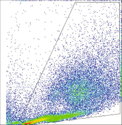
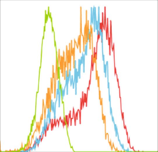
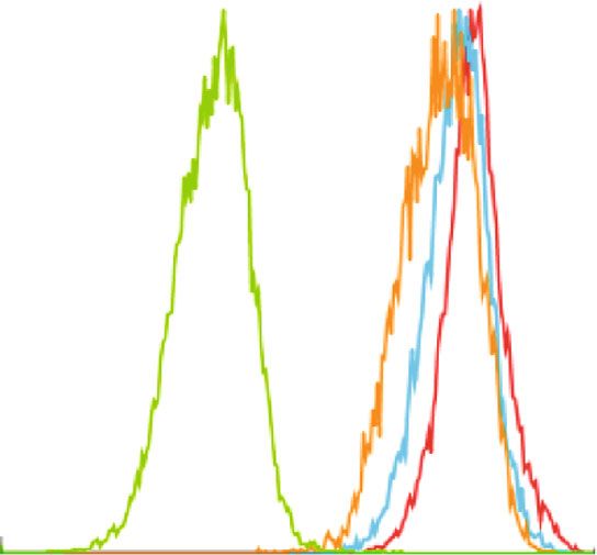
BioMed Research International 3

and was kept throughout the infection. The inhibitor was 3.2. Resident Macrophages Downregulate CD73 Expression In
dissolved in PBS. Vitro. We found that CD39 and CD73 enzymes were abun-
 In all conditions, coverslips were removed 3 h, 24 h, and dantly present in macrophages ex vivo, but the characteris-
48 h post infection from macrophage culture plates. Cover- tics of these molecules in in vitro were yet to be elucidated.
slips were then fixed in methanol for 10 min (Vetec Fine The CD39 and CD73 expressions in resident macrophages
Chemistry), dried, and stained using Panótico Rápido kit were, therefore, characterized at different time points after
(Renylab química e farmacêutica, MG, Brazil) following harvest from the peritoneum. For this, cells were incubated
manufacturer’s instructions. Coverslips were analyzed using for 24, 48, and 72 h in the absence of any external stimulus.
an Olympus BX50 optical microscope (Olympus, Center As shown in Figure 2, although the percentage of macro-
Valley, PA, USA). A minimum of 200 macrophages per cov- phages remained constant during the incubation period
erslip was examined, and the number of uninfected, infected, (Figure 2(b)), macrophages spontaneously downregulated
and amastigotes in infected macrophages was recorded. CD73 without altering their CD39 expression in vitro
 (Figures 2(c)–2(f)). The level of the CD73 expression gradu-
2.5. Flow Cytometry. All samples were washed twice with ally decreased over the incubation period. This result sug-
PBS (210×g, 4°C, 10 min). Cells were then resuspended in gests that culture conditions may have an important role
0.2% bovine serum albumin (0.2% BSA/PBS) and Fc- in the expression of CD73 in vitro. Alternatively, the expres-
blocked (purified rat anti-mouse CD16/CD32, clone 2.4G2, sion of CD73 could have been activated by the harvesting
BD Pharmingen) for 15 min in ice. The cells were then procedure and then gradually returned to a steady state
washed and stained with anti-mouse F4/80PE-CY7 antibody condition.
(clone BM8, BioLegend), anti-mouse CD73PE antibody
(clone Ty/11.8, eBiosciences), and anti-mouse CD39Alexa 3.3. L. amazonensis Increases CD73 Expression in Rested
Fluor 647 (clone 24DMS1, eBiosciences) antibodies in ice Macrophages but Does Not Affect Cytokine and NO
for 30 min. The cells were washed with PBS and then fixed Production. Having shown that the rested resident macro-
in 250 μl fixation solution (1% paraformaldehyde, 47.7 mM phages decrease CD73 expression upon incubation, our next
sodium cacodylate, and 113 mM NaCl; pH 7.2). Samples objective was to observe if the infection by L. amazonensis
were analyzed using a BD FACSCaliburTM flow cytometer. induces any change in CD39 and CD73 surface expressions
All cytometric analyses were performed by using Flow Jo in macrophages. We exposed 72 h rested macrophages to
version 7.6.5 (Tree Star, Ashland, OR, USA). CFSE-labeled metacyclics of L. amazonensis. Interestingly,
 it was found that although the percentage of CD39+ cells
2.6. Cytokine and Nitric Oxide Measurements. TNF-α and
 did not alter after infection (Figure 3(a)), a significant
IL-10 in cell culture supernatants were determined by ELISA
 increase in the CD73 expression was observed in this group
kits (Mouse TNF-α DuoSet catalogue DY410, Mouse IL-10
 (Figure 3(a)). LPS treatment did not affect either CD39 or
Duoset catalogue DY417E from R&D system). Assays were
 CD73 expression, suggesting that LPS-activated macro-
performed according to the manufacturer’s instructions.
 phages do not upregulate CD73. Figure 3(c) demonstrates
Nitric oxide in cell culture supernatants was measured by
 that the combined expression of CD39 and CD73 is higher
spectrophotometric assay based on the Griess reaction [28].
 amongst L. amazonensis-infected macrophages when com-
2.7. Statistical Analysis. Data were expressed as mean ± SD. pared to unstimulated or LPS-treated cells. Furthermore,
Several group data were analyzed by one-way analysis of var- when the infection was prolonged to 48 h of incubation after
iance (ANOVA) followed by Bonferroni posttest or by 72 h rested period, we found that the infected macrophages
Newman-Keuls multiple comparison test. Two group com- still kept CD73 expression higher than the control groups
parisons were performed by paired Student’s test. p value (data not shown).
4 BioMed Research International

 ⁎

 100 100 ⁎

 80 80

 % CD73+
 % CD39+

 60 60

 40 40

 20 20

 0 0
 F 4/80+ F 4/80– F 4/80+ F 4/80–
 (a) (b)

 100 ⁎

 80
 % CD39+CD73+

 60

 40

 20

 0
 F 4/80+ F 4/80–
 (c)

Figure 1: Expression of CD39 and CD73 in naïve macrophages in in vivo. Resident macrophages were harvested from the peritoneum of
naïve C57BL/6 mice using ice-cold 10 ml PBS. After centrifugation and resuspension in 0.2% bovine serum albumin (0.2% BSA/PBS) and
Fc-blocked (purified rat anti-mouse CD16/CD32, clone 2.4G2, BD Pharmingen), the harvested cells were labeled with anti-murine F4/80,
anti-CD39, and anti-CD73 antibodies and then were analyzed by flow cytometry. Indicated in the figure, the percentage of cells
expressing (a) CD39, (b) CD73, and (c) CD39CD73 in F4/80+ and F4/80- cells from total peritoneal population. This result is the mean ± SD
of at least 3 independent experiments. ∗ p < 0:05 means the statistical difference in the expression of CD39 and CD73 between F4/80+ and F4/
80- cell populations using paired two-tailed Student’s t-test.

of L. amazonensis-infected macrophages was demonstrated, on the activity of CD73 surface enzymes, rather than
we sought to determine whether the activity of these cytokine-mediated NO production.
enzymes is crucial for the survival of the parasites. For this, Our results demonstrate that L. amazonensis upregulates
resident macrophages were seeded and were rested for 72 h CD73 in resident macrophages and its survival within the
prior to infection in the presence of inhibitor of CD73. Since cell is strictly dependent on CD73 enzymatic activity.
the use of inhibitors at the time of infection could interfere
with similar enzymes present on the surface of these para- 4. Discussion
sites [20], APCP, inhibitor for CD73, was added after the
parasites had been incubated with the macrophages for 3 h Macrophages and Leishmania have a complex relationship.
and subsequently removed by washing. There was no death In the presence of an adequate immune response, macro-
or changing in cellular viability concerning the incubation phages can be activated and kill intracellular amastigote
with β-methyleneadenosine 5 ′ -diphosphate sodium salt forms of Leishmania [29]. However, as in the case of L.
inhibitor. Our data showed that already after 24 h of infec- amazonensis infection, even in the presence of a Th1
tion, treatment of infected macrophages with APCP reduced response, capable of eliminating other Leishmania species,
parasitism (data not shown) and by 48 h of incubation with the parasite survives within the infected macrophages, indi-
this inhibitor, the percentage of infected macrophages and cating its ability to control macrophages activation [4, 6, 7].
the number of amastigotes per 100 macrophages were signif- Several studies have pointed out that CD39 and CD73,
icantly decreased (Figures 4(a) and 4(b)). Interestingly, the which are present in many immune cells, play an important
reduction in parasite survival in macrophages that had been role in the infections [30–33], inflammation [34], and
treated with ectonucleotidase inhibitors was not associated immune modulation [16, 35–38].
with alterations in the levels of TNF-α, IL-10, or NO produc- Interestingly, it was observed that all freshly harvested
tion as shown in Figures 4(c)–4(e), suggesting that survival resident peritoneal macrophages (F4/80+) expressed CD39
of L. amazonensis within infected macrophages is dependent which is in agreement with the previous studies showing that
BioMed Research International 5

 1K
 F4/80+

 800 Resident cell population

 600

 Count
 SSC
 400

 200

 0 0
 0 200 400 600 800 1K 100 101 102 103 104
 FSC F4/80
 24 h (Resting) 72 h (Resting)
 48 h (Resting) Isotypes

 (a) (b)
 Count

 Count

 0 0
 100 101 102 103 104 100 101 102 103 104
 CD39 CD73
 24 h (Resting) 72 h (Resting) 24 h (Resting) 72 h (Resting)
 48 h (Resting) Isotypes 48 h (Resting) Isotypes

 (c) (d)
 100 100 ⁎

 80 80
 % CD39+

 % CD73+

 60 60

 40 40

 20 20

 0 0
 24 h (Resting)

 48 h (Resting)

 72 h (Resting)

 24 h (Resting)

 48 h (Resting)

 72 h (Resting)

 (e) (f)

Figure 2: Resident macrophages down regulate CD73 expression in in vitro. Resident macrophages were harvested from naïve C57BL/6
mice. Total peritoneal cell population was counted, and viability of the cells was determined by trypan blue. 5 × 10⁵ cells were cultured
in vitro at 37°C and was left for 24 h, 48 h, and 72 h of resting and subsequently incubated for 24 h at 33°C/5%CO2 before analysis by
flow cytometry (a). Size and granularity for total peritoneal population (b). F4/80+ cells were first gated from total peritoneal population
and then cells expressing CD39 and CD73 in F4/80+ population from 24 h, 48 h, and 72 h rested macrophages were overlaid in the
histograms (c and d). The percentage of cells expressing (e) CD39 and (f) CD73 is represented in bar diagrams for macrophages. This
result is representative of 3 independent experiments. ∗ p < 0:05 indicates the statistical difference using paired two-tailed Student’s t-test.
6 BioMed Research International

 80
 100 ⁎

 60

 % CD73+
 % CD39+ 40
 50

 20

 0 0
 (a) (b)
 80 1500
 ⁎
 ⁎
 60
 % CD39+CD73+

 TNF- (pg/ml)
 1000
 40

 500
 20

 0 0

 Uninfected MФ

 Infected MФ

 LPS MФ
 (c) (d)
 800 15 ⁎
 ⁎

 600
 IL-10 (pg/ml)

 10
 NO ( M)

 400

 5
 200

 0 0
 Uninfected MФ

 Infected MФ

 LPS MФ

 Uninfected MФ

 Infected MФ

 LPS MФ

 (e) (f)

Figure 3: Leishmania amazonensis increases CD73 expression but does not alter cytokines and nitric oxide (NO) production. Resident cell
population was collected from naïve C57BL/6 mice and rested for 72 h prior to infection as previously discussed in the methodology. The
cells were infected with CFSE-tagged metacyclics of L. amazonensis, and additionally, another group was treated with 5 μg/ml of LPS. The
cells were further incubated for 24 h at 33°C/5%CO2. Supernatant from all groups was collected for the measurement of TNF-α, IL-10, and
NO. Macrophages (MФ) expressing (a) CD39, (b) CD73, (c) CD39CD73, and immunoassays from culture supernatants for (d) TNF-α, (e)
IL-10, and (f) nitric oxide production in treated macrophages are shown in bar diagrams. This result is the mean ± SD of at least 3
independent experiments. ∗ p < 0:05 indicates the statistical significance between infected and control groups using one-way analysis of
variance (ANOVA) followed by Bonferroni posttest.

CD39 is one of the predominant markers for mature macro- tion in culture medium for 72 hr. The expression of CD73
phages [39]. The CD73 expression, on the other hand, was in resident peritoneal macrophages has been described in
limited only to 72% of the F4/80+ population which suggests the previous studies [40, 41]; however, to the best of our
that the expression of this enzyme is dependent on other fac- knowledge, no previous study has evaluated the expression
tors such as the activation state of the cell or that it is of this enzyme after an extended period of incubation such
restricted to a certain subpopulation of macrophages. as the one used in the present study.
Remarkably, it was observed that the expression CD73 in During infection, macrophages secrete ATP via pannexin
resident macrophages decreased significantly with incuba- channels [42–44] or P2X7 receptors [45]. Accumulation of
BioMed Research International 7

 100 400
 ⁎ ⁎

 Amastigotes (100 MФ)
 80
 % of infected cells
 300
 60
 200
 40

 100
 20

 0 0
 Control 3 h

 Control 48 h

 APCP

 Control 3 h

 Control 48 h

 APCP
 Control 3 h Control 3 h
 Control 48 h Control 48 h
 APCP 48 h APCP 48 h
 (a) (b)
 400 400

 300 300
 TNF- (pg/ml)

 IL-10 (pg/ml)

 200 200

 100 100

 0 0
 Control 48 h Control 48 h
 APCP 48 h APCP 48 h
 (c) (d)
 10

 8
 NO ( M)

 6

 4

 2

 0
 Control 48 h
 APCP 48 h
 (e)

Figure 4: CD73 activity determines survival of L. amazonensis. Resident macrophages were obtained from naïve mice by injecting 10 ml of
ice-cold PBS into the peritoneal cavity and rested for 72 h at 37°C. The cells were then infected with the metacyclic forms of the parasites in a
ratio of 1 : 3 and allowed for the parasites to interact for 3 h at 33°C/5%CO2. Extracellular parasites were washed away, and the inhibitor α, β-
methyleneadenosine 5 ′ -diphosphate sodium salt (APCP) was added against CD73 at a concentration of 200 μM. This treatment was left
throughout the period of infection. Supernatant was collected from these groups after 48 h. The percentage of infection (a) and the
amastigote number per 100 macrophages (b) were shown compared with 3 h and 48 h of incubation with or without inhibitor.
Production of cytokines, (c) TNF-α, (d) IL-10, and (e) nitric oxide (NO) in treated groups is also illustrated here. Data are the mean ±
SD from 3 independent experiments. ∗ p < 0:05 is the statistical difference between control and treated groups using repeated measures of
ANOVA followed by Newman-Keuls multiple comparison test.
8 BioMed Research International

extracellular ATP in the surrounding environment may mainly of the CD73, the inhibition of adenosine production/
induce excessive inflammatory reactions at the site of infec- activity inhibits parasite survival within the infected macro-
tion, and in addition, it may also induce apoptosis of the cells phage. In perspective, defining the role of adenosine produc-
that produce them [46–48]. Cohen et al. [15] showed that in tion on the control of the host immune response may
the absence of CD39 activity, the accumulation of extracellular present new alternatives for the control of Leishmania and/
ATP secreted by TLR stimulated macrophages leads to an or other trypanosomatids.
exacerbated inflammatory response that can be harmful for
the host. Our results showed that while CD39 expression in Data Availability
L. amazonensis-infected macrophages was not altered with
the parasite, CD73 was significantly upregulated. The fact that The data of this manuscript will be made available by the
infection by L. amazonensis increases CD73 expression, our corresponding author on a reasonable request.
result suggests that the infected macrophages would present
a higher regulatory capacity than that of LPS-treated macro- Disclosure
phages. The increase in the CD73 expression by infected mac-
rophages corroborates the previous findings from our The Center for Health and Disease Studies-Nepal, Kath-
laboratory which demonstrated that the CD39 and CD73 mandu, Nepal, is the current address of Bijay Bajracharya
expressions are increased in infected dendritic cells [23] indi- and Deena Shrestha.
cating that the upregulation of ectonucleotidase expression is
a conserved mechanism of inhibiting the establishment of Conflicts of Interest
immune response by the parasite on various cell types.
 The importance of adenosine production by the infected The authors declare no competing interests.
macrophage in the regulation of cellular activation was
established by the decreased parasite survival in cells treated Acknowledgments
with CD73 inhibitor (Figure 4). Macrophages treated with
APCP were capable of, at least partially, control parasite sur- This work was supported by grants from the CAPES,
vival within the infected cell. Similar findings were also FAPEMIG (CBB-APQ-01172-09, CBB-APQ-01419-14, and
observed in L. donovani-infected macrophages when CBB-APQ-02326-17), CNPq, Rede Mineira de Bioterismo/
enzymes CD39 and CD73 were blocked by the inhibitors FAPEMIG, Rede de Pesquisa em Doenças Infecciosas Huma-
at concentrations similar to those used in the present study nas e Animais do Estado de Minas Gerais - RMPDI/FAPE-
[44]. The same study shows that the enzyme activity (release MIG, and UFOP. B.B. thanks the CNPq/TWAS and
of Pi by ATP and AMP degradation) in the presence of an Programa de Pós-Graduação em Ciências Biológicas for the
inhibitor decreased significantly when CD39 and CD73 support concerning his PhD thesis (http://www.repositorio
enzyme activities were inhibited [44]. All this evidence sup- .ufop.br/handle/123456789/3697). L.C.C.A. (Process 307695/
ports the hypothesis that parasite survival is dependent on 2017-4) and A.T (Process # 305634/2017-8) are fellow
the enzyme activity of CD39 and CD73. researchers from CNPq. The authors also thank Tiago D
 The end result of the combined activity of CD39 and Serafim, Pauline M Leite, Rodrigo S Gomes, Amanda B
CD73 is the production of extracellular adenosine which, Figueiredo, Hellem Damazo, Leandro H. Santos, and
by acting on the A2a or A2b receptors, will downmodulate Marcorelio D. Souza for the guidance, support, and technical
macrophage microbicidal mechanisms such as NO and assistance. We would also like to thank Dr. Bilon Khambu
ROS production. We did not address in the present study for his input in this project.
which adenosine receptor was involved in the downmodula-
tion of the microbicidal activity and cytokine production of References
infected macrophages. However, the involvement of A2a
and A2b receptors in L. donovani infection in macrophages [1] M. Olivier, D. J. Gregory, and G. Forget, “Subversion mecha-
was highlighted in a recent study [44]. Furthermore, in the nisms by which Leishmania parasites can escape the host
same study, it was demonstrated that if these receptors were immune response: a signaling point of view,” Clinical Microbi-
blocked by specific antagonists, parasite survival was moder- ology Reviews, vol. 18, no. 2, pp. 293–305, 2005.
ately decreased [44]. These results implicate that adenosine [2] B. M. Scorza, E. M. Carvalho, and M. E. Wilson, “Cutaneous
production rather than decrease in the levels of ATP, which manifestations of human and murine leishmaniasis,” Interna-
 tional Journal of Molecular Sciences, vol. 18, no. 6, p. 1296,
could activate the macrophage if its hydrolysis was impaired,
 2017.
is the important step in parasite growth restriction. More-
 [3] F. de Oliveira Cardoso, C. da Silva Freitas de Souza,
over, previous findings [49] from our laboratory in J774 cells
 V. Gonçalves Mendes, A. L. Abreu-Silva, S. C. G. da Costa,
infected with L. amazonensis supported the evidence that and K. da Silva Calabrese, “Immunopathological studies
when A2b receptors were blocked by MRS1754, parasite sur- ofLeishmania amazonensisInfection in resistant and in suscep-
vival within macrophages decreased significantly. tible mice,” The Journal of Infectious Diseases, vol. 201, no. 12,
 In summary, our results demonstrated that upon infection pp. 1933–1940, 2010.
by L. amazonensis, macrophages differentiate into regulatory [4] L. C. Afonso and P. Scott, “Immune responses associated with
cells and were capable of hydrolyzing extracellular ATP and susceptibility of C57BL/10 mice to Leishmania amazonensis,”
produce adenosine. By activation of the CD39CD73 enzymes, Infection and Immunity, vol. 61, pp. 2952–2959, 1993.
BioMed Research International 9

 [5] P. Scott and F. O. Novais, “Cutaneous leishmaniasis: immune [21] P. M. Leite, R. S. Gomes, A. B. Figueiredo et al., “Ecto-nucleo-
 responses in protection and pathogenesis,” Nature Reviews. tidase activities of promastigotes from Leishmania (Viannia)
 Immunology, vol. 16, no. 9, pp. 581–592, 2016. braziliensis relates to parasite infectivity and disease clinical
 [6] J. Ji, J. Sun, and L. Soong, “Impaired expression of inflamma- outcome,” PLoS Neglected Tropical Diseases, vol. 6, no. 10, arti-
 tory cytokines and chemokines at early stages of infection with cle e1850, 2012.
 Leishmania amazonensis,” Infection and Immunity, vol. 71, [22] T. U. Maioli, E. Takane, R. M. Arantes, J. L. Fietto, and L. C.
 pp. 4278–4288, 2003. Afonso, “Immune response induced by New World Leish-
 [7] S. M. Christensen, A. T. Belew, N. M. El-Sayed, W. L. Tafuri, mania species in C57BL/6 mice,” Parasitology Research,
 F. T. Silveira, and D. M. Mosser, “Host and parasite responses vol. 94, no. 3, pp. 207–212, 2004.
 in human diffuse cutaneous leishmaniasis caused by L. amazo- [23] A. B. Figueiredo, T. D. Serafim, E. A. Marques-da-Silva, J. R.
 nensis,” PLoS Neglected Tropical Diseases, vol. 13, no. 3, Meyer-Fernandes, and L. C. Afonso, “Leishmania amazonen-
 p. e0007152, 2019. sis impairs DC function by inhibiting CD40 expression via
 [8] M. Martínez-López, M. Soto, S. Iborra, and D. Sancho, “Leish- A2B adenosine receptor activation,” European Journal of
 mania hijacks myeloid cells for immune escape,” Frontiers in Immunology, vol. 42, no. 5, pp. 1203–1215, 2012.
 Microbiology, vol. 9, p. 883, 2018. [24] A. B. Figueiredo, M. C. Souza-Testasicca, T. W. P. Mineo, and
 [9] M. F. Horta, B. P. Mendes, E. H. Roma et al., “Reactive oxygen L. C. C. Afonso, “Leishmania amazonensis-induced cAMP
 species and nitric oxide in cutaneous leishmaniasis,” Journal of triggered by adenosine A2B receptor is important to inhibit
 Parasitology Research, vol. 2012, Article ID 203818, 11 pages, dendritic cell activation and evade immune response in
 2012. infected mice,” Frontiers in Immunology, vol. 8, p. 849, 2017.
[10] C. L. Meier, M. Svensson, and P. M. Kaye, “Leishmania- [25] G. F. Spath and S. M. Beverley, “A Lipophosphoglycan-
 induced inhibition of macrophage antigen presentation ana- Independent Method for Isolation of Infective _Leishmania_
 lyzed at the single-cell level,” Journal of Immunology, Metacyclic Promastigotes by Density Gradient Centrifuga-
 vol. 171, pp. 6706–6713, 2003. tion,” Parasitology, vol. 99, no. 2, pp. 97–103, 2001.
[11] V. Ralevic and G. Burnstock, “Receptors for purines and [26] R. Goncalves, E. R. Vieira, M. N. Melo, K. J. Gollob, D. M.
 pyrimidines,” Pharmacological Reviews, vol. 50, pp. 413–492, Mosser, and W. L. Tafuri, “A sensitive flow cytometric meth-
 1998. odology for studying the binding of L. chagasi to canine peri-
[12] N. Riteau, L. Baron, B. Villeret et al., “ATP release and puriner- toneal macrophages,” BMC Infectious Diseases, vol. 5, no. 1,
 gic signaling: a common pathway for particle-mediated p. 39, 2005.
 inflammasome activation,” Cell Death & Disease, vol. 3, [27] X. Zhang, R. Goncalves, and D. M. Mosser, “The isolation and
 no. 10, article e403, 2012. characterization of murine macrophages,” Current Protocols in
[13] I. Hide, M. Tanaka, A. Inoue et al., “Extracellular ATP triggers Immunology, vol. 83, p. 14, 2008.
 tumor necrosis factor-α release from rat microglia,” Journal of [28] L. C. Green, D. A. Wagner, J. Glogowski, P. L. Skipper, J. S.
 Neurochemistry, vol. 75, no. 3, pp. 965–972, 2000. Wishnok, and S. R. Tannenbaum, “Analysis of nitrate, nitrite,
[14] G. G. Yegutkin, “Nucleotide- and nucleoside-converting and [15N] nitrate in biological fluids,” Analytical Biochemistry,
 ectoenzymes: important modulators of purinergic signalling vol. 126, pp. 131–138, 1982.
 cascade,” Biochimica et Biophysica Acta, vol. 1783, pp. 673– [29] J. Mauel, “Macrophage-parasite interactions in Leishmania
 694, 2008. infections,” Journal of Leukocyte Biology, vol. 47, pp. 187–
[15] H. B. Cohen, K. T. Briggs, J. P. Marino, K. Ravid, S. C. Robson, 193, 1990.
 and D. M. Mosser, “TLR stimulation initiates a CD39-based [30] A. L. J. Leite, D. S. Oliveira, L. R. W. Mota et al., “Ectonucleo-
 autoregulatory mechanism that limits macrophage inflamma- tidases from trypomastigotes from different sources and vari-
 tory responses,” Blood, vol. 122, pp. 1935–1945, 2013. ous genetic backgrounds of _Trypanosoma cruzi_ potentiate
[16] L. Antonioli, P. Pacher, E. S. Vizi, and G. Hasko, “CD39 and their infectivity and host inflammation,” Cytokine, vol. 136,
 CD73 in immunity and inflammation,” Trends in Molecular article 155255, 2020.
 Medicine, vol. 19, pp. 355–367, 2013. [31] M. Nikolova, M. Carriere, M. A. Jenabian et al., “CD39/aden-
[17] K. M. Dwyer, S. Deaglio, W. Gao, D. Friedman, T. B. Strom, osine pathway is involved in AIDS progression,” PLoS Patho-
 and S. C. Robson, “CD39 and control of cellular immune gens, vol. 7, article e1002110, 2011.
 responses,” Purinergic Signal, vol. 3, pp. 171–180, 2007. [32] T. Russo-Abrahao, D. Cosentino-Gomes, M. T. Gomes et al.,
[18] F. M. Sansom, S. C. Robson, and E. L. Hartland, “Possible “Biochemical properties of Candida parapsilosis ecto-5'-nucleo-
 effects of microbial ecto-nucleoside triphosphate dipho- tidase and the possible role of adenosine in macrophage interac-
 sphohydrolases on host-pathogen interactions,” Microbiol- tion,” FEMS Microbiology Letters, vol. 317, pp. 34–42, 2011.
 ogy and Molecular Biology Reviews, vol. 72, pp. 765–781, [33] V. Thammavongsa, J. W. Kern, D. M. Missiakas, and
 2008. O. Schneewind, “Staphylococcus aureus synthesizes adenosine
[19] J. C. de Almeida Marques-da-Silva, A. B. de Oliveira, J. D. to escape host immune responses,” The Journal of Experimen-
 Figueiredo et al., “Extracellular nucleotide metabolism in tal Medicine, vol. 206, pp. 2417–2427, 2009.
 _Leishmania_ : influence of adenosine in the establishment [34] J. Reutershan, I. Vollmer, S. Stark, R. Wagner, K. C. Ngamsri,
 of infection,” Microbes and Infection, vol. 10, no. 8, pp. 850– and H. K. Eltzschig, “Adenosine and inflammation: CD39 and
 857, 2008. CD73 are critical mediators in LPS-induced PMN trafficking
[20] M. C. de Souza, E. A. de Assis, R. S. Gomes et al., “The influ- into the lungs,” The FASEB Journal, vol. 23, pp. 473–482, 2009.
 ence of ecto-nucleotidases on _Leishmania amazonensis_ [35] L. Antonioli, C. Blandizzi, P. Pacher, and G. Hasko, “Immu-
 infection and immune response in C57B/6 mice,” Acta Tro- nity, inflammation and cancer: a leading role for adenosine,”
 pica, vol. 115, no. 3, pp. 262–269, 2010. Rev. Cancer, vol. 13, no. 12, pp. 842–857, 2013.
10 BioMed Research International

[36] K. E. Barletta, K. Ley, and B. Mehrad, “Regulation of neutro-
 phil function by adenosine,” Arteriosclerosis, Thrombosis, and
 Vascular Biology, vol. 32, pp. 856–864, 2012.
[37] N. Mizumoto, T. Kumamoto, S. C. Robson et al., “CD39 is the
 dominant Langerhans cell-associated ecto-NTPDase: modula-
 tory roles in inflammation and immune responsiveness,”
 Nature Medicine, vol. 8, pp. 358–365, 2002.
[38] A. B. de Figueiredo, M. C. Souza-Testasicca, and L. C. C.
 Afonso, “Purinergic signaling and infection by _Leishmania_
 : A new approach to evasion of the immune response,” Biomed
 J., vol. 39, no. 4, pp. 244–250, 2016.
[39] S. A. Levesque, F. Kukulski, K. Enjyoji, S. C. Robson, and
 J. Sevigny, “NTPDase1 governs P2X7-dependent functions in
 murine macrophages,” European Journal of Immunology,
 vol. 40, pp. 1473–1485, 2010.
[40] D. Eichin, J. P. Laurila, S. Jalkanen, and M. Salmi, “CD73 activ-
 ity is dispensable for the polarization of M2 macrophages,”
 PLoS One, vol. 10, no. 8, article e0134721, 2015.
[41] P. Murphy, J. Wang, S. Bhagwat et al., “CD73 regulates anti-
 inflammatory signaling between apoptotic cells and
 endotoxin-conditioned tissue macrophages,” Cell Death and
 Differentiation, vol. 24, no. 3, pp. 559–570, 2017.
[42] F. B. Chekeni, M. R. Elliott, J. K. Sandilos et al., “Pannexin 1
 channels mediate 'find-me' signal release and membrane per-
 meability during apoptosis,” Nature, vol. 467, pp. 863–867,
 2010.
[43] Y. Qu, S. Misaghi, K. Newton et al., “Pannexin-1 is required for
 ATP release during apoptosis but not for inflammasome acti-
 vation,” Journal of Immunology, vol. 186, pp. 6553–6561, 2011.
[44] M. Basu, P. Gupta, A. Dutta, K. Jana, and A. Ukil, “Increased
 host ATP efflux and its conversion to extracellular adenosine
 is crucial for establishingLeishmaniainfection,” Journal of Cell
 Science, vol. 133, no. 7, 2020.
[45] C. Marques-da-Silva, M. M. Chaves, S. P. Chaves et al., “Infec-
 tion with Leishmania amazonensis upregulates purinergic
 receptor expression and induces host-cell susceptibility to
 UTP-mediated apoptosis,” Cellular Microbiology, vol. 13,
 pp. 1410–1428, 2011.
[46] S. C. Chow, G. E. Kass, and S. Orrenius, “Purines and their
 roles in apoptosis,” Neuropharmacology, vol. 36, no. 9,
 pp. 1149–1156, 1997.
[47] S. P. Chaves, E. C. Torres-Santos, C. Marques et al., “Modula-
 tion of P2X (7) purinergic receptor in macrophages by Leish-
 mania amazonensis and its role in parasite elimination,”
 Microbes and Infection, vol. 11, pp. 842–849, 2009.
[48] L. M. Zheng, A. Zychlinsky, C. C. Liu, D. M. Ojcius, and J. D.
 Young, “Extracellular ATP as a trigger for apoptosis or pro-
 grammed cell death,” The Journal of Cell Biology, vol. 112,
 pp. 279–288, 1991.
[49] R. S. Gomes, L. C. de Carvalho, V. R. de Souza, J. L. Fietto, and
 L. C. Afonso, “E-NTPDase (ecto-nucleoside triphosphate
 diphosphohydrolase) of Leishmania amazonensis inhibits
 macrophage activation,” Microbes and Infection, vol. 17,
 no. 4, pp. 295–303, 2015.
You can also read