The effect of hyperbaric oxygenation on cardiodynamics and oxidative stress in rats with sepsis
←
→
Page content transcription
If your browser does not render page correctly, please read the page content below
Vojnosanit Pregl 2021; 78(8): 825–833.           VOJNOSANITETSKI PREGLED                                                   Page 825

ORIGINAL ARTICLE                                                                                       UDC: 616.94-085.835-092.9
                                                                                     DOI: https://doi.org/10.2298/VSP191026142J

The effect of hyperbaric oxygenation on cardiodynamics and oxidative
stress in rats with sepsis
        Efekti hiperbarične oksigenacije na kardiodinamiku i oksidacioni stres kod
                                                                pacova sa sepsom

                   Aleksandar Jevtić*, Vladimir Živković†, Milica Milinković†, Željko Mijailović‡,
                        Nevena Draginić‡, Marijana Andjić‡, Andjela Milojević Šamanović§,
                                     Sergey Bolevich||, Vladimir Jakovljević†||

                        *Institute for Orthopedic Surgery “Banjica”, Belgrade, Serbia; University of
                     Kragujevac, Faculty of Medical Sciences, †Department of Physiology, ‡Department of
                     Pharmacy, §Department of Dentistry, Kragujevac, Serbia; ||1st Moscow State Medical
                            University IM Sechenov, Department of Human Pathology, Moscow,
                                                    Russian Federation

Abstract                                                            following parameters of heart function were continuously
                                                                    recorded: maximum and minimum rate of left ventricular
Background/Aim. Dysfunctions at the cellular, tissue, and           pressure development (dp/dt max, dp/dt min); systolic and
organ level, which can result in death, are caused by metabol-      diastolic left ventricular pressure (SLVP and DLVP); heart
ic changes and affection on the regulation of gene transcrip-       rate (HR). Coronary flow (CF) was measured flowmetrically.
tion and micro- and macrocirculation. The aim of the present        Following oxidative stress markers were measured: nitrites
study was to assess the impact of hyperbaric oxygenation            (NO2−), superoxide anion radical (O2−), hydrogen peroxide
(HBO) on isolated heart as well as on the oxidative status of       (H2O2), index of lipid peroxidation (TBARS), activity of su-
rats with sepsis. Methods. The investigation included male          peroxide dismutase (SOD) and catalase (CAT) and the level
Wistar albino rats classified into three groups: the first group    of reduced glutathione (GSH). Results. There were no sig-
was a control group (CTRL); the second group included ani-          nificant differences in dp/dt max, dp/dt min, SLVP and HR
mals exposed only to the induction of sepsis without HBO            between the groups. CF was statistically significantly higher (p
treatment (the Sepsis group), while the third group included        < 0.01) in the sepsis group. The values of all cardiac oxidative
animals treated with HBO after the induction of sepsis (the         markers were lower in the sepsis + HBO group (p < 0.05),
Sepsis + HBO group). For the induction of sepsis, fecal peri-       while systemic pro-oxidative and antioxidative parameters
tonitis model was used (3 mL/kg of fecal suspension admin-          were unchanged. Conclusion. Our results showed that HBO
istered intraperitoneally). After the induction of sepsis, the      treatment was not associated with improved cardiac function
rats were exposed twice a day (on 12 hours) to HBO treat-           and coronary perfusion, while expressed promising beneficial
ment at 2.8 atmospheres absolute (ATA) for 90 minutes over          effects on cardiac oxidative stress.
a period of 3 days. 72 h after the confirmation of sepsis, the
animals were sacrificed and the hearts were retrogradely per-       Key words:
fused on the Langendorff apparatus at a gradually increased         hyperbaric oxygenation; oxidative stress; sepsis; heart;
coronary perfusion pressure (CPP = 40–120 cm H2O). The              rats.

Apstrakt                                                            izložene samo sepsi bez HBO tretmana (grupa Sepsa), dok
                                                                    su u trećoj grupi životinje tretirane HBO nakon indukcije
Uvod/Cilj. Disfunkcija na nivou ćelije, tkiva ili organa, koja      sepse (grupa Sepsa + HBO). Za indukciju sepse korišćen je
može imati za posledicu smrtni ishod, javlja se usled               model fekalnog peritonitisa (3 mL/kg fekalne suspenzije, in-
metaboličkih promena i poremećaja regulacije transkripcije          traperitonealno). Posle indukcije sepse, pacovi su bili
gena i mikro- i makro cirkulacije. Cilj ove studije bio je da se    izloženi dva puta dnevno (tokom 12 sati) HBO tretmanu sa
proceni uticaj hiperbarične oksigenacije (HBO) na izolo-            2,8 apsolutnih atmosfera (ATA) tokom 90 minuta u periodu
vano srce, kao i na oksidacioni status pacova sa sepsom.            od 3 dana. 72 h nakon potvrđivanja sepse, životinje su
Metode. Istraživanjem su obuhvaćeni mužjaci Wistar albi-            žrtvovane, a srca su retrogradno perfundovana na Langen-
no pacova klasifikovani u tri grupe: prva grupa je bila             dorfovom aparatu, pri postepenom povećanju koronarnog
kontrolna grupa (CTRL), drugu grupu su činile životinje             perfuzionog pritiska (CPP = 40–120 cm H2O). Sledeći par

Correspondence to: Vladimir Živković, University of Kragujevac, Faculty of Medical Sciences, Department of Physiology, Svetozara
Markovića 69, 34 000 Kragujevac, Serbia. E-mail: vladimirziv@gmail.com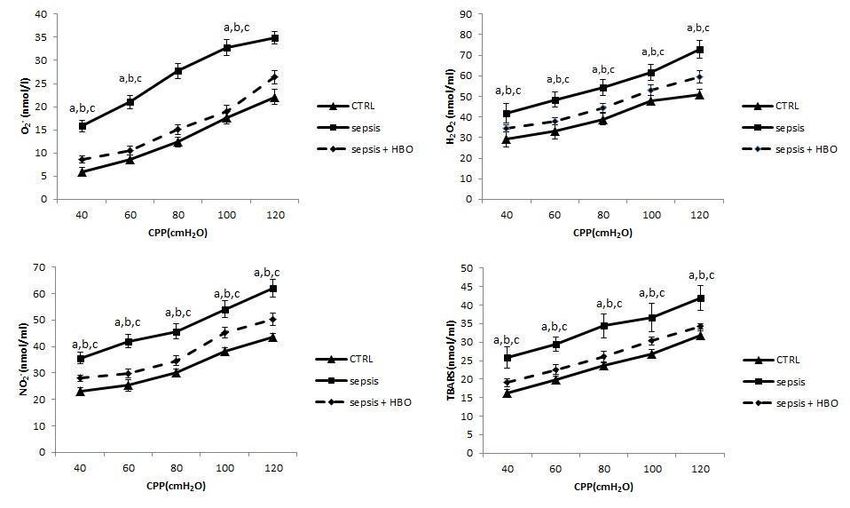
Page 826                                         VOJNOSANITETSKI PREGLED                                                Vol. 78, No. 8

ametri srčane funkcije su kontinuirano mereni: maksimalna i         0,01) u grupi sa sepsom. Vrednosti svih srčanih oksida-
minimalna stopa promene pritiska u levoj komori (dp/dt              cionih markera bile su niže u grupi sepsa + HBO (p < 0,05),
max, dp/dt min); sistolni i dijastolni pritisak leve komore         dok su sistemski pro-oksidacioni i antioksidacioni parametri
(SLVP i DLVP) i srčana frekvenca (HR). Koronarni protok             bili nepromenjeni. Zaključak. Naši rezultati su pokazali da
(CF) je meren floumetrijski. Određivani su sledeći markeri          HBO tretman nije bio povezan sa poboljšanom funkcijom
oksidacionog stresa: nitriti (NO2−), superoksid anjon radikal       srca i koronarnom perfuzijom, dok je ostvario obećavajući
(O2−), vodonik peroksid (H2O2), indeks lipidne peroksida-           korisne efekte na oksidacioni status u srcu pacova.
cije (TBARS), aktivnost superoksid dismutaze (SOD) i kata-
laze (CAT) i nivo redukovanog glutationa (GSH). Rezulta-            Ključne reči:
ti. Nije bilo značajne razlike u dp/dt max, dp/dt min, SLVP         hiperbarička oksigenacija; stres, oksidativni; sepsa;
i HR između grupa. CF je bio statistički značajno veći (p <         srce; pacovi.

     Introduction                                                   tricle, an increase in the maximum and minimum rate of left
                                                                    ventricular pressure development, bradycardia and a de-
      A life-threatening organ dysfunction caused by a dis-         crease in cardiac output 11, 12. The mechanisms underlying
rupted response of the host organism to infection is sepsis, a      this cardiovascular response are linked to baroreceptor-
condition whose global epidemiological significance is diffi-       mediated regulation 11.
cult to assess, and which places a heavy financial burden on              HBO is a potential intervention for the prevention of
health systems. Dysfunctions at the cellular, tissue, and or-       septic shock and can also be used in the treatment of severe
gan level, which can result in death, are caused by metabolic       pancreatitis, diabetic foot ulcer, carbon monoxide poisoning
changes and affection on the regulation of gene transcription       and other conditions 13. Studies addressing the use of HBO in
and micro- and macrocirculation 1–3. Much progress has been         the treatment of sepsis have primarily addressed the benefits
made in the knowledge of the pathogenetic mechanisms                of HBO in the domain of cytokine production, especially
mentioned above in the last few decades, but extensive trials       about the increased expression of interleukin (IL)-10 and the
are still needed to implement newer modalities of therapy in-       decreased levels of IL-6 13. More importantly, due to its
to sepsis treatment.                                                mechanism of action, HBO can affect the production of oxi-
      In addition to immune and metabolic disorders, cardio-        dative stress biomarkers and thus change redox homeostasis
vascular disorders develop. The two manifestations of cardi-        in sepsis, which could be responsible for the potential posi-
ac dysfunction are the hyperdynamic (warm shock) and the            tive impact of this procedure 14.
hypodynamic (cold shock) phase 4, 5. The hyperdynamic                     Having in mind that there are almost no studies that in-
phase is characterized by the elevated vascular tone and low        vestigate the effects of hyperbaric oxygenation on cardiac
cardiac output, while in the hypodynamic phase we have a            function and coronary circulation during sepsis, we aimed to
reverse situation – decreased vascular tone and increased           assess the potential impact of this therapeutic approach on
cardiac output, and a difference in the form of manifestation       isolated heart as well as on cardiac and systemic oxidative
in adults and the pediatric population was observed 5. Tests        status of rats with sepsis.
on the isolated cardiomyocyte revealed that with the passage
of time from the onset of sepsis, an increase in heart rate was          Methods
observed, as well as a decrease in the maximum and mini-
                                                                         Animals and study design
mum rate of left ventricular pressure development and a de-
crease in diastolic pressure in the left ventricle 5–7. The basis         The study was carried out on eighteen male Wistar albino
of these changes is the production of cytokines, whose indi-        rats, 8 weeks old, body weight 200 ± 30 g. The animals were
rect influence is reflected by the increased production of          kept in an artificial 12-h light–dark cycle (8:00 a.m–8:00 p.m.)
vasoactive mediators 5. The association of Ca2+ receptors,          at room temperature (22 ± 1°C). Water and food were availa-
which control Na+/K+-ATPase function in cardiomyocytes,             ble ad libitum. The animals were housed in their respective
with the onset of cardiac dysfunction in sepsis in an animal        groups in a collective cage and received water and standard la-
model has also been demonstrated 4.                                 boratory chow. The animals were classified into three groups.
      Hyperbaric oxygenation (HBO) is the process of expos-               The first group (n = 6) was the control group (CTRL).
ing the whole organism to 100% oxygen at elevated pressure          The second group (n = 6) included animals exposed only to the
[greater than 1 atmosphere absolute – ATA (760 mmHg)] at            induction of sepsis without HBO treatment (the Sepsis group),
different time intervals. The effect of this treatment, whose       while the third group (n = 6) included animals treated with
effects can be divided into primary and secondary, is based         HBO after the induction of sepsis (the Sepsis + HBO group).
on the principles of Henry and Dalton's law. The primary ef-
fect is the percentage increase in dissolved oxygen in the cir-          Ethical standards
culation, while the secondary effects are vasoconstriction,
neovascularization, and a decrease in gas volume 8–10. Vaso-             This research was carried out in the Laboratory for Car-
constriction leads to an increase in vascular resistance and        diovascular Physiology of the Faculty of Medical Sciences,
arterial pressure, and an increase in pressure in the left ven-     University of Kragujevac, Serbia. The study protocol was ap-

                                                                                 Jevtić A, et al. Vojnosanit Pregl 2021; 78(8): 825–833.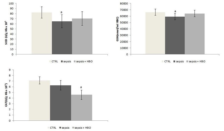
Vol. 78, No. 8                                      VOJNOSANITETSKI PREGLED                                               Page 827

proved by the Ethical Committee for the welfare of experi-        xylazine (5 mg/kg) and sacrificed by decapitation. The hearts
mental animals of the Faculty of Medical Sciences, University     were then rapidly isolated and retrogradely perfused on
of Kragujevac, Serbia. All experiments were performed ac-         Langendorff apparatus (Langendorff apparatus, Experimetria
cording to EU Directive for the welfare of laboratory animals     Ltd, 1062 Budapest, Hungary) through the ascending aorta at a
(86/609/EEC) and the principles of Good Laboratory Practice.      gradually increased coronary perfusion pressure (CPP = 40–
                                                                  120 cm H2O). The hearts were perfused with Krebs–Henseleit
     Sepsis induction protocol                                    solution, while a transducer was inserted in the left ventricle to
                                                                  continuously record the following parameters of myocardial
     Firstly, fresh feces were collected from a heterogeneous     function: maximum and minimum rate of left ventricular pres-
group of rats. The feces were then dissolved in saline and        sure development (dp/dt max, dp/dt min), systolic and diastolic
this mixture was homogenized and filtered through gauze to        left ventricular pressure (SLVP and DLVP), and heart rate
obtain fecal suspension. Animals were anesthetized by intra-      (HR). Coronary flow (CF) was measured flowmetrically. The
peritoneal administration of ketamine and xylazine (10            perfusion started at CPP = 70 cm H2O and hearts were allowed
mg/kg, 5 mg/kg, respectively) and 3 mL/kg of fecal suspen-        to equilibrate until HR and contractility reached steady-state.
sion was administered intraperitoneally. For the confirmation     After stabilization (approximately 30 min), the CPP was grad-
of sepsis, the previously established clinical rat scoring sys-   ually increased from 40, 60, 80, 100 and 120 cm H2O to esti-
tem (Table 1) was used for each animal as well as rectal          mate coronary autoregulation. At each value of CPP, the coro-
body temperature and biochemical indicators of sepsis [C re-      nary venous effluent was collected for the determination of ox-
active protein (CRP) and procalcitonin (PCT) values]. Ani-        idative stress parameters.
mals from both groups were monitored for the next 72 hours
before sacrifice 15.                                                   Evaluation of systemic oxidative stress
Table 1                                                                At the moment of sacrificing animals, blood samples
      Clinical rat scoring system for the confirmation            were collected from jugular vein in order to estimate follow-
             of sepsis and the severity of sepsis                 ing pro-oxidants in plasma: nitrites (NO2−), superoxide anion
                                                                  radical (O2−), hydrogen peroxide (H2O2), index of lipid pe-
    Characteristic                             Scoring range
                                                                  roxidation (thiobarbituric acid reactive substances, TBARS)
                                                                  and parameters of antioxidative defense system in erythro-
    Hunched                                          0–1
                                                                  cytes samples: activity of superoxide dismutase (SOD and
                                                                  catalase (CAT) and the level of reduced glutathione (GSH).
    Bloated                                          0–1
                                                                  All parameters were measured on the spectrophotometer ap-
                                                                  paratus (Shimadzu UV 1800, Japan).
    Conjunctival injection/mucky eyes                0–1

    Piloerection                                     0–1               Evaluation of cardiac oxidative stress

                                                                       The coronary venous effluent from each value of CPP
    Lack of movement                                 0–2
                                                                  (40–120 cm H2O) was collected for the determination of oxi-
                                                                  dative stress parameters from the isolated rat heart. The fol-
    Lack of alertness                                0–2
                                                                  lowing oxidative stress parameters were determined spectro-
      Legend for scoring: absence (0), presence (1) or            photometrically: NO2−, O2−, H2O2 and TBARS.
      marked presence (2).
      The total score of 0 to 3 denotes mild sepsis and                Markers of oxidative stress
      ≥ 4 severe sepsis.
                                                                         Nitric oxide decomposes rapidly to form stable metabolite
     HBO treatment protocol                                       nitrite/nitrate products. NO2− level was measured spectrophoto-
                                                                  metrically at a wavelength of 543 nm and used as an index of ni-
     HBO treatment was carried out in a specially construct-      tric oxide (NO) production using the Griess reagent as previous-
ed hyperbaric chamber for rats (HYB-C 300). After the in-         ly described by Green et al. 17. The measurement of H2O2 is
duction of sepsis, the rats were exposed twice a day (on 12       based on oxidation of Phenol Red by hydrogen peroxide, in a
hours) to 100% O2 at 2.8 ATA for 90 minutes for 3 days. To        reaction catalyzed by horseradish peroxidase (HRPO) at 610
avoid the effects of diurnal rhythm variation, the HBO ses-       nm, as previously described by Pick and Keisari 18. The index of
sion started at the same time, each day 16.                       lipid peroxidation was estimated by measuring of TBARS using
                                                                  1% thiobarbituric acid (TBA) in 0.05 NaOH incubated with the
     Ex vivo assessment of heart function                         plasma as previously described 19. The concentration of O2− was
                                                                  measured by Nitro Blue Tetrazolium (NBT) reaction in hy-
      72h after the confirmation of sepsis, animals from all      droxymethylaminomethane (TRIS) buffer with a plasma sample
groups were anesthetized with short-term narcosis induced by      at 530 nm as previously described by Auclair and Voisin 20. The
intraperitoneal application of ketamine (10 mg/kg) and            whole analysis was determined using the spectrophotometrical

Jevtić A, et al. Vojnosanit Pregl 2021; 78(8): 825–833.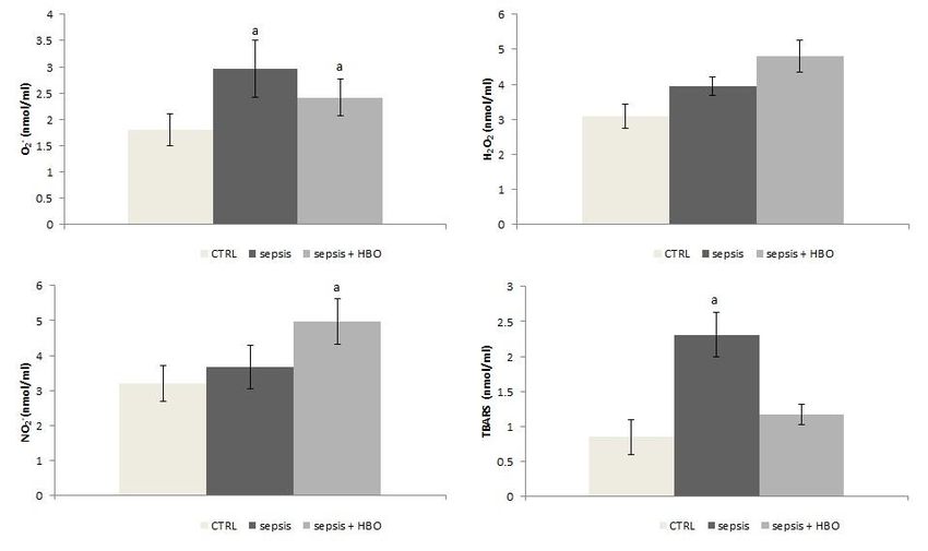
Page 828 VOJNOSANITETSKI PREGLED Vol. 78, No. 8
method (UV-1800 UV – Vis Spectrophotometer by Shimadzu addition, a whole group of animal’s biochemical parameters
Scientific Instruments Inc). of sepsis, as well as rectal temperature confirmed the state of
sepsis (Table 3).
Antioxidative enzymes
Table 2
For the determination of antioxidant parameters, isolat- Average clinical rat sepsis score for
ed erythrocytes were prepared according to McCord and Fri- the whole group of animals (n = 12)
dovich 21. SOD activity was determined by the epinephrine
Animals with sepsis (%) Score
method described by Misra and Fridovich 22. A 100 μL lysate
and 1 mL carbonate buffer were mixed, and then 100 μL of
epinephrine was added. Detection was performed at 470 nm. 22 3 (mild sepsis)
CAT activity was determined according to Beutler 23. Ly-
sates were diluted with distilled water (1:7 v/v) and treated 78 4–6 (severe sepsis)
with chloroform-ethanol (0.6:1 v/v) to remove hemoglobin
and then, 50 μL CAT buffer, 100 μL sample, and 1 mL 10
mM H2O2 were added to the samples. Detection was per- Table 3
formed at 360 nm. The level of reduced glutathione (GSH) Average rectal temperature and values of biochemical
was determined based on GSH oxidation with 5.5-dithio-bis- indicators of sepsis for the whole group of animals before the
6.2-nitrobenzoic acid, as previously described by Beutler 24. induction of sepsis and at the moment of sacrificing (n = 12)
Measuring was performed at 420 nm. At the moment
Before sepsis
Parameter of sacrificing
mean ± SD
mean ± SD
Determination of CRP and PCT values
Rectal temperature (°C) 36.9 ± 0.4 38.6 ± 0.8
Serum CRP and PCT were detected using specific en-
zyme-linked immunoassay kits according to the manufactur-
er’s instructions 25. CRP (ng/mL) 325.74 ± 23.46 527.86 ± 20.67
The measurement of rectal temperature PCT (pg/mL) 76.35 ± 10.84 200.54 ± 38.21
The rectum temperature was continuously monitored CRP – C-reactive protein; PCT – procalcitonin.
using a digital thermometer (PIC solutions, Artsana S.p.A.,
Grandate, Italy).
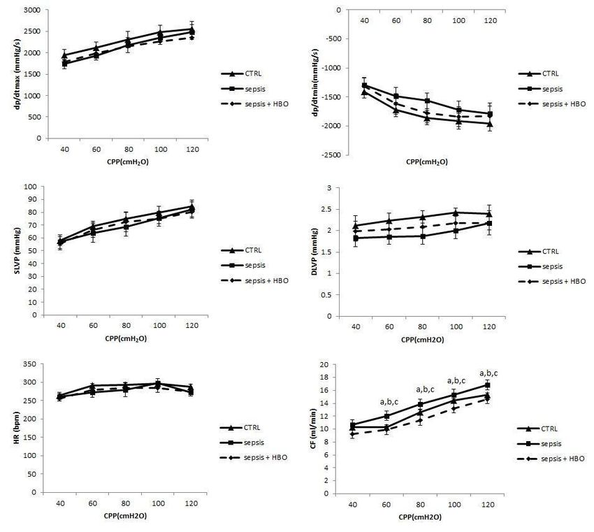
Cardiac function ex vivo
Drugs Cardiodynamic parameters for the assessment of car-
diac function in ex vivo model are presented in Figure 1. As
All kits, reagents and substances used in the study were it can be seen, there was no significant difference in dp/dt
purchased from Sigma-Aldrich Chemie GmbH Eschenstrasse max during all CPPs among the groups. Dp/dt min was
5, 82024 Taufkirchen, Germany. higher in the control group (CPPs = 60 – 100 cm H2O) than
in the Sepsis and Sepsis + HBO group but without statisti-
Statistical analysis cal significance. The SLVP and HR values were almost
similar in all groups, while the DLVP was insignificantly
IBM SPSS Statistics 20.0 Desktop for Windows was higher in the control group. On the other hand, CF was sta-
used for statistical analysis. The distribution of data was tistically significantly higher (p < 0.05) in the Sepsis group
checked by the Shapiro-Wilk test. Where distribution between than in the Sepsis + HBO group and also in the control
groups was normal, statistical comparisons were performed group in comparison to other two groups at almost all CPPs
using the one-way analysis of variance (ANOVA) tests with a (60–120 cm H2O).
Tukey’s post hoc test for multiple comparisons. Kruskal-
Wallis test was used for the comparison between groups when
Cardiac oxidative stress
the distribution of data was different from normal. Values of p
< 0.05 were considered to be statistically significant.
Biomarkers from coronary venous effluent as indica-
tors of cardiac oxidative stress are presented in Figure 2.
Results The values of O2- were significantly lower in the Sepsis +
HBO group compared to the Sepsis group (at CPPs = 40,
Confirmation of sepsis and scoring of sepsis severity
60 and 80 cm H2O, p < 0.05), while between the control
The distribution of the septic score was presented in and the Sepsis + HBO group there were no statistical dif-
Table 2. Of the septic animals surviving to sacrifice (n = 12), ferences. Similarly, the H2O2 values were lower in the Sep-
22% were scored mild and 78% severe sepsis (Table 2). In- sis + HBO group compared to the Sepsis group with strong
Jevtić A, et al. Vojnosanit Pregl 2021; 78(8): 825–833.
Vol. 78, No. 8                                      VOJNOSANITETSKI PREGLED                                                Page 829

          Fig. 1 – Parameters of cardiac function ex vivo (data are presented as mean values ± standard deviation)
             dp/dtmax – maximum rate of pressure development; dp/dtmin – minimum rate of pressure development;
   SLVP – systolic left ventricular pressure; DLVP – diastolic left ventricular pressure; HR – heart rate; CF – coronary flow;
                                                  CPP – coronary perfusion pressure.
   Statistically significant differences (p < 0.05) among the groups at the same coronary perfusion pressure (CPP) are marked
       as follows: a – comparison between the control group (CTRL) and the group of rats with sepsis (the Sepsis group);
   b – comparison between the control group (CTRL) and the group of rats with sepsis treated with HBO (the Sepsis + HBO
    group); c – comparison between the group of rats with sepsis (the Sepsis group) and the group of rats with sepsis treated
                                                 with HBO (the Sepsis + HBO group).

           Fig. 2 – Parameters of cardiac oxidative stress (data are presented as mean values ± standard deviation).
    O2− – superoxide anion radical; H2O2 – hydrogen peroxide; NO2− – nitrites; TBARS – thiobarbituric acid reactive substances
                                                        (index of lipid peroxidation).
   Statistically significant differences (p < 0.05) between the groups at the same coronary perfusion pressure (CPP) are marked as
            follows: a – comparison between the control group (CTRL) and the group of rats with sepsis (the Sepsis group);
     b – comparison between the control group (CTRL) and the group of rats with sepsis treated with HBO (the Sepsis + HBO
    group); c – comparison between the group of rats with sepsis (the Sepsis group) and the group of rats with sepsis treated with
                                                       HBO (the Sepsis + HBO group.
Jevtić A, et al. Vojnosanit Pregl 2021; 78(8): 825–833.
Page 830 VOJNOSANITETSKI PREGLED Vol. 78, No. 8
statistical significance at all CPPs (p < 0.01), without sig- oxidants, a significant difference between the Sepsis group
nificant differences between the control and the Sepsis + and the Sepsis + HBO group was not observed. Namely, the
HBO group. The same trend in the Sepsis + HBO group values of NO2- and H2O2 were insignificantly lower in the
had values of NO2- with statistical significance at all CPPs Sepsis group, while the values of O2- and TBARS were in-
(p < 0.05). As in previous cases, TBARS values were also significantly lower in the Sepsis + HBO group. Compared to
significantly lower in the Sepsis + HBO group (CPPs = 40– the control group, the values of pro-oxidant were significant-
100 cm H2O (p < 0.05)) compared to the Sepsis group ly higher in both, the Sepsis and Sepsis + HBO groups. In
without significant differences between the control and the addition, the values of antioxidant enzymes from erythrocyte
Sepsis + HBO group. lysate are presented in Figure 4. Although it can be seen that
SOD and GSH values were higher in the Sepsis + HBO
Systemic oxidative stress group, there was no statistical confirmation. The CAT values
were also without significant difference between the Sepsis
The values of pro-oxidants from plasma of both groups and the Sepsis + HBO group, but compared to the control
of rats are presented in Figure 3. Unlike cardiac oxidative group, the values of this antioxidant enzyme were signifi-
stress markers, in the values of all measured systemic pro- cantly lower in the Sepsis + HBO group.
Fig. 3 – The values of pro-oxidants in plasma samples (data are presented as mean values ± standard deviation).
O2− – superoxide anion radical; H2O2 – hydrogen peroxide; NO2− – nitrites; TBARS – thiobarbituric
acid reactive substances (index of lipid peroxidation).
Statistically significant differences (p < 0.05) are marked as follows:
a – statistical significance difference compared to the control group (CTRL).
Fig. 4 – The values of antioxidant enzymes in erythrocyte lysate samples (data
are presented as mean values ± standard deviation).
SOD – superoxide dismutase; CAT – catalase; GSH – glutathione.
Statistically significant differences (p < 0.05) are marked as follows:
a – statistical significance difference compared to the control group (CTRL).
Jevtić A, et al. Vojnosanit Pregl 2021; 78(8): 825–833.Vol. 78, No. 8 VOJNOSANITETSKI PREGLED Page 831
Discussion sepsis and estimated the role of oxidative stress in achieved
effects. As expected, cardiac oxidative markers were the
The present study aimed to assess the effects of HBO lowest in healthy animals indicating correlation between
on cardiac function as well as cardiac and systemic oxidative sepsis and increased oxidative stress within the heart. In
state in rats with sepsis. The estimation of pro-oxidative and terms of septic conditions, our findings showed the evident
antioxidative markers was chosen to estimate a potential role and strong depressed release of all investigated pro-
of oxidative stress in the changes of cardiac function during oxidants in isolated rat hearts (Figure 2) after HBO treat-
sepsis and after HBO pretreatment. The function of the heart ment, indicating powerful protective influence of this pro-
from aspect of cardiodynamics in sepsis and a possible im- cedure. When considering these results in light of rat cardi-
pact of HBO were almost uninvestigated, with poor and het- odynamics, it can be assumed that beneficial effects of
erogeneous data in the literature. HBO are firstly seen on molecular levels and that for func-
Cardiac dysfunction is a consequence of severe sepsis tional improvement, longer time of HBO exposure is need-
and is characterized by impaired contractility, diastolic dys- ed. The mechanisms of positive HBO impact on the cardiac
function, as well as reduced cardiac index and ejection frac- oxidative status are not easy to explain. It seems that in the
tion (EF) 26. The mechanisms involved in adverse effects of heart, exogenously derived hyperbaric oxygen is some
sepsis on myocardial function are little-known. Inflammatory which succeeded in suppressing the production of endoge-
mediators through impaired calcium turnover can lead to the nous reactive oxygen species, but this claim is difficult to
alteration of cardiomyocyte contraction 26. Other factors im- prove.
ply diminished β-adrenergic stimulation or reduced ATP Unlike this, we did not find any changes in systemic ox-
production, which both cause cardiomyocyte dysfunc- idative status (Figures 3 and 4). However, although insignifi-
tion 27, 28. cant, noticed trend of increased activity of SOD and GSH
On the other hand, HBO was introduced in recent years level as well as a drop in the production of O2- and TBARS,
as a promising tool in the treatment of sepsis. The beneficial pointed out that perhaps for more prominent results longer
influence of HBO on sepsis is based on the enhancement of duration of exposure to HBO and/or higher ATA could be
killing capacity of leukocytes, which substantially depends applied.
on the amount of oxygen 29. Although some studies high- Most of the literature data covering the impact of HBO
lighted the positive effects of HBO pretreatment on kidney on oxidative damages during sepsis are gathered from animal
and liver injury in a model of rat sepsis 30, there are no data models focused on liver and kidney. It is previously docu-
regarding the heart tissue. mented that hyperbaric oxygen applied at 2 ATA for 60 min
In this study we evaluate the function of the isolated and 1, 4, 9, and 24 h after the induction of sepsis, reduces
rat heart through the whole set of different cardiac parame- synthesis of free radicals and mortality in rats 32. The differ-
ters. In that manner, our results showed that dp/dt max, as ence in comparison with results of our study could be the
an indirect indicator of the inotropic properties of the heart, fact that authors used a lipopolysaccharide model of sepsis
was changed neither after sepsis nor after the HBO therapy. and a different protocol of HBO therapy. Moreover, we fol-
Similarly, dp/dt min, as an indirect indicator of lusitropic lowed the animals for 72 hours after the induction of sepsis
properties of the heart, was also unchanged in sepsis with a with intense and frequent HBO sessions.
slight improvement after HBO, but still not enough for sta- Others investigated the effects of HBO on rat renal
tistical confirmation. The same results were noticed with all damage and oxidative stress markers after the induction of
other parameters, except CF. Namely, coronary endothelial sepsis with an intraperitoneal injection of Escherichia coli
response in healthy animals and after HBO treatment was cells (2.1 × 10⁹) while HBO treatment was conducted
weaker (Figure 1). However, coronary perfusion was in all through five sessions of 2 ATA at intervals of 6 h 33. It was
groups in physiological range for this kind of protocol. found that hyperbaric oxygen increased SOD and CAT activ-
These findings pointed out that hyperbaric oxygenation ity and consequently reduced oxidative damages of the kid-
limited the effect on coronary endothelium and not on car- ney induced by sepsis 33.
diomyocytes. In addition, it can be assumed that the longer Oter et al. 34 assessed the effects of HBO on liver func-
time of exposure or different HBO protocols may achieve tion and morphology as well as oxidative status in rats with
other effects. sepsis caused by intraperitoneal application of Escherichia
The possible explanation for these results could be the coli cells (2.1 × 10⁹). In that study, HBO (which applied as
fact that septic animals often develop tachycardia that pro- six sessions at 2 ATA for 90 min at 6h intervals) in combina-
gressed, especially in non-survivors. This includes mecha- tion with cefepime reverses sepsis-induced both histopatho-
nisms such as sympathetic overstimulation, despite the use of logical and functional changes of liver potentially through
continuous opioid analgesia to manage pain 31. It was previ- improved antioxidant activity.
ously reported that stroke volume and heart rate could be Interestingly, the newest researches support the ap-
good predictors of the early phase of sepsis in a 3-day rat proach that for the best antioxidative results in sepsis, HBO
model of fecal peritonitis, which is the same design we should be used along with antibiotic therapy 35. All of these
used 31. studies partially correlate with the present investigation
In the other part of the study we evaluated the possible along with the fence that studies differ in sepsis model and
impact of HBO on cardiac and systemic redox state during HBO protocols.
Jevtić A, et al. Vojnosanit Pregl 2021; 78(8): 825–833.Page 832 VOJNOSANITETSKI PREGLED Vol. 78, No. 8
Conclusion while it expressed promising beneficial effects on cardiac
oxidative stress. A deeper assessment of this topic includ-
To the best of our knowledge, this is one of only few ing underlying molecular mechanisms requires further in-
studies that estimate the influence of hyperbaric oxygena- vestigations.
tion on cardiac function and coronary circulation during
septic conditions. In that sense, the findings of the present Acknowledgment
research may be an important basis for designing future ex-
periments, as well as clinical investigations. In the present This work was supported by the Junior project No
study, we showed that HBO treatment was not associated 03/19 of the Faculty of Medical Sciences, University of Kra-
with improved cardiac function and coronary perfusion, gujevac, Kragujevac, Serbia.
R E F E R E N C E S
1. Gyawali B, Ramakrishna K, Dhamoon AS. Sepsis: The evolution 17. Green LC, Wagner DA, Glogowski J, Skipper PL, Wishnok JS,
in definition, pathophysiology, and management. SAGE Open Tannenbaum SR. Analysis of nitrate, nitrite, and [15N] nitrate in
Med 2019; 7: 2050312119835043. biological fluids. Anal Biochem 1982; 126(1): 131–8.
2. Manfredini A, Constantino L, Pinto MC, Michels M, Burger H, Kist 18. Pick E, Keisari Y. A simple colorimetric method for the meas-
LW, еt al. Mitochondrial dysfunction is associated with long- urement of hydrogen peroxide produced by cells in culture. J
term cognitive impairment in an animal sepsis model. Clin Sci Immunol Methods 1980; 38(1‒2): 161–70.
(Lond) 2019; 133(18): 1993–2004. 19. Ohkawa H, Ohishi N, Yagi K. Assay for lipid peroxides in ani-
3. Rello J, Valenzuela-Sánchez F, Ruiz-Rodriguez M, Moyano S. Sepsis: mal tissues by thiobarbituric acid reaction. Anal Biochem
A review of advances in management. Adv Ther 2017; 34(11): 1979; 95(2): 351–8.
2393–411. 20. Auclair C, Voisin E. Nitroblue tetrazolium reduction. In: Green-
4. Fattahi F, Ward PA. Complement and sepsis-induced heart vvald RA, editor. Handbook of methods for oxygen radical re-
dysfunction. Mol Immunol 2017; 84: 57–64. search. Boca Raton: CRC Press; 1985. p. 123–32.
5. Gupta A, Brahmbhatt S, Kapoor R, Loken L, Sharma AC. Chronic 21. McCord JM, Fridovich I. The utility of superoxide dismutase in
peritoneal sepsis: myocardial dysfunction, endothelin and sig- studying free radical reactions. I. Radicals generated by the in-
naling mechanisms. Front Biosci 2005; 10: 3183–205. teraction of sulfite, dimethyl sulfoxide, and oxygen. J Biol
6. Chopra M, Sharma AC. Distinct cardiodynamic and molecular Chem 1969; 244(22): 6056–63.
characteristics during early and late stages of sepsis-induced 22. Misra HP, Fridovich I. The role of superoxide-anion in the au-
myocardial dysfunction. Life Sci 2007; 81(4): 306–16. tooxidation of epinephrine and a simple assay for superoxide
7. Chopra M, Sharma AC. Apoptotic cardiomyocyte hypertrophy dismutase. J Biol Chem 1972; 247(10): 3170–5.
during sepsis and septic shock results from prolonged expo- 23. Beutler E. Catalase. In: Beutler E editor. Red cell metabolism, a
sure to endothelin precursor. Front Biosci 2007; 12: 3052– manual of biochemical methods. New York, NY, USA: Grune
60. and Stratton; 1982. p. 105–6.
8. Babul S, Rhodes EC. The role of hyperbaric oxygen therapy in 24. Beutler E. Reduced glutathione (GSH). In: Beutler E editor. Red
sports medicine. Sports Med 2000; 30(6): 395–403. cell metabolism, a manual of biochemical methods. New York,
9. Poff AM, Kernagis D, D'Agostino DP. Hyperbaric environment: NY, USA: Grune and Stratton; 1975. p. 112–4.
oxygen and cellular damage versus protection. Compr Physiol 25. Zhai X, Yang Z, Zheng G, Yu T, Wang P, Liu X, et al. Lactate as
2016; 7(1): 213–34. a potential biomarker of sepsis in a rat cecal ligation and punc-
10. Bennett M, Best TM, Babul S, Taunton J, Lepawsky M. Hyperbaric ture model. Mediators Inflamm 2018; 2018: 8352727.
oxygen therapy for delayed onset muscle soreness and closed 26. Drosatos K, Lymperopoulos A, Kennel PJ, Pollak N, Schulze PC,
soft tissue injury. Cochrane Database Syst Rev 2005; (4): Goldberg IJ. Pathophysiology of sepsis-related cardiac dysfunc-
CD004713. tion: driven by inflammation, energy mismanagement, or both?
11. Demchenko IT, Zhilyaev SY, Moskvin AN, Krivchenko AI, Pian- Curr Heart Fail Rep 2015; 12(2): 130–40.
tadosi CA, Allen BW. Baroreflex-mediated cardiovascular re- 27. de Montmollin E, Aboab J, Mansart A, Annane D. Bench-to-
sponses to hyperbaric oxygen. J Appl Physiol 2013; 115(6): bedside review: beta-adrenergic modulation in sepsis. Crit Care
819–28. 2009; 13(5): 230.
12. Bergo GW, Tyssebotn I. Cardiovascular effects of hyperbaric ox- 28. Drosatos K, Khan RS, Trent CM, Jiang H, Son NH, Blaner WS, et
ygen with and without addition of carbon dioxide. Eur J Appl al. Peroxisome proliferator-activated receptor-γ activation pre-
Physiol Occup Physiol 1999; 80(4): 264–75. vents sepsis-related cardiac dysfunction and mortality in mice.
13. Halbach JL, Prieto JM, Wang AW. Early hyperbaric oxygen ther- Circ Heart Fail 2013; 6(3): 550–62.
apy improves survival in a model of severe sepsis. Am J Phys- 29. Yang ZJ, Bosco G, Montante A, Ou XI, Camporesi EM. Hyperbaric
iol Regul Integr Comp Physiol 2019; 317(1): R160–8. O2 reduces intestinal ischemia-reperfusion-induced TNF-alpha
14. Tai PA, Chang CK, Niu KC, Lin MT, Chiu WT, Lin JW. Reduc- production and lung neutrophil sequestration. Eur J Appl
tion of ischemic and oxidative damage to the hypothalamus by Physiol 2001; 85(1‒2): 96–103.
hyperbaric oxygen in heatstroke mice. J Biomed Biotechnol 30. Chang KY, Tsai PS, Huang TY, Wang TY, Yang S, Huang CJ.
2010; 2010: 609526. HO-1 mediates the effects of HBO pretreatment against sep-
15. Zolfaghari PS, Pinto BB, Dyson A, Singer M. The metabolic phe- sis. J Surg Res 2006; 136(1): 143–53.
notype of rodent sepsis: cause for concern? Intensive Care 31. Rudiger A, Dyson A, Felsmann K, Carré JE, Taylor V, Hughes S, et
Med Exp 2013; 1(1): 25. al. Early functional and transcriptomic changes in the myocar-
16. Ozturk А, Yamanel L, Ozenc S, Ince M, Simsek K, Comert B, et al. dium predict outcome in a long-term rat model of sepsis. Clin
Comparison of the effects of hyperbaric oxygen and normo- Sci (Lond) 2013; 124(6): 391–401.
baric oxygen on sepsis in rats. Arch Clin Exp Surg 2016; 5(1): 32. Lin HC, Wan FJ, Wu CC, Tung CS, Wu TH. Hyperbaric oxy-
7–12. gen protects against lipopolysaccharide-stimulated oxidative
Jevtić A, et al. Vojnosanit Pregl 2021; 78(8): 825–833.Vol. 78, No. 8 VOJNOSANITETSKI PREGLED Page 833
stress and mortality in rats. Eur J Pharmacol 2005; 508(1‒3): 35. Bektas A, Ulusoy M, Mas MR. Do late phase hyperbaric and
249–54. normobaric oxygen therapies have effect on liver damage? An
33. Edremitlioğlu M, Kiliç D, Oter S, Kisa U, Korkmaz A, Coşkun O, et experimental sepsis model. Gen Med (Los Angel) 2019; 7(1):
al. The effect of hyperbaric oxygen treatment on the renal 324.
functions in septic rats: relation to oxidative damage. Surg To-
day 2005; 35(8): 653–61.
34. Oter S, Edremitlioglu M, Korkmaz A, Coskun O, Kilic D, Kisa U, et Received on October 26, 2019
al. Effects of hyperbaric oxygen treatment on liver functions, Revised on December 9, 2019
oxidative status and histology in septic rats. Intensive Care Accepted December 10, 2019
Med 2005; 31(9): 1262–8. Online First December, 2019
Jevtić A, et al. Vojnosanit Pregl 2021; 78(8): 825–833.You can also read