THE IRAQI DOMINANT RABBIT (ORYCTOLAGUS CUNICULUS) UNDER A STOMACH-BASED HISTOLOGICAL STUDY - EURASIAN JOURNAL OF BIOSCIENCES
←
→
Page content transcription
If your browser does not render page correctly, please read the page content below
EurAsian Journal of BioSciences
                Eurasia J Biosci 14, 3031-3034 (2020)

                The Iraqi dominant rabbit (Oryctolagus cuniculus) under
                a stomach-based histological study
                Sawsan Gafoori Ahmed 1*, Shaymaa K. Jaifar 1, Masart Swadi Medakel 2
                1
                 Middle Technical University, Technical Medical Institute, Baghdad, IRAQ
                2
                 Department of Anatomy and Histology, College of Veterinary Medicine, University of Baghdad, IRAQ
                *Corresponding author: Sawsan Gafoori Ahmed

                    Abstract
                    Background: The wild rabbit (Oryctolagus cuniculus) is highly present in Iraqi rural regions.
                    Objectives: Due to no histological information regarding the histological characterization of this local
                    rabbit, this study was conducted to identify histological characteristic features of its stomach.
                    Materials and Methods: According to that, ten stomach tissue samples were collected from ten
                    rabbits. These samples were tissue-section-processed, stained using Harris hematoxylin and eosin
                    dyes, and visualized and photo-taken under a light microscope. Results: The gastric wall of the rabbit
                    consisted of a quadric-tunic layer, in to out; mucosa, submucosa, muscularis, and serosa. The inside
                    surface of the stomach was lined by mucosal cells that made an appearance as a tall simple columnar
                    epithelium spread through the pits of the stomach. Moreover, the mucosa displayed three
                    histologically distinct regions based on glandular tissue types (branch-, tubule-, and coil-like
                    appearance) in which these glands were short in the cardiac and pyloric regions. The fundic gland
                    region showed simple long straight branch-like tubular features and revealed mostly mucous
                    secreting cells and less frequent parietal cells in the cardiac and pyloric glands. Furthermore, the
                    fundic gland region consists of different kinds of cells; however, the parietal and chief cells are the
                    highly common cells in this region. Conclusion: Characteristic regions are presents in the gastric
                    mucosa of the rabbit with high distribution of parietal and chief cells in the fundic gland region.

                    Keywords: Oryctolagus cuniculus, rabbit stomach, stomach histopathology

                    Ahmed SG, Jaifar GS, Medakel MS (2020) The Iraqi dominant rabbit (Oryctolagus cuniculus) under
                    a stomach-based histological study. Eurasia J Biosci 14: 3031-3034.

                    © 2020 Ahmed et al.
                    This is an open-access article distributed under the terms of the Creative Commons Attribution License.

   INTRODUCTION                                                          The O. cuniculus is widely present in different
                                                                    countries of Europe and Asia. The rabbit represents a
    In the wild, European rabbits sometimes reside in               range of diverse groups that has different evolutional
broad families, in which a wide-spread buck might share             history (Ferrand and Branco, 2007; Parveen, et al,
a region around with a different entranced burrow                   2014)). The rabbit has unique mental and behavioral
structure called Warren, with many females and male                 structures that make the animal gets affected by
subordinates. The sociability of rabbits and hares is               different levels of stressors (Hansen and Berthelsen,
special, and European rabbits are the only                          2000; DiVincenti and Rehrig 2017). These animals are
leporid animals documented to shape cohesive                        very important especially when they are recruited to take
communities. These species wild communities split into              a major role in laboratory experiments that involve
social classes and may involve several subgroups. For               inventing, discovering, and examining different system
example, one community of free-living rabbits composed              pathways similar to those in humans and other animal
of (Püschel et al. 2010; Duranthon et al. 2012) breeding            species. The rabbit could also be enrolled in great pre-
groups, 89% of which were males, and 96% of which                   clinical trials of testing drug and vaccine candidates
were females living in a social group with at least one             against a vary groups of diseases in humans and
other adult of the same sex. In both genders of these               animals (Duranthon et al. 2012 ; Yamada et al. 2016).
communities, a rigid linear hierarchy evolves. For these                 Due to the importance of this animal model and the
groups, a dominant buck regularly patrols the territory.            little- or no-information presence regarding the gastric
The submissiveness of all other members, both males                 histology of these rabbits present in Iraq, this study was
and females, communicating this very same
space. These submissive actions are reflected in
dependent animals which are backing away from or                                                                Received: May 2019
fleeing the predominant buck (DiVincenti and Rehrig,                                                           Accepted: April 2020
2016).                                                                                                      Printed: September 2020

                                                                                                                              3031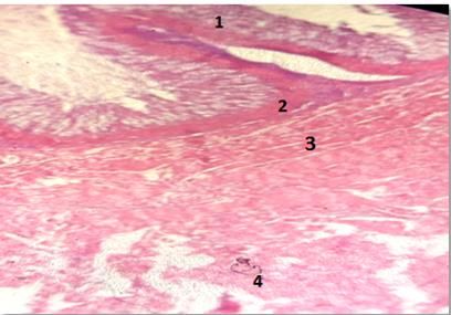
EurAsian Journal of BioSciences 14: 3031-3034 (2020) Ahmed et al.
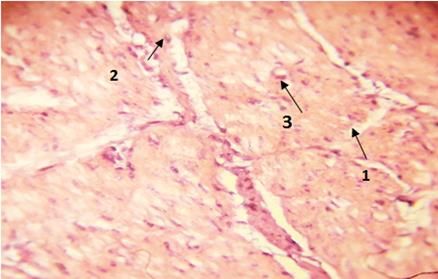
Fig. 4. Section of the pyloric gland region (stomach) that
displays the lining cells of Iraqi rabbit; 1- Glands 2- Mucous
Fig. 1. Section of fundic gland region (stomach) in Iraqi secreting cells, 3- Parietal cells (H&E) X40
rabbit that displays gastric layers; 1. Mucosa, 2.
Submucosa, 3. Muscularis, and 4. Serosa. (H&E) 40X
MATERIAL AND METHODS
Ten stomach tissue samples were collected from ten
rabbits. These samples were tissue-section-processed
by 10%-formalin fixation, washing, dehydration,
infiltration, embedding, sectioning into slices, making
slides, staining using Harris hematoxylin and eosin dyes,
and visualized and photo-taken under a light
microscope. The procedure protocols were followed
from (Bancroft, 2013).
RESULTS
The gastric wall of the rabbit consisted of a quadric-
Fig. 2. Section of pyloric gland region (stomach) in Iraqi tunic layer, in to out; mucosa, submucosa, muscularis,
rabbit that displays gastric layers; 1. Mucosa, 2.
Submucosa, 3. Muscularis, and 4. Serosa. (H&E) 400X
and serosa (Fig.1 and 2).
The inside surface of the stomach was lined by
mucosal cells that made an appearance as a tall simple
columnar epithelium spread through the pits of the
stomach (Fig. 3).
Moreover, the mucosa displayed three histologically
distinct regions based on glandular tissue types (branch-
, tubule-, and coil-like appearance) in which these
glands were short in the cardiac and pyloric regions. The
fundic gland region showed simple long straight branch-
like tubular features and revealed mostly mucous
secreting cells and less frequent parietal cells in the
cardiac and pyloric glands. Furthermore, the fundic
gland region consists of different kinds of cells; however,
the parietal and chief cells are the highly common cells
in this region (Figs. 4, 5, & 6).
Fig. 3. Section of the cardiac gland region (stomach) that
displays the lining cells (surface) of Iraqi rabbit; 1. Simple DISCUSSION
columnar epithelium, 2. Pits, 3. Lamina propria, 4. Glands, The stomach of the local rabbit showed histological
and 5. Muscularis mucosa. (H&E) 400X
features.(Khalel and Ghafi, 2012) found, in a study that
they performed on the local rabbit, that The Tunica
conducted to identify histological characteristic features mucosa appears as a ground lining cell which appears
of the animal stomach. as high-column mucus cells, extending into reasonably
long stomach pits.A lamina propria, a connective tissue
which appears only under the epithelial surface and
3032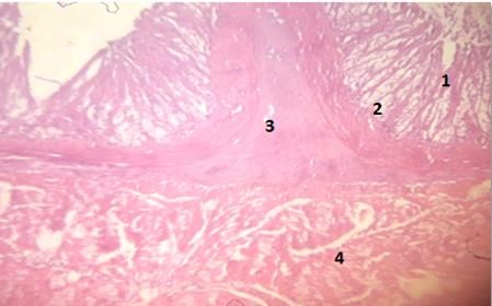
EurAsian Journal of BioSciences 14: 3031-3034 (2020)                                                           Ahmed et al.

                                                                 propria show up to the lower part of the rather long stand
                                                                 as a short branched tube, coiled gland.
                                                                     The stomach mucosa in rabbits in the region of the
                                                                 fundic glands is cast in prominent longitudinal folds or
                                                                 rugs into which the core of these folds consists of
                                                                 submucosa, the lumen from the contracted stomach.
                                                                 The epithelial cells on the surface are a simple high
                                                                 column epithelial extending out and lining up the short
                                                                 gastric     pits.    The      lamina Proria        is    a
                                                                 great connective tissue, and it is so hard to distinguish
                                                                 between the fundic gland numerous simple, ramified
Fig. 5. Section of the cardiac gland region (stomach) that       tubular glands, which are organized almost in parallel
displays the lining cells of Iraqi rabbit; 1- Glands 2- Mucous   with each other and are perpendicular to the mucous
secreting cells, 3- Parietal cells (H&E) X1000 (oil)             surface lining. In the glands of the cardiac area and
                                                                 fundic areas, lining epithelium appears a s a loose
                                                                 connective tissue with the lamina propria demonstrates
                                                                 more than in fundic region, inhabited with pyloric glands
                                                                 weakly loaded. The glands in the pyloric region are
                                                                 ramified and allow access at the base of long pits as
                                                                 short pyloric glands. The muscularis mucosa occurs as
                                                                 layers from the submucosa of smooth muscle fibers.
                                                                 Tunica submucosas are built up with a loose connective
                                                                 tissue and have many blood supplies in the pyloric
                                                                 glands area which are as same as in the cardiac and
                                                                 fundic glands (Khalel and Ghafi, 2012).
Fig. 6. Section of the fundic gland region (stomach) that
displays the lining cells of Iraqi rabbit; 1- Glands 2- Mucous      CONCLUSION
secreting cells, 3- Parietal cells (H&E) X1000 (oil)                Regions are presents in the gastric mucosa of the
                                                                 rabbit with high distribution of parietal and chief cells in
occupies the cardiac glands, is the second element of            the fundic gland region.
mucoa. The cardiac glands which occupy the lamina

                                                      REFERENCES
Bancroft J (2013) Histochemical Techniques. 2nd ed. Histochemical Techniques. Elsevier;.
Cabezas S, Blas J, Marchant TA, Moreno S (2007) Physiological stress levels predict survival probabilities in wild
  rabbits.    Horm     Behav     [Internet]. [cited  2020      Jul     13];51(3):313–20.      Available      from:
  https://pubmed.ncbi.nlm.nih.gov/17258747/
Deflers H, Gandar F, Bolen G, Farnir F, Marlier D (2018) Influence of a single dose of buprenorphine on rabbit
   (Oryctolagus cuniculus) gastrointestinal motility. Vet Anaesth Analg [Internet]. [cited 2020 Jul 13];45(4):510–9.
   Available from: https://pubmed.ncbi.nlm.nih.gov/29880277/
DiVincenti L, Rehrig A (2017) Social Behavior of Adult Male New Zealand White Rabbits Housed in Groups or Pairs
  in the Laboratory. J Appl Anim Welf Sci [Internet]. [cited 2020 Jul 13];20(1):86–94. Available from:
  http://www.ncbi.nlm.nih.gov/pubmed/27827538
DiVincenti L, Rehrig AN (2016) The social nature of european rabbits (oryctolagus cuniculus). J Am Assoc Lab Anim
   Sci [Internet]. [cited 2020 Jul 13];55(6):729–36. Available from: /pmc/articles/PMC5113872/?report=abstract
Duranthon V, Beaujean N, Brunner M, Odening KE, Santos AN, Kacskovics I, et al (2012) On the emerging role of
   rabbit as human disease model and the instrumental role of novel transgenic tools. Transgenic Res [Internet].
   [cited 2020 Jul 13];21(4):699–713. Available from: https://pubmed.ncbi.nlm.nih.gov/22382461/
Ferrand N, Branco M (2007) The evolutionary history of the European rabbit (Oryctolagus cuniculus): Major patterns
   of population differentiation and geographic expansion inferred from protein polymorphism. In: Phylogeography
   of Southern European Refugia: Evolutionary Perspectives on the Origins and Conservation of European
   Biodiversity [Internet]. Springer Netherlands; [cited 2020 Jul 13]. p. 207–35. Available from:
   https://link.springer.com/chapter/10.1007/1-4020-4904-8_7

                                                                                                                        3033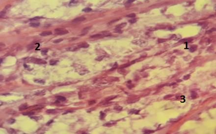
EurAsian Journal of BioSciences 14: 3031-3034 (2020)                                                  Ahmed et al.

Hansen LT, Berthelsen H (2000) The effect of environmental enrichment on the behaviour of caged rabbits
  (Oryctolagus cuniculus). Appl Anim Behav Sci [Internet]. [cited 2020 Jul 13];68(2):163–78. Available from:
  https://pubmed.ncbi.nlm.nih.gov/10771324/
Khalel EM, Ghafi HD (2012) Anatomical and histological study of stomach in adult local rabbits Oryctolagus
  cuniculus. Al- Mustansiriyah J Sci. 23(7):1–22.
Morgaz J, Navarrete R, Muñoz-Rascón P, Domínguez JM, Fernández-Sarmiento JA, Gómez-Villamandos RJ, et al
  (2013) Postoperative analgesic effects of dexketoprofen, buprenorphine and tramadol in dogs undergoing
  ovariohysterectomy. Res Vet Sci [Internet]. [cited 2020 Jul 13];95(1):278–82. Available from:
  http://www.ncbi.nlm.nih.gov/pubmed/23562407
Parveen, S., Khattak, S. G., & Saleem, Z. (2014). Effect of Different Types of Surfectants on Zinc Efficiency in
   Spinach Yield. International Journal of Sustainable Agricultural Research, 1(2), 45-51.
Püschel B, Daniel N, Bitzer E, Blum M, Renard JP, Viebahn C (2010) The rabbit (Oryctolagus cuniculus): A model
  for mammalian reproduction and early embryology. Cold Spring Harb Protoc [Internet]. [cited 2020 Jul 13];5(1).
  Available from: https://pubmed.ncbi.nlm.nih.gov/20150104/
Student PD, Benazzo A (2010) ISCRA Application form Curriculum Vitae. Mol Biol Evol [Internet]. [cited 2020 Jul
   13];23(10):1–7. Available from: http://feelsynapsis.com/pg/file/read/35739/natural-selection-on-the-influenza-
   virus-genome
Swennes AG, Alworth LC, Harvey SB, Jones CA, King CS, Crowell-Davis SL (2011) Human handling promotes
  compliant behavior in adult laboratory rabbits. J Am Assoc Lab Anim Sci [Internet]. [cited 2020 Jul 13];50(1):41–
  5. Available from: http://www.ncbi.nlm.nih.gov/pubmed/21333162
Weaver LA, Blaze CA, Linder DE, Andrutis KA, Karas AZ (2010) A model for clinical evaluation of perioperative
  analgesia in rabbits (Oryctolagus cuniculus). J Am Assoc Lab Anim Sci [Internet]. [cited 2020 Jul 13];49(6):845–
  51. Available from: /pmc/articles/PMC2994053/?report=abstract
Yamada S, Koike T, Nakagawa T, Kuniyoshi N, Ying Y, Itabe H, et al (2016) Morphological features of coronary
  plaques in WHHLMI rabbits (Oryctolagus cuniculus), an animal model for familial hypercholesterolemia. Exp
  Anim [Internet]. [cited 2020 Jul 13];66(2):145–57. Available from: https://pubmed.ncbi.nlm.nih.gov/28025424/
Yang J, Pospisil R, Ray S, Milton J, Mage RG (2009) Investigations of a rabbit (Oryctolagus cuniculus) model of
  Systemic Lupus Erythematosus (SLE), BAFF and its receptors. PLoS One [Internet]. [cited 2020 Jul 13];4(12).
  Available from: https://pubmed.ncbi.nlm.nih.gov/20041151/

                                                   www.ejobios.org

3034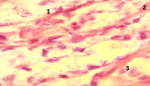
You can also read