THE PROTEOME OF FROZEN-THAWED PIG SPERMATOZOA IS DEPENDENT ON THE EJACULATE FRACTION SOURCE - DIVA PORTAL
←
→
Page content transcription
If your browser does not render page correctly, please read the page content below

www.nature.com/scientificreports

                         OPEN       The proteome of frozen-thawed pig
                                    spermatozoa is dependent on the
                                    ejaculate fraction source
Received: 19 July 2018              Cristina Pérez-Patiño1, Junwei Li1, Isabel Barranco1, Emilio A. Martínez1, Heriberto Rodriguez-
Accepted: 20 November 2018          Martínez 2, Jordi Roca 1 & Inmaculada Parrilla1
Published: xx xx xxxx
                                    The preservation of sperm functional parameters and fertility post-cryopreservation largely varies in the
                                    porcine, a species with a fractionated ejaculate. Although intrinsic individual differences have primarily
                                    been linked to this variation, differences in protein abundance among frozen-thawed (FT)-spermatozoa
                                    are far more relevant. This study, performed in two experiments, looked for proteomic quantitative
                                    differences between FT-sperm samples differing in post-thaw viability, motility, apoptosis, membrane
                                    lipid peroxidation and nuclear DNA fragmentation. The spermatozoa were either derived from the
                                    sperm-rich ejaculate fraction (SRF) or the entire ejaculate (Experiment 1) or from the first 10 mL of the
                                    SRF, the remaining SRF and the post-SRF (Experiment 2). Quantitative sperm proteomic differences
                                    were analysed using a LC-ESI-MS/MS-based SWATH approach. In Experiment 1, FT-spermatozoa from
                                    the SRF showed better preservation parameters than those from the entire ejaculate, with 26 Sus scrofa
                                    proteins with functional sperm relevance showing relative quantitative differences (FC ≥ 1.5) between
                                    sperm sources. In Experiment 2, FT-spermatozoa from the first 10 mL of the SRF and the remaining SRF
                                    were qualitatively better than those from the post-SRF, and 187 proteins showed relative quantitative
                                    differences among the three ejaculate sources. The results indicate that quantitative proteome
                                    differences are linked to sperm cryosurvival.

                                    Improving fertility outcomes of frozen-thawed (FT) spermatozoa remains a pending challenge for some livestock
                                    species, including the porcine1. In spite of the valuable progress in cryopreserving boar spermatozoa in recent
                                    years2, parameters defining relevant post-thaw sperm attributes are still variable and affect fertility, which remains
                                    considerably lower for FT-semen compared to liquid-stored semen3,4. This status of variable cryosurvival impairs
                                    the efficient inclusion of FT-spermatozoa in commercial artificial insemination (AI)-programs5 and is not exclu-
                                    sive to pigs since it also occurs in other species, such as the ovine6 or humans7. However, it is especially relevant
                                    for porcine commercial husbandry, where the magnitude of such variability classifies AI-boars as either good or
                                    bad sperm freezers8, sometimes impairing the efficient AI-use of genetically superior boars.
                                        The usually consistent variability in sperm cryosurvival among boars and among ejaculates within boars is in
                                    praxis compensated for by matching the numbers of cryosurviving FT-spermatozoa to those of the viable sper-
                                    matozoa used in AI-doses of liquid stored semen, e.g., increasing the number of FT-spermatozoa per dose. This
                                    process obviously implies up to a 4-fold increase in the total number of FT-sperm per AI-dose5, thus consuming
                                    more spermatozoa per boar. This accommodating and inefficient practice does, moreover, not match the fertility
                                    of FT-spermatozoa to that of the liquid stored semen, nor minimizes the variability between boars/ejaculates
                                    in AI-fertility9. Roca et al.10 noted that in vitro fertility outcomes of FT-boar spermatozoa from semen samples
                                    showing bad sperm freezability were lower than those showing good sperm freezability, even after inseminations
                                    with similar numbers of cryosurvived spermatozoa. This background suggests that there may be putative differ-
                                    ences in molecular arrangement affecting fertilizing capacity between the FT-spermatozoa from semen samples
                                    differing in sperm freezability. Since proteins are involved in most critical sperm functions, including fertilizing
                                    ability11, the present study attempts to clarify this issue by analysing the proteome of FT-spermatozoa with doc-
                                    umented freezability. It is currently known that cryopreservation remodels the proteome of boar spermatozoa12,
                                    but it is yet unknown if the proteome of FT-spermatozoa varies with the source of the spermatozoa, i.e., derived
                                    from semen sources with clearly different sperm freezability.

                                    1
                                     Department of Medicine and Animal Surgery, Faculty of Veterinary Science, University of Murcia, Murcia, Spain.
                                    2
                                     Department of Clinical & Experimental Medicine (IKE), Linköping University, Linköping, Sweden. Correspondence
                                    and requests for materials should be addressed to J.R. (email: roca@um.es)

        SCIEntIFIC REPOrTs |    (2019) 9:705 | DOI:10.1038/s41598-018-36624-5                                                                           1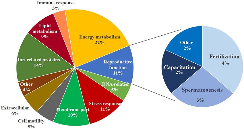
www.nature.com/scientificreports/

                                                                     Sperm source
                                                                     Sperm rich
                           Sperm attributes (%)                      ejaculate fraction   Entire ejaculate
                           Total motility                            49.02 ± 1.79a        36.13 ± 1.16b
                           Progressive motility                      40.93 ± 1.55   a
                                                                                          31.87 ± 1.83b
                           Viability                                 54.05 ± 1.65   a
                                                                                          41.53 ± 1.38b
                           Viable sperm with early apoptosis signs    4.17 ± 0.35a        10.74 ± 0.84b
                           Viable sperm with lipid peroxidation       4.76 ± 0.27a        12.00 ± 0.80b
                           Sperm with fragmented nuclear DNA          1.64 ± 0.24          2.07 ± 0.25

                           Table 1. Attributes (as percentage mean ± SEM) of frozen-thawed boar spermatozoa derived from the sperm
                           rich ejaculate fraction or the entire ejaculate (15 ejaculates from 5 boars). a,bP ≤ 0.001.

                               The boar ejaculate is emitted in fractions, and the so-called sperm-rich fraction (SRF) and the post-SRF are
                           the two main fractions13. Currently, pig AI-centres are moving from selectively collecting the SRF, by using the
                           gloved-hand method, towards collection of the entire ejaculate (EE), using semi-automatic methods5. This change
                           in method for ejaculate collection is mainly motivated by sanitary and labour cost reasons. However, it does not
                           advance animal welfare and jeopardizes sperm cryosurvival, since the post-thawing functional attributes of sper-
                           matozoa from the EE are worse than those from the SRF14,15. The current study, split into two experiments, aimed
                           therefore to compare the proteome of FT-spermatozoa derived from the SRF and the EE (Experiment 1) and to
                           compare that of the ejaculate fractions with clear differences in sperm freezability15,16, specifically, the first 10 mL
                           of the SRF, the remaining SRF and the post-SRF (Experiment 2).

                           Results
                           Sperm proteome profile.           A total of 93,457 spectra corresponding to 16,777 distinct peptides and 1,157
                           proteins were identified when assuming an FDR ≤ 1% at the protein level. Of the latter, 673 belonged to Sus scrofa
                           taxonomy. The complete list of the 1,157 sperm-proteins identified, including their unused score, UniProt acces-
                           sion number, protein name, species, % of sequence coverage and matched peptides, is provided in Supplementary
                           Table S1. The SWATH approach allowed the quantification of 1,094 sperm-proteins, of which 670 belong to Sus
                           scrofa taxonomy (Supplementary Table S2). All the quantified proteins were present in FT-spermatozoa from the
                           different sources evaluated, specifically the EE, the entire SRF, the first 10 mL of the SRF, the remaining SRF and
                           the post-SRF.

                           Proteins with different relative abundance in FT-spermatozoa among semen sources.                        The
                           results comparing the entire SRF and the EE and those comparing the different portions of the ejaculate (first
                           10 mL of the SRF, the remaining SRF and the post-SRF) are separately shown. In addition, the results of the
                           post-thaw assessment of sperm attributes are also included.

                           Differences between FT-spermatozoa retrieved from the SRF or the EE. Concerning post-thaw sperm attributes,
                           sperm source (P < 0.001) and boar (P < 0.01) influenced all sperm parameters evaluated 30 min after thawing,
                           except for proportion of fragmented nuclear DNA. The interaction between the FT-sperm source and boar was
                           not significant for any of the sperm attributes evaluated. Therefore, the data of the five boars were averaged for
                           each sperm source. Post-thawing measured sperm attributes were better (P < 0.001) in spermatozoa from the
                           SRF source than those from the EE (Table 1). Concerning the total FT-sperm proteins quantified, the two first
                           components of the PCA explained 94.4% of total variance, and the PC1 (explaining 85.2% of total variance) dis-
                           criminated between the three replicates of the SRF from those of the EE (Fig. 1). A total of 34 proteins belonging
                           to Sus scrofa taxonomy showed quantitative differences (P < 0.01) between the FT-sperm from the SRF and those
                           from the EE. The quantitative value of these proteins, after data normalization for each one of the two FT-sperm
                           sources and the FC estimation of the groups after log2 transformation, is shown in Supplementary Table S3.
                           Twenty-six of these 34 proteins showed an FC ≥ 1.50, and 11 of them were more abundant in the FT-spermatozoa
                           retrieved from the SRF, whereas the other 15 were more abundant in those derived from the EE (Table 2). The
                           heat-map of Fig. 2 showed that the three technical replicates of each FT-sperm source were grouped in a same
                           cluster merged at a short distance; evidenced further by the dendrogram with a large distance between the clus-
                           ter of SRF and that of EE. Regarding the allocation of the differentially quantified proteins into the functional
                           categories contemplated in UniProt KB and the DAVID database and specifically focused on categories related
                           to sperm and reproductive functions, most proteins showing quantitative differences were closely related to the
                           cell membrane (20%, 10 proteins), energy metabolism (14%, 7), cell motility (12%, 6), and reproduction (12%,
                           6). The six differentially quantified proteins framed into reproductive function were equally distributed among
                           spermatogenesis, sperm capacitation and fertilization (Fig. 3). Of note, some of the proteins showing quantitative
                           differences were related to the immune response (10%, 5), apoptosis (6%, 3), the stress response (6%, 3) and DNA
                           (2%, 1).

                           Differences among FT-spermatozoa retrieved from three specific ejaculate portions. Concerning post-thaw sperm
                           attributes, the sperm source and boar influenced (P < 0.01) all of the attributes, except for fragmented nuclear
                           DNA. The interaction between the FT-sperm source and boar was not significant for any of the sperm attributes
                           evaluated. Therefore, the data of the five boars were averaged for each sperm source. The post-thawing measured

SCIEntIFIC REPOrTs |   (2019) 9:705 | DOI:10.1038/s41598-018-36624-5                                                                             2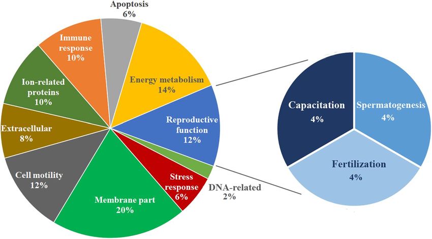
www.nature.com/scientificreports/

                           Figure 1. Principal Component Analysis (PCA) relative to the proteins quantified in frozen-thawed
                           spermatozoa retrieved from the sperm rich fraction (SRF) of the ejaculate or from the entire ejaculate (EE).
                           The points represent the three technical replicates for each sperm source and are based on the relative amounts
                           quantified from each source.

                           attributes were better (P < 0.01) in FT-spermatozoa from the first 10 mL of the SRF and the remaining SRF than
                           those from the post-SRF (Table 3). Regarding the total FT-sperm proteins quantified, the two first components
                           of the PCA explained 92.1% of the total variance. The PC1 (explaining 58.3% of total variance) best descriminate
                           best among the three ejaculate portions/sources (Fig. 4). Accordingly, the FT-sperm samples were separated into
                           two groups: the first one included samples from the first 10 mL of the SRF and the remaining SRF and the second
                           group included samples from the post-SRF. A total of 257 proteins belonging to Sus scrofa showed quantitative
                           differences (P < 0.01) among the FT-spermatozoa derived from the three ejaculate portions/sources. The relative
                           amount of these proteins, following data normalization for each one of the three FT-sperm sources and the FC
                           estimation of the groups after log2 transformation, is shown in Supplementary Table S4. A total of 187 of the
                           differentially quantified proteins showed an FC ≥ 1.50 (Supplementary Table S5), most of them differing between
                           FT-spermatozoa from the post-SRF and those from the two-other ejaculate portion/sources. Specifically, 173
                           FT-sperm proteins quantitatively differed between the post-SRF and the first 10 mL of SRF, while 165 proteins
                           differed between the post-SRF and the remaining SRF. In addition, 90 FT-sperm proteins from the post-SRF were
                           quantitatively higher than in the first 10 mL of the SRF, while 103 FT-sperm proteins from the post-SRF were
                           quantitatively higher compared to the remaining SRF. The heat-map in Fig. 5 showed that the three technical
                           replicates of FT-spermatozoa from the post-SRF were clustered together, clearly separated from those of the
                           FT-spermatozoa derived from either the first 10 mL of SRF or the remaining SRF, grouped into a same cluster.
                           Regarding the allocation of the differentially quantified proteins into the functional categories in UniProt KB and
                           the DAVID database and specifically focused on those related to sperm and reproductive functions, most of the
                           proteins were related to energy metabolism (22%, 66 proteins), ions (14%, 43), stress response (11%, 35), and
                           reproduction (11%, 33). Most of the differentially quantified proteins framed into reproductive function were
                           related to fertilization (4%, 12), spermatogenesis (3%, 11) and sperm capacitation (2%, 6) (Fig. 6). Some of these
                           differentially quantified proteins were also related to the cell membrane (10%, 31), lipid metabolism (9%, 27), cell
                           motility (5%, 16) and DNA (5%, 15).

                           Discussion
                           The results of the present study showed quantitative differences of proteins with relevance for sperm function
                           among FT-spermatozoa. The results showed differences in freezability and sperm cryosurvival from different
                           portions/fractions of the pig ejaculate. The results highlight that the spermatozoa derived from the EE would be
                           less functional post-thaw than those retrieved from the SRF because of the differential relative abundance of some
                           proteins, a matter which could seriously impair sperm fertilizing capacity.
                               The results from the post-thaw sperm confirm previous reports, demonstrating that boar spermatozoa from
                           the SRF cryosurvive better than those retrieved from the EE14,15. Cryosurviving spermatozoa derived from the EE
                           showed higher proportions of dysfunctions (lower sperm motility and higher rates of apoptosis and membrane
                           lipid peroxidation) compared to FT-spermatozoa derived from the SRF. Molecular derangement, involving pro-
                           teins, probably underlies these differences in sperm freezability. Subtle changes in relative protein abundance may
                           compromise the reproductive performance of spermatozoa since proteins are involved in membrane remodelling,
                           capacitation, oocyte zona binding, acrosome reaction and fusion to the oolemma17. Moreover, sperm proteins,
                           together with those of the seminal plasma, play a critical role in the immunological reaction of the internal sow
                           genital tract tissues after insemination18.

SCIEntIFIC REPOrTs |   (2019) 9:705 | DOI:10.1038/s41598-018-36624-5                                                                         3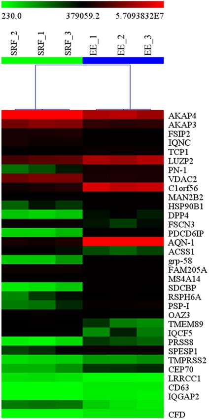
www.nature.com/scientificreports/

                           Protein Name                                                  Accession    Gene Name            FC
                           A-kinase anchoring protein 4                                  A0A286ZWH7   AKAP4                 3.46
                           Fibrous sheath interacting protein 2                          F1RYK8       FSIP2                 2.43
                           Uncharacterized protein                                       A0A287AI93   IQCN                  1.83
                           Nexin-1                                                       Q8WNW8       PN-1                 −4.49
                           Voltage-dependent anion-selective channel protein 2           F1S2F6       VDAC2                 2.01
                           Chromosome 1 open reading frame 56                            F1SSA1       C1orf56              −2.84
                           Endoplasmin                                                   Q29092       HSP90B1 GRP94 TRA1   −2.38
                           Dipeptidyl peptidase 4                                        A0A2C9F3H7   DPP4                 −2.07
                           Fascin                                                        Q2I373       FSCN3                 2.90
                           Programmed cell death 6 interacting protein                   F1RRD6       PDCD6IP              −2.93
                           Carbohydrate-binding protein AQN-1                            P26322       AQN-1                −3.80
                           Protein disulfide-isomerase                                   E1CAJ5       grp-58               −2.58
                           Family with sequence similarity 205 member A                  I3L912       FAM205A               1.83
                           Uncharacterized protein                                       F1RT83       SDCBP                −4.20
                           Radial spoke head 6 homolog A                                 A0A287AGL6   RSPH6A               −1.54
                           Spermadhesin PSP-I                                            Q4R0H6       PSP-I                −3.34
                           Ornithine Decarboxylase Antizyme 3                            I3LTK6       OAZ3                  1.68
                           Transmembrane protein 89                                      F1SKK4       TMEM89                1.91
                           IQ motif containing F5                                        A0A287A4D1   IQCF5                 1.73
                           Protease, serine 8                                            A0A286ZNI7   PRSS8                −2.69
                           Uncharacterized protein                                       A0A287B423   SPESP1               −2.99
                           Leucine rich repeat and coiled-coil centrosomal 1 protein 1   I3LBJ0       LRRCC1                1.83
                           Tetraspanin CD63                                              F1SPK8       CD63                 −1.88
                           Ras GTPase-activating-like protein IQGAP2                     K9J6M1       IQGAP2               −2.16
                           Uncharacterized protein                                       A0A286ZN09                         1.82
                           Complement factor D preproprotein                             B4F449       CFD                  −3.18

                           Table 2. Differentially abundant Sus scrofa proteins with a fold change (FC) ≥ 1.50 between frozen-thawed boar
                           spermatozoa from the rich ejaculate fraction and the entire ejaculate.

                               A total of 26 proteins encoded by Sus scrofa with a relationship to sperm function were proportionally differ-
                           ent, with an FC ≥ 1.5, between FT-spermatozoa retrieved from the SRF and those from the EE. Eleven of these
                           proteins, including two still uncharacterized by current databases, were most abundant in FT-spermatozoa from
                           the SRF, which had the best sperm attributes post-thaw. Many of these proteins are structural components of the
                           sperm tail, playing essential roles in the activation of the flagellum19–21. A-kinase anchoring protein (AKAP)-
                           4, the most abundant cytoskeletal glycoprotein of the sperm fibrous sheath, Fibrous sheath interacting protein
                           2, Fascin, Ornithine decarboxylase antizyme 3 and Leucine rich repeat and coiled-coil centrosomal protein 1
                           were among the detected proteins. A lower abundance of these proteins could indicate damage in the tail and/or
                           the mitochondrial sheath of spermatozoa and would explain why FT-spermatozoa from the EE have decreased
                           motility parameters than spermatozoa derived from the SRF. Similarly, the higher abundance of the other four
                           proteins could also clarify the improved functionality shown by FT-spermatozoa from the SRF. In this sense, the
                           mitochondrial outer membrane porin protein voltage-dependent anion-selective channel protein 2 (VDAC2)
                           is considered a positive biomarker of boar sperm freezability22, actively participating in the regulation of sperm
                           mitochondrial function23. Family with sequence similarity 205-member A (FAM205A) is overexpressed in sub-
                           populations of ejaculated human viable spermatozoa with low nuclear DNA fragmentation24. Finally, the other
                           two more abundant proteins in FT-spermatozoa from the pig SRF, specifically the Transmembrane protein 89
                           and IQ motif containing F5, are proteins of the sperm plasma membrane involved in membrane stabilization and
                           permeability regulation25.
                               Fifteen proteins were more abundant in FT-spermatozoa retrieved from the EE. Such relatively higher abun-
                           dance could explain why FT-spermatozoa from the EE were less functional than those retrieved from the SRF.
                           For instance, one of these proteins, the dipeptidyl peptidase IV (DPP-IV or CD26), a mitochondrial-associated
                           protein26 related to sperm motility27, could be involved in inducing premature acrosome reaction when present in
                           excess on the sperm surface. This protein is transferred to the sperm surface from sperm-binding seminal plasma
                           vesicles28, and studies clearly demonstrated that human and bovine spermatozoa reach acrosome reaction if cul-
                           tured with seminal plasma vesicles rich in DPP-IV29,30. Endoplasmin, better known as tumour rejection antigen
                           gp96, a chaperone involved in the rebuilding of the sperm surface during capacitation31, was more abundant in
                           sperm that underwent stress conditions such as cryopreservation32. In addition, endoplasmin was also compara-
                           tively more abundant in cryopreserved bull spermatozoa with low motility parameters33. Spermadhesin AQN1, a
                           seminal plasma protein that binds to the sperm plasma membrane over the acrosomal domain during ejaculation,
                           is involved in the formation of the oviductal sperm reservoir34–36. It is maintained in this location before capac-
                           itation, to disappear once spermatozoa are capacitated37,38. However, many spermatozoa surviving cryoinjury

SCIEntIFIC REPOrTs |   (2019) 9:705 | DOI:10.1038/s41598-018-36624-5                                                                        4
www.nature.com/scientificreports/
Figure 2. Heat-map with dendrograms representing the differentially abundant Sus scrofa proteins between
frozen-thawed spermatozoa derived from either the sperm-rich ejaculate fraction (SRF) or the entire ejaculate
(EE). The hierarchical clustering tree of sperm samples is shown at the top. The relative abundance level of each
protein is shown on a colour scale from green (lowest level) to red (highest level).
often exhibit the phenomenon named “cryocapacitation”, characterized by capacitation-like changes in sperm
membrane phospholipids39,40. In these “cryocapacitated” spermatozoa, the AQN1 remains present in the sperm
surface35. The sperm equatorial segment protein 1 (SPESP1), a protein involved in sperm-oocyte binding41, is
particularly exposed in acrosome-reacted spermatozoa42, being found comparatively more abundant in cryopre-
served than in fresh pig spermatozoa12. In view of this, a larger amount of SPESP1 would suggest the presence of a
larger number of acrosome-reacted spermatozoa, cells that would hardly fertilize oocytes following insemination.
The higher abundance of some of these 15 proteins in FT-spermatozoa retrieved from the EE would entail
that their fertilizing capacity could be compromised. For instance, Nexin 1, Spermadhesins PSPI, Tetraspanin
CD63 (CD63), Complement Factor D (CFD) and Ras GTPase-activating-like protein IQGAP2 (IQGAP2) have
been related to fertility losses when found comparatively more abundant in either spermatozoa or in seminal
plasma. A higher abundance of PSPI and Nexin 1 was related to lower fertility outcomes in artificially insemi-
nated sows43,44. Similarly, CD63, CFD and IQGAP2 were most abundant in fresh human spermatozoa with poor
embryo developmental capacity after ICSI45,46. Other of the more abundant proteins found in FT-spermatozoa
SCIEntIFIC REPOrTs | (2019) 9:705 | DOI:10.1038/s41598-018-36624-5 5www.nature.com/scientificreports/
Figure 3. Functional distribution of Sus scrofa sperm proteins into the categories “sperm functions” and
“reproductive functions” available on the UniProtKB/Swiss-Prot website (www.uniprot.org) and from DAVID
Bioinformatics Resources 6.8 (https://david.ncifcrf.gov).
Figure 4. Principal Component Analysis (PCA) of proteins quantified in frozen-thawed spermatozoa retrieved
from three identifiable portions of the pig ejaculate (the 10 first mL of the sperm-rich ejaculate fraction (SRF),
the remaining SRF or the post-SRF). The points represent the three technical replicates for each sperm source
and are based on the relative amounts quantified for each source.
Sperm source
Sperm attributes (%) First 10 ml of SRF Rest of SRF Post SRF
Total motility 53.67 ± 1.98a 47.40 ± 2.35a 20.87 ± 1.50b
Progressive motility 44.33 ± 1,74a 40.87 ± 2.27a 15.73 ± 0.84b
Viability 58.50 ± 1.99 a
52.55 ± 2.54 a
26.09 ± 1.88b
Viable sperm with early apoptosis signs 4.26 ± 0.47 a
4.69 ± 0.44 a
19.32 ± 1.36b
Viable sperm with lipid peroxidation 3.73 ± 0.36a 4.69 ± 0.70a 16.63 ± 1.27b
Sperm with fragmented nuclear DNA 1.62 ± 0.24 1.82 ± 0.25 1.78 ± 0.22
Table 3. Attributes (percentage mean ± SEM) of frozen-thawed boar spermatozoa retrieved from three clearly
identifiable ejaculate fractions (the first 10 mL of sperm-rich ejaculate fraction [SRF], the remaining SRF and
the post-SRF; 15 ejaculates from 5 boars). a,bP ≤ 0.01.
SCIEntIFIC REPOrTs | (2019) 9:705 | DOI:10.1038/s41598-018-36624-5 6www.nature.com/scientificreports/
Figure 5. Heat-map with dendrograms representing the differentially abundant Sus scrofa proteins among
frozen-thawed spermatozoa from three identifiable portions of the pig ejaculate (the 10 first mL of the sperm-
rich ejaculate fraction (SRF), the remaining SRF or the post-SRF). The hierarchical clustering tree of sperm
samples is shown at the top. The relative abundance level of each protein is shown on a colour scale from green
(lowest level) to red (highest level).
retrieved from the EE are related to the immune response either through complement and T-cell activation or
cytokine modulation, specifically Syndecan binding protein (also named Syntenin, SDCBP), the DPP-IV and
the programmed cell death 6 interacting protein47–49. Their higher abundance could make FT-spermatozoa from
the EE particularly susceptible to phagocytosis once inseminated in the internal genital tract of the sow50,51. The
SCIEntIFIC REPOrTs | (2019) 9:705 | DOI:10.1038/s41598-018-36624-5 7www.nature.com/scientificreports/
Figure 6. Functional distribution of Sus scrofa sperm proteins into the categories “sperm funcrions” and
“reproductive functions” available on the UniProtKB/Swiss-Prot website (www.uniprot.org) and from DAVID
Bioinformatics Resources 6.8 (https://david.ncifcrf.gov).
consequences on FT-sperm performance by the four remaining more abundant proteins is less clear. The higher
abundance of protein disulfide-isomerase could be related to cryocapacitation52. Radial spoke head 6 homolog A
(RSPH6A), a structural protein of the sperm fibrous sheath that is involved in sperm motility53, was more abun-
dant in FT- than fresh ovine spermatozoa6. Protease serine 8 is an enzyme involved in semen coagulation54, and
chromosome 1 open reading frame 56 was recently identified in boar sperm chromatin55. Their relationship to
FT-sperm fertilizing capacity requires further studies.
It was interesting to note the conspicuous relationship of many of the above-mentioned abundant proteins
with the seminal plasma. However, whereas none of the more abundant proteins in the FT-spermatozoa from the
SRF were identified in the seminal plasma of this fraction of the ejaculate, 11 of the 15 most abundant proteins in
the EE-derived FT-spermatozoa were present in the seminal plasma44. These findings would indicate that most of
the more abundant proteins in the FT-spermatozoa from the EE would come from the seminal plasma, binding
to the sperm surface either during ejaculation or in overnight storage before freezing ex situ. Moreover, as these
proteins were also more abundant in the seminal plasma from the post-SRF56, it is reasonable to consider they
could primarily bind to the spermatozoa present in the post-SRF fraction. This assumption would also explain
why there were not quantitative changes between the FT-spermatozoa retrieved from the SRF (the first 10 mL of
the SRF vs the remaining SRF). The assumption would suggest that the binding of the proteins to the spermatozoa
had already occurred during ejaculation when in contact with seminal plasma. Spermatozoa of the three main,
identifiable, ejaculate fractions, specifically the first 10 mL of the SRF, the remaining SRF and the post-SRF, were
separately cryopreserved and the proteome of FT-spermatozoa was evaluated to clarify the above assumptions.
The post-thaw sperm attributes agree with those previously reported by Alkmin et al.14 and Li et al.15, con-
firming that spermatozoa derived from the post-SRF were those with the worst freezability. The results prove that
distinguishable sperm populations with clear differences in freezability are present in a single porcine ejaculate,
suggesting biological reasons for the presence of a fractionated ejaculate in this species. Taken together, the results
indicate that the worse freezability of spermatozoa from the EE compared to those from the SRF would be due
to the negative concerted contribution of spermatozoa and seminal plasma from the post-SRF in building the
EE. The clear quantitative differences in the proteome between the FT-spermatozoa from the post-SRF fraction
and those from the other two ejaculate fractions could explain the poor functionality of FT-spermatozoa from
the post-SRF source, as they showed a relative overabundance of many proteins directly or indirectly involved
in sperm dysfunctionality. As expected, the 15 proteins that were more abundant in the FT-spermatozoa from
the EE than in the SRF were among the most abundant proteins in the FT-spermatozoa from the post-SRF.
Most of the more abundant proteins in the FT-spermatozoa from the post-SRF were previously identified as
abundant in its seminal plasma56. This scenario would also support the notion that seminal plasma proteins bind
most likely to the spermatozoa during ejaculation rather than during overnight in vitro sperm storage, a step in
cryopreservation.
In conclusion, the present study showed that relative differences in abundance of specific proteins could
explain why spermatozoa retrieved from the EE were less functional post-thaw than those derived from the SRF,
a matter with clear biological and practical implications. The decreased functionality of the FT-spermatozoa
derived from the EE could be caused by the negative contribution of the FT-spermatozoa from the post-SRF,
coated with several seminal plasma proteins whose overabundance could negatively influence freezability and
sperm cryosurvival. These results should be taken into careful consideration by technicians responsible for col-
lecting and cryopreserving porcine ejaculates, to the extent that they showed that FT-spermatozoa from the EE
were less functional than those from the SRF.
SCIEntIFIC REPOrTs | (2019) 9:705 | DOI:10.1038/s41598-018-36624-5 8www.nature.com/scientificreports/
Methods
Boars, ejaculates and sperm sources. All procedures involving boars and semen samples were per-
formed following international guidelines (Directive 2010/63/EU) and were approved by the Bioethics Committee
of the University of Murcia (research code: 639/2012). The chemicals used for elaborate semen extenders and the
fluorescent probes used for sperm evaluation were purchased from Sigma-Aldrich Co. (St. Louis, MO, USA).
Five healthy, young (12- to 18-months-old), sexually mature and fertility proven boars (two Large White,
two of Duroc and one of Landrace breeds) were used as semen donors. The boars were housed in a commercial
AI-centre (Topigs Norsvin España, Madrid, Spain) under controlled environmental conditions (16 h of light and
15–25 °C) and subjected to regular ejaculate collections (two ejaculates per week). A total of 15 ejaculates (three
per boar) were manually collected using the gloved-hand method. All the ejaculates met the minimum require-
ments stablished by the AI-centre for the preparation of insemination doses, specifically more than 200 × 106
spermatozoa/mL with 75% and 80% of spermatozoa displaying normal motility and morphology. The ejaculates
were collected in three separate fractions, specifically the first 10 mL of the SRF, the remaining SRF and the
post-SRF. Volumes of the first 10 mL of the SRF and the remaining SRF were proportionally mixed to generate
semen samples simulating entire SRFs. Similarly, volumes of the three ejaculate fractions were mixed for gen-
erating semen samples simulating EEs. The five resulting semen samples of each ejaculate were extended (1:1
vol:vol) in Beltsville Thawing Solution (BTS), cooled at 17 °C, placed into a Styrofoam box and transported to the
Andrology Laboratory of the Veterinary Teaching Hospital (VTH) of the University of Murcia (trip < 2 h). Once
in the VHT, the BTS-extended semen samples were stored overnight at 17 °C to be frozen the next morning.
Semen cryopreservation and post-thaw sperm evaluation. Semen samples were frozen using the
straw freezing protocol described by Alkmin et al.14. Briefly, semen samples were centrifuged (2,400 xg for 3 min;
Megafuge 1.0 R, Heraeus, Hanau, Germany), and the resulting sperm pellets were extended to 1.5 × 10⁹ sperma-
tozoa/mL in a Tris-citric acid-glucose extender supplemented with egg yolk (80:20, vol:vol). Then, the extended
spermatozoa were cooled to 5 °C and re-extended to 1.0 × 10⁹ spermatozoa/mL in the same extender supple-
mented with 9% glycerol and 1.5% Equex STM (v:v) (Nova Chemical Sales, Scituate, MA, USA). The spermato-
zoa were then packed into 0.5 mL polyvinyl chloride French straws (Minitüb, Tiefenbach, Germany) and frozen
on a metal rack 3 cm above liquid nitrogen (LN2) for 20 min (Freezing unit, Minitüb). After being stored in an
LN2-tank (GT40, Air Liquide, Paris, France) for at least a week, the straws (two per sperm sample/source) were
thawed in a circulating water bath at 37 °C for 20 s, the contents were extended in BTS (1:1, vol:vol) and the mix-
ture was incubated at 37 °C for 30 min before sperm analyses.
Post-thaw total and progressive sperm motility was evaluated using a computer-assisted sperm analyser
®
(ISASV1 CASA, Proiser R + D, Paterna, Spain) and sperm viability was evaluated using a BD FACS Canto II
flow cytometer (Becton Dickinson & Company, Franklin Lakes, NJ, USA) after labelling the spermatozoa (100-µL
with 3 × 10⁶ spermatozoa) with 3 µL Hoechst 33342 (H-42; 0.05 mg/mL in PBS), 2 µL propidium iodide (PI,
0.5 mg/mL in PBS), and 2 µL fluorescein-conjugated peanut agglutinin (PNA-FITC, 100 µg/mL in PBS). Viable
sperm were categorized as those that were H-42 positive, PI negative and PNA-FITC negative. Protocols are
described at length in Li et al.15.
Further functional attributes of cryosurvived spermatozoa were cytometrically evaluated. The early events
of apoptosis and membrane lipid peroxidation (100-µL with 2 × 10⁶ spermatozoa labelled with 2 µL H-42 and
10 µL of PI) were evaluated using the fluorescent probes Annexin V-FITC (3 µL) and BODIPY 581/591 C11
(2.5 µL), respectively57. Viable spermatozoa (H-42 positive and PI negative) showing either early signs of apopto-
sis (Annexin V-FITC positive) or membrane lipid peroxidation (BODIPY positive) were recorded. Lastly, nuclear
®
DNA fragmentation was assessed using the Sperm-Sus-Halomax kit (Halotech DNA SL, Madrid, Spain) follow-
ing the method reported by Alkmin et al.58.
Sperm proteomics. The proteomics analyses were carried out in the Proteomics Unit of the University of
Valencia, Valencia, Spain (member of the PRB2-ISCIII ProteoRed Proteomics Platform).
Preparation of sperm samples. Three straws for each one of the 75 sperm sample/source (five semen samples of
each one of the three ejaculates from each of the five boars) were thawed a 37 °C and their contents were centri-
fuged at 600 xg for 20 min at room temperature (rt) (Megafuge 1.0 R, Heraeus) on a Percoll monolayer gradient
®
(45% in PBS [v:v], Percoll P4937; Sigma-Aldrich Co.) to separate the spermatozoa from other putative cells and
debris, including egg-yolk remnants. The sperm pellets were extended in PBS (1:10, v:v) and centrifuged again
(300 xg, 10 min at rt). Then, the resulting sperm pellets were extended in PBS to a concentration of 1,000 × 106
sperm/mL, and stored in cryotubes (2 mL Cryogenic vial, Fisher Scientific, Madrid, Spain) that were preserved at
−80 °C (Ultra Low Freezer; Haier, Schomberg, Ontario, Canada) until protein analysis.
During proteomics analysis, sperm samples were thawed at room temperature and the fifteen sperm samples
of each one of the five semen sources (first 10 mL of the SRF, the remaining SRF, the entire SRF, the post-SRF and
the EE) were pooled, generating five single sperm pools (one pool per source). Each sperm pool was in turn split
into three aliquots generating three technical replicates per sperm sample. Thus, a total of 15 sperm samples were
analysed.
Protein extraction. Sperm samples were centrifuged at 14,000 xg for 10 min (Eppendorf 5424 R, Eppendorf
AG, Hamburg, Germany) and the supernatant was discarded. The total protein from the resulting sperm pellets
was extracted using 200 µL of UTC buffer [7 M Urea, 2 M thiourea, 4% 3-[(3-cholamidopropyl) dimethylammo-
nio]-1-propanesulfonate, (CHAPS)], supplemented with protease inhibitor cocktail (1%, v:v), followed by vigor-
ous stirring (one hour at 5 °C). The concentration of extracted protein was quantified by RC_DC Lowry (Bio-Rad,
SCIEntIFIC REPOrTs | (2019) 9:705 | DOI:10.1038/s41598-018-36624-5 9www.nature.com/scientificreports/
Richmond, CA, USA) following the manufacturer’s instructions. Then, a total of 30 µg of final protein extract was
taken for one dimensional sodium dodecyl sulfate-polyacrylamide gel electrophoresis (1D SDS-PAGE) loading to
remove the remaining UTC and other interferences for tandem mass spectrometry (MS/MS) analysis.
SWATH analysis. In-gel digestion processing: The 1D SDS-PAGE portion containing proteins was digested at
37 °C using 500 ng of sequencing grade trypsin (V511, Promega Co., Madison, WI, USA) following the protocol
used by Shevchenko et al.59. The trypsin digestion was stopped with 10% trifluoroacetic (TFA) and the superna-
tant, containing the non-extracted digests, was carefully removed, leaving behind the sliced gels in the Eppendorf
tube. For peptide extraction, 200 µL of pure acetonitrile (ACN) was then added, followed by incubation at 37 °C
in a shaker for 15 min. The new supernatant containing the peptide mixture was carefully withdrawn and dried
in a speed vacuum (ISS 110 SpeedVac System, Thermo Savant, ThermoScientific, Langenselbold, Germany) for
20 min and then re-suspended in 25 µL of 2% ACN and 0.1% TFA.
Liquid chromatography and tandem mass spectrometry (LC-MS/MS) analyses: For the spectral library acqui-
sition, a mixture of all digested samples (2 µL of each sample) was examined by liquid chromatography (LC) using
a NanoLC Ultra 1D plus Eksigent (Eksigent Technologies, Dublin, CA, USA), which was directly connected to
an AB SCIEX TripleTOF 5600 mass spectrometer (AB SCIEX, Framingham, MA, USA) in direct injection mode.
Five µL of the digested sample was loaded on a trap NanoLC pre-column (3 μm particles size C18-CL, 350 µm
diameter × 0.5 mm long; Eksigent Technologies) and desalted with 0.1% TFA at 3 µL/min for 5 min. Then, the
peptides were separated using an analytical LC column (3 μm particles size C18-CL, 75 µm diameter × 12 cm
®
long, Nikkyo Technos Co , Tokyo, Japan) equilibrated in 5% ACN and 0.1% formic acid (FA) (Fisher Scientific).
Peptide elution was performed by applying a linear gradient from 5% to 35% of ACN containing 0.1% FA at a
constant flow rate of 300 nL/min over 180 min.
The TripleTOF was operated in data-dependent mode, in which a time of flight (TOF) MS scan occurred
from 350 to 1250 m/z, accumulated for 250 ms TOF followed by 150 ms TOF with the same scan range for MS.
The 25 most abundant multiply charged (2+, 3+, 4+ or 5+) precursor peptide ions were automatically selected.
Ions with 1+ and unassigned charge states were rejected from the MS/MS analysis. The rolling collision energy
equations were automatically set by the instrument according to the equation |CE| = (slope) × (m/z) + (intercept)
with Charge = 2; Slope = 0.0575 and Intercept = 9.
LC-SWATH-MS acquisition. To determine quantitative differences between the SRF and the EE and among the
first 10 mL of the SRF, the remaining SRF and the post-SRF, the sequential window acquisition of all theoretical
spectra (SWATH) analysis of individual sperm samples was performed following the procedure described by
Perez-Patiño et al.56, tuning the TripleTOF 5600 (AB SCIEX) as described by Gillet et al.60 for SWATH-MS-based
experiments. In this way, 5 µL of one of the three technical replicates from each sample was randomly loaded
onto a trap column (NanoLC Column, 3 µm C18-CL, 75 μm × 15 cm; Eksigent Technologies) and flushed for
5 min with 0.1% TFA at 3 µL/min to remove salts. Peptide separation was achieved using an analytical column
®
(LC Column, 3 µm C18-CL, 75 µm × 12 cm, Nikkyo Technos Co ) equilibrated in 5% ACN and 0.1% FA, eluted
with a linear gradient from 5 to 40% ACN for 90 min at a flow rate of 300 nL/min. The analysis of eluted peptides
was carried out in the spectrometer nanoESI-qQTOF (SCIEX TripleTOF 5600) and the TripleTOF operated in
SWATH mode, in which a 0.050 s TOF MS scan from 350 to 1250 m/z was performed, followed by 0.080 s product
ion scans from 230 to 1800 m/z in the 37 defined windows (3.05 sec/cycle). The collision energy for each window
was calculated for 2+ charged ion at the centre of each SWATH block with a collision energy spread of 15 eV. The
MS was always operated in high sensitivity mode.
Protein identification, validation and quantification. After the LC-MS/MS, the resulting SCIEX.wiff
data-files were processed by the ProteinPilot v5.0 search engine (AB SCIEX) and the Paragon algorithm (4.0.0.0,
4767) was used to search against the UniProt_mammals database with the following parameters: trypsin speci-
ficity, cys-alkylation and the search effort set to through. To avoid using the same spectral evidence for more than
one protein, the identified proteins were grouped based on MS/MS spectra by the Protein-Pilot Pro Group
algorithm, regardless of the peptide sequence assigned. The protein within each group that could explain the
™
most spectral data with confidence was depicted as the primary protein of the group. The SCIEX.wiff data-files
®
obtained from SWATH experiment were analysed by PeakView (v2.1, AB SCIEX) and peaks from SWATH were
extracted with a peptide confidence threshold of 95% confidence. A false discovery rate (FDR) less than 1% and 6
transitions per peptide were required to quantify one peptide. The extracted ion chromatograms were integrated
and the area under the curve (AUC) was used to calculate total protein. A normalization of the calculated AUCs
was done by total sum, and the sum of all areas was equalized for all the samples.
Gene ontology and bioinformatics analysis. The bioinformatics of differentially expressed sperm pro-
teins was manually annotated using two comprehensive bioinformatic tools, specifically UniProt KB and DAVID.
The UniProt KB database (www.uniprot.org), downloaded 19/04/2018, contains 115,678,811 total entries with
47,499 entries encoded in the Sus scrofa taxonomy. The DAVID Database for Annotation, Visualization and
Integrated Discovery (DAVID Bioinformatics Resources 6.8; https://david.ncifcrf.gov/61,62) integrates numerous
public sources of protein annotation and, consequently, contains information from more than 1.5 million genes
and more than 65,000 species.
Statistical analysis. A mixed ANOVA test (IBM SPSS v24.0 software, IBM Spain, Madrid), using ejaculate
as a random effect, was performed to evaluate the influence of the semen source (entire SRF vs EE or comparing
the first 10 mL of the SRF, the remaining SRF and the post-SRF) and the boar (n = 5) on the post-thaw sperm
®
attributes. In proteomics, the quantitative data obtained by PeakView were analysed using MarkerView (v1.2, ®
SCIEntIFIC REPOrTs | (2019) 9:705 | DOI:10.1038/s41598-018-36624-5 10www.nature.com/scientificreports/
AB SCIEX) and the peak areas were normalized by the sum of peak areas of all identified peptides. Principal
Component Analysis (PCA) was performed to evaluate the discrimination ability of FT-sperm proteins between
semen sources from Experiment 1 and 2. Then, the Multiexperiment Viewer (MeV) software (version 4.8) (http://
www.tm4.org/mev.html) was used to identify changes in relative proteins abundance among FT-sperm sources.
Once identified, differences in protein abundance were statistically evaluated using a Student’s t-test and ANOVA
in Experiment 1 and 2, respectively. Differentially abundant proteins among FT-sperm sources were defined using
an adjusted P-value < 0.01, and those with a fold change (FC) ≥ 1.50 after log2 transformation were highlighted.
The explanatory ability of the resulting selected proteins in both experiments was illustrated by mean of heat
maps after z-score normalization using Euclidean distances.
Data Availability
All data generated in the experiments are provided in supplementary files.
References
1. Yeste, M., Rodríguez-Gil, J. E. & Bonet, S. Artificial insemination with frozen-thawed boar sperm. Mol. Reprod. 84, 802–813 (2017).
2. Yeste, M. Recent Advances in Boar Sperm Cryopreservation: State of the Art and Current Perspectives. Reprod. Domest. Anim. 50,
71–79 (2015).
3. Knox, R. V. The Fertility of Frozen Boar Sperm When used for Artificial Insemination. Reprod. Domest. Anim. 50, 90–97 (2015).
4. Yeste, M. Sperm cryopreservation update: Cryodamage, markers, and factors affecting the sperm freezability in pigs. Theriogenology.
85, 47–64 (2016).
5. Roca, J., Parrilla, I., Bolarin, A., Martinez, E. A. & Rodriguez-Martinez, H. Will AI in pigs become more efficient? Theriogenology.
86, 187–193 (2016).
6. Pini, T. et al. Cryopreservation and egg yolk medium alter the proteome of ram spermatozoa. J. Proteomics. 181, 73–82 (2018).
7. Marlea, D. S., Nicoletta, T., Marco, N. & Andrea, B. Human Sperm Cryopreservation: Update on Techniques, Effect on DNA
Integrity, and Implications for ART. Adv. Urol. 2012, 854837, https://doi.org/10.1155/2012/854837 (2012).
8. Thurston, L. M., Siggins, K., Mileham, A. J., Watson, P. F. & Holt, W. V. Identification of amplified restriction fragment length
polymorphism markers linked to genes controlling boar sperm viability following cryopreservation. Biol. Reprod. 66, 545–554
(2002).
9. Bolarín, A. et al. Dissimilarities in sows ovarian status at the insemination time could explain differences in fertility between farms
when frozen-thawed semen is used. Theriogenology. 65, 669–80 (2006).
10. Roca, J., Martinez-Alborcia, M. J., Gil, M. A., Parrilla, I. & Martinez, E. A. Dead spermatozoa in raw semen samples impair in vitro
fertilization outcomes of frozen-thawed spermatozoa. Fertil. Steril. 100, 875–881 (2013).
11. Gadella, B. M. & Boerke, A. An update on post-ejaculatory remodeling of the sperm surface before mammalian fertilization.
Theriogenology. 85, 113–24 (2016).
12. Chen, X. et al. Identification of differentially expressed proteins in fresh and frozen-thawed boar spermatozoa by iTRAQ-coupled
2D LC-MS/MS. Reproduction. 147, 321–330 (2014).
13. Rodríguez-Martínez, H. et al. The physiological roles of the boar ejaculate. Soc. Reprod. Fertil. 66, 1–21 (2009).
14. Alkmin, D. V. et al. Boar sperm cryosurvival is better after exposure to seminal plasma from selected fractions than to those from
entire ejaculate. Cryobiology. 69, 203–210 (2014).
15. Li, J. et al. Seminal plasma antioxidants are directly involved in boar sperm cryotolerance. Theriogenology. 107, 27–35 (2018).
16. Saravia, P. et al. Exposure to the seminal plasma of different portions of the boar ejaculate modulates the survival of spermatozoa
cryopreserved in MiniFlatPacks. Theriogenology. 71, 662–675 (2009).
17. Gadella, B. M. Sperm membrane physiology and relevance for fertilization. Anim. Reprod. Sci. 107, 229–236 (2008).
18. Rozeboom, K. J., Troedsson, M. H., Molitor, T. W. & Crabo, B. G. The effect of spermatozoa and seminal plasma on leukocyte
migration into the uterus of gilts. J. Anim. Sci. 77, 2201–2206 (1999).
19. Ruan, Y., Cheng, M., Ou, Y., Oko, R. & van der Hoorn, F. A. Ornithine decarboxylase antizyme Oaz3 modulates protein phosphatase
activity. J. Biol. Chem. 286, 29417–29427 (2011).
20. Hashemitabar, M., Sabbagh, S., Orazizadeh, M., Ghadiri, A. & Bahmanzadeh, M. A proteomic analysis on human sperm tail:
comparison between normozoospermia and asthenozoospermia. J. Assist. Reprod. Genet. 32, 853–63 (2015).
21. Inaba, K. & Mizuno, K. Sperm dysfunction and ciliopathy. Reprod. Med. Biol. 15, 77–94 (2015).
22. Vilagran, I. et al. Relationship of sperm small heat-shock protein 10 and voltage-dependent anion channel 2 with semen freezability
in boars. Theriogenology. 82, 418–426 (2014).
23. Menzel, V. A. et al. Molecular and functional characterization of VDAC2 purified from mammal spermatozoa. Biosci. Rep. 29,
351–362 (2009).
24. Intasqui, P. et al. Unraveling the sperm proteome and post-genomic pathways associated with sperm nuclear DNA fragmentation. J.
Assist. Reprod. Genet. 30, 1187–1202 (2013).
25. Daskalova, D., Kukov, A., Kirilova, I. & Ivanova-Kicheva, M. Protein analysis of boar seminal plasma proteins with protective effect
during low-temperature storage of spermatozoa. Biotechnol. Biotechnol. Equip. 28, 716–720 (2014).
26. An, T. et al. Comparative analysis of proteomes between diabetic and normal human sperm: Insights into the effects of diabetes on
male reproduction based on the regulation of mitochondria-related proteins. Mol. Reprod. Dev. 85, 7–16 (2018).
27. Shaw, M. & Nath, D. Characterization of a Dipeptidyl Peptidase and its Role in Motility of Rat Epididymal Maturing Spermatozoa.
Reproductive Biology Insights. 8, 9–16 (2015).
28. Arienti, G., Polci, A., Carlini, E. & Palmerini, C. A. Transfer of CD26/dipeptidyl peptidase IV (E.C. 3.5.4.4) from prostasomes to
sperm. FEBS. Lett. 410, 343–346 (1997).
29. Agrawal, Y. & Vanha-Perttula, T. Effect of secretory particles in bovine seminal vesicle secretion on sperm motility and acrosome
reaction. J. Reprod. Fertil. 79, 409–419 (1987).
30. Palmerini, C. A., Saccardi, C., Carlini, E., Fabiani, R. & Arienti, G. Fusion of prostasomes to human spermatozoa stimulates the
acrosome reaction. Fertil. Steril. 80, 1181–1184 (2003).
31. Mitchell, L. A., Nixon, B. & Aitken, R. J. Analysis of chaperone proteins associated with human spermatozoa during capacitation.
Mol Hum Reprod. 13, 605–613 (2007).
32. Naresh, S. & Atreja, S. K. The protein tyrosine phosphorylation during in vitro capacitation and cryopreservation of mammalian
spermatozoa. Cryobiology. 70, 211–216 (2015).
33. D’Amours, O. et al. Proteomic Markers of Functional Sperm Population in Bovines: Comparison of Low- and High-Density
Spermatozoa Following Cryopreservation. J. Proteome. Res. 17, 177–188 (2018).
34. Sanz, L. et al. The complete primary structure of the boar spermadhesin AQN-1, a carbohydrate-binding protein involved in
fertilization. Eur. J. Biochem. 205, 645–652 (1992).
35. Sanz, L., Calvete, J. J., Jonáková, V. & Töpfer-Petersen, E. Boar spermadhesins AQN-1 and AWN are sperm-associated acrosin
inhibitor acceptor proteins. FEBS Lett. 300, 63–66 (1992).
SCIEntIFIC REPOrTs | (2019) 9:705 | DOI:10.1038/s41598-018-36624-5 11www.nature.com/scientificreports/
36. Yi, Y. J. et al. Ubiquitin-activating enzyme (UBA1) is required for sperm capacitation, acrosomal exocytosis and sperm-egg coat
penetration during porcine fertilization. Int. J. Androl. 35, 196–210 (2012).
37. Calvete, J. C. et al. Monoclonal antibodies against boar sperm zona pellucida-binding protein AWN-1. Characterization of a
continuous antigenic determinant and immunolocalization of AWN epitopes in inseminated sows. Biol. Reprod. 57, 735–742 (1997).
38. Rodríguez-Martínez, H., Iborra, A., Martínez, P. & Calvete, J. J. Immunoelectronmicroscopic imaging of spermadhesin AWN
epitopes on boar spermatozoa bound in vivo to the zona pellucida. Reprod. Fertil. Dev. 10, 491–497 (1998).
39. Pini, T., Leahy, T. & de Graaf, S. P. Sublethal sperm freezing damage: Manifestations and solutions. Theriogenology. 118, 172–181
(2018).
40. Kumaresan, A., Siqueira, A. P., Hossain, M. S. & Bergqvist, A. S. Cryopreservation-induced alterations in protein tyrosine
phosphorylation of spermatozoa from different portions of the boar ejaculate. Cryobiology. 63, 137–44 (2011).
41. Fujihara, Y. et al. Sperm equatorial segment protein 1, SPESP1, is required for fully fertile sperm in mouse. J. Cell. Sci. 123, 1531–6
(2010).
42. Suryavathi, V. et al. Dynamic Changes in Equatorial Segment Protein 1 (SPESP1) Glycosylation During Mouse Spermiogenesis. Biol.
Reprod. 92, 129 (2015).
43. Novak, S., Ruiz-Sánchez, A., Dixon, W. T., Foxcroft, G. R. & Dyck, M. K. Seminal plasma proteins as potential markers of relative
fertility in boars. J. Androl. 31, 188–200 (2010).
44. Pérez-Patiño, C. et al. New In-Depth Analytical Approach of the Porcine Seminal Plasma Proteome Reveals Potential Fertility
Biomarkers. J. Proteome Res. 17, 1065–1076 (2018).
45. García-Herrero, S. et al. Differential transcriptomic profile in spermatozoa achieving pregnancy or not via ICSI. Reprod. Biomed.
Online. 22, 25–36 (2011).
46. McReynolds, S. et al. Toward the identification of a subset of unexplained infertility: a sperm proteomic approach. Fertil. Steril. 102,
692–699 (2014).
47. Lambeir, A. M., Durinx, C., Scharpé, S. & De Meester, I. Dipeptidyl-peptidase IV from bench to bedside: an update on structural
properties, functions, and clinical aspects of the enzyme DPP IV. Crit. Rev. Clin. Lab. Sci. 40, 209–294 (2003).
48. Carlsson, L., Ronquist, G., Nilsson, B. O. & Larsson, A. Dominant prostasome immunogens for sperm-agglutinating autoantibodies
of infertile men. J. Androl. 25, 699–705 (2004).
49. Roche, S. et al. Comparative analysis of protein expression of three stem cell populations: models of cytokine delivery system in vivo.
Int. J. Pharm. 440, 72–82 (2013).
50. Matthijs, A., Harkema, W., Engel, B. & Woelders, H. In vitro phagocytosis of boar spermatozoa by neutrophils from peripheral blood
of sows. J. Reprod. Fertil. 120, 265–73 (2000).
51. Rodríguez-Martínez, H. et al. Boar spermatozoa in the oviduct. Theriogenology. 63, 514–35 (2005).
52. Wong, C. W. et al. The roles of protein disulphide isomerase family A, member 3 (ERp57) and surface thiol/disulphide exchange in
human spermatozoa-zona pellucida binding. Hum. Reprod. 32, 733–742 (2017).
53. Pereira, R., Sá, R., Barros, A. & Sousa, M. Major regulatory mechanisms involved in sperm motility. Asian J. Androl. 19, 5–14 (2017).
54. Yu, J. X., Chao, L. & Chao, J. Molecular cloning, tissue-specific expression, and cellular localization of human prostasin mRNA. J.
Biol. Chem. 270, 13483–13489 (1995).
55. Mendonça, A. G. et al. Isolation and identification of proteins from swine sperm chromatin and nuclear matrix. Animal
Reproduction. 14, 418–428 (2017).
56. Perez-Patiño, C. et al. Characterization of the porcine seminal plasma proteome comparing ejaculate portions. J. Proteomics. 142,
15–23 (2016).
57. Li, J. et al. Is boar sperm freezability more intrinsically linked to spermatozoa than to the surrounding seminal plasma? Anim.
Reprod. Sci. 195, 30–37 (2018).
58. Alkmin, D. V. et al. The nuclear DNA longevity in cryopreserved boar spermatozoa assessed using the Sperm-Sus-Halomax.
Theriogenology. 79, 1294–300 (2013).
59. Shevchenko, A. et al. Linking genome and proteome by mass spectrometry: large-scale identification of yeast proteins from two
dimensional gels. Proc. Natl. Acad. Sci. 93, 14440–14445 (1996).
60. Gillet, L. C. et al. Targeted data extraction of the MS/MS spectra generated by data-independent acquisition: a new concept for
consistent and accurate proteome analysis. Mol. Cell. Proteomics. 11, O111.016717, https://doi.org/10.1074/mcp.O111.016717
(2012).
61. Huang, D. W., Sherman, B. T. & Lempicki, R. A. Systematic and integrative analysis of large gene lists using DAVID Bioinformatics
Resources. Nature. Protoc. 4, 44–57 (2009).
62. Huang, D. W., Sherman, B. T. & Lempicki, R. A. Bioinformatics enrichment tools: paths toward the comprehensive functional
analysis of large gene lists. Nucleic. Acids. Res. 37, 1–13 (2009).
Acknowledgements
The authors are grateful to AIM Iberica (Topigs Norsvin Iberica) for supplying the boar ejaculates. This
experimental study was supported by MINECO (Spain) and FEDER funds (EU) (AGL2015-69738-R), and Seneca
Foundation Murcia, Spain (19892/GERM-15); FORSS (745971) and The Swedish Research Council FORMAS
(2017-00946), Stockholm, Sweden. C. Perez-Patiño, I. Barranco and J. Li were financially supported by Seneca
Foundation (Murcia, Spain), MECD (Madrid, Spain) and the China Scholarship Council, respectively.
Author Contributions
J. Roca, E.A. Martinez and H. Rodriguez-Martinez secured funding. I. Parrilla and J. Roca conceived and directed
the study. C. Perez-Patiño, I. Barranco, J. Li and I. Parrilla designed and performed the experiments. J. Roca and
H. Rodriguez-Martinez analysed and interpreted the data. C. Perez-Patiño and I. Barranco wrote the first draft
of the manuscript, being revised by J. Roca, E.A. Martinez, I. Parrilla and H. Rodriguez-Martinez. All authors
approved the final manuscript and the version to be published.
Additional Information
Supplementary information accompanies this paper at https://doi.org/10.1038/s41598-018-36624-5.
Competing Interests: The authors declare no competing interests.
Publisher’s note: Springer Nature remains neutral with regard to jurisdictional claims in published maps and
institutional affiliations.
SCIEntIFIC REPOrTs | (2019) 9:705 | DOI:10.1038/s41598-018-36624-5 12www.nature.com/scientificreports/
Open Access This article is licensed under a Creative Commons Attribution 4.0 International
License, which permits use, sharing, adaptation, distribution and reproduction in any medium or
format, as long as you give appropriate credit to the original author(s) and the source, provide a link to the Cre-
ative Commons license, and indicate if changes were made. The images or other third party material in this
article are included in the article’s Creative Commons license, unless indicated otherwise in a credit line to the
material. If material is not included in the article’s Creative Commons license and your intended use is not per-
mitted by statutory regulation or exceeds the permitted use, you will need to obtain permission directly from the
copyright holder. To view a copy of this license, visit http://creativecommons.org/licenses/by/4.0/.
© The Author(s) 2019
SCIEntIFIC REPOrTs | (2019) 9:705 | DOI:10.1038/s41598-018-36624-5 13You can also read