Trichoscopic Features of a Folliculotropic Mycosis Fungoides: A Case Report
←
→
Page content transcription
If your browser does not render page correctly, please read the page content below
ISSN: 2474-3682
Abdelmouttalib et al. Clin Med Img Lib 2021, 7:170
DOI: 10.23937/2474-3682/1510170
Volume 7 | Issue 2
Clinical Medical Image Library
Open Access
IMAGE ARTICLE
Trichoscopic Features of a Folliculotropic Mycosis Fungoides:
A Case Report
Asmae Abdelmouttalib*, Mariame Meziane and Karima Senouci
Check for
Department of Dermatology and Venereology, Mohammed V University in Rabat, Morocco updates
*Corresponding author: Asmae Abdelmouttalib, Department of Dermatology and Venereology, Mohammed V University
in Rabat, Morocco, Tel: 00212613508184
Abstract preferential infiltration of the follicular epithelium by
atypical T lymphocytes, with or without follicular mu-
Mycosis fungoides represents the majority of the primary
cinosis or infiltration of the intervening epidermis [1].
cutaneous T-cell lymphomas. Folliculotropic Mycosis Fun-
goides (FMF) is an aggressive variant of Mycosis Fungoides It mainly affects the face and scalp and lead to alopecia
(MF) with a tropism at the follicular epithelium. Trichoscopic that is a common feature of FMF and can be scarring
features of FMF are rarely studied in the literature. A case or non-scarring. Trichoscopic features of FMF are rarely
of FMF of the scalp in a 38-year-old female is discussed. studied in the literature. A case of FMF of the scalp in a
38-year-old female is discussed.
Letter to Editor
A 38-year-old woman presented with a 3-months
Folliculotropic Mycosis Fungoides (FMF) is an ag- history of pruritic alopecic plaques on the scalp. Histo-
gressive variant of mycosis fungoides characterized by pathology and immunohistochemistry confirmed the
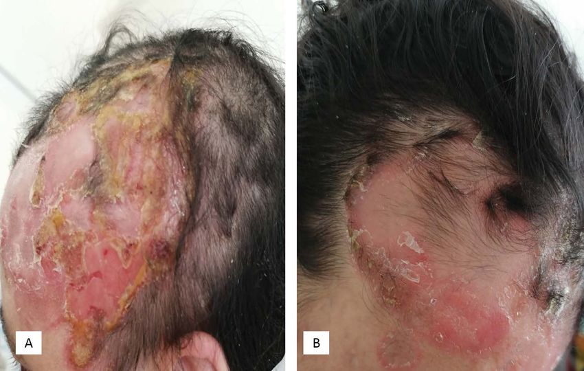
Figure 1: (A) Alopecic plaques of the scalp; (B) Erosive plaques of the scalp surmounted by yellowish crusts with circled
edges.
Citation: Abdelmouttalib A, Meziane M, Senouci K (2021) Trichoscopic Features of a Folliculotropic
Mycosis Fungoides: A Case Report. Clin Med Img Lib 7:170. doi.org/10.23937/2474-3682/1510170
Accepted: February 08, 2021; Published: February 10, 2021
Copyright: © 2021 Abdelmouttalib A, et al. This is an open-access content distributed under the terms
of the Creative Commons Attribution License, which permits unrestricted use, distribution, and repro-
duction in any medium, provided the original author and source are credited.
Abdelmouttalib et al. Clin Med Img Lib 2021, 7:170 • Page 1 of 3 •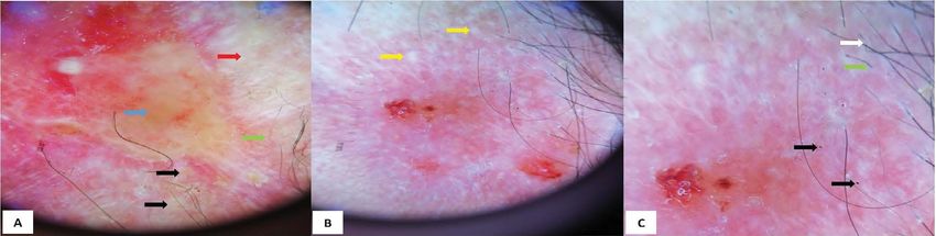
DOI: 10.23937/2474-3682/1510170                                                                                       ISSN: 2474-3682

   Figure 2: Dermoscopic features of scalp FMF (A) Yellow-orange areas (blue arrow), white areas without structure (red arrow),
   milky white globules (black arrow) and linear vessels (green arrow); (B) White dots (yellow arrows) with disappearance of
   hair follicles in the center of the lesion and persistence of some fluffy hairs in the periphery; (C) Zigzag hairs (white arrow),
   exclamation point hairs (green arrow), black dots/broken hair (black arrow).

     Figure 3: Evolution of the lesions of the scalp after 4 weeks of RePUVA therapy: Almost total healing of the erosions
     with scarring alopecia.

diagnosis of CD8+ mycosis fungoides. On clinical exam-               and white dots with radial lines that replaced the hair
ination, there were several alopecic plaques at the frontal          follicles [2]. Zigzag hair, short hair with split-end, short
and temporal areas of the scalp. Pull test from different            hair with triangular-shape end, broken hair and pigtail
areas of the scalp (lesional and nonlesional skin) showed            appearance hair are also reported [2]. Others have de-
anagen hairs with intact root sheaths. Plaques were ery-             scribed the presence of milky-red globules, orange-yel-
thematous and ulcerated with yellowish crusts and a cir-             low patchy areas and the vascular granular well-mar-
cinate border (Figure 1). On dermoscopy, there were or-              gined milky-red areas surrounded by normal skin in MF
ange-yellow areas, unstructured white areas, milky white             patients with scalp involvement [3]. Another observa-
globules and white and black dots. The vessels were                  tion with comedonal lesions [4] and a spiky follicular
linear and some hairs were in zigzag and in exclamation              Mycosis Fungoides were also reported [5].
point (Figure 2). The bacteriological sampling of the scalp
                                                                         Mycosis fungoides is a great imitator and can simu-
was negative and the patient received retinoids at 25 mg/
                                                                     late a wide variety of benign inflammatory skin disor-
day and PUVA therapy with a healing start of the lesions
                                                                     ders. Trichoscopy can help in clinical diagnosis of FMF
after 4 weeks but with scarring alopecia (Figure 3).
                                                                     on the scalp but should be followed by histopathology
   Various trichoscopic aspects of FMF have been re-                 and immunohistochemical study to definite diagnosis.
ported in the literature such as the presence of milky-              Further studies are necessary to determine the sensitiv-
white globules, yellow and white dots, white scales,                 ity and specificity of each trichoscopic feature.

Abdelmouttalib et al. Clin Med Img Lib 2021, 7:170                                                                       • Page 2 of 3 •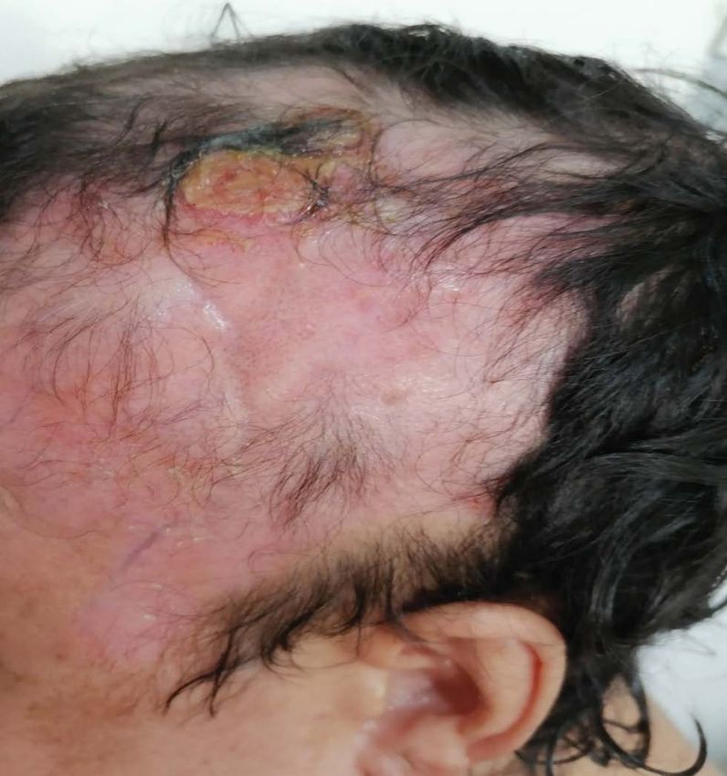
DOI: 10.23937/2474-3682/1510170                                                                                   ISSN: 2474-3682

Competing Interests                                               2. Sławińska M, Sobjanek M, Olszewska B, Nowicki R,
                                                                     Sokołowska -Wojdyło M (2018) Trichoscopic spectrum of
   The authors declare no competing interest.                        folliculotropic mycosis fungoides. J Eur Acad Dermatol Ve-
                                                                     nereol 32: 107-108.
Authors’ Contributions                                            3. Rudnicka L, Rakowska A, Olszewska M (2012) Trichos-
    All the co-authors contributed to the realization of             copy in General Medicine. In: Rudnicka L, Olszewska M,
this work.                                                           Rakowska A, Atlas of Trichoscopy. Springer, London, 483-
                                                                     493.
Source of Support                                                 4. Trüeb RM (2012) Systemic Lymphoproliferative Diseas-
                                                                     es. In: Rudnicka L, Olszewska M, Rakowska A, Atlas of
   Nil.                                                              Trichoscopy. Springer, London, 2012: 475-480.
Conflict of Interest                                              5. Souissi A, Ben Lagha I, Jendoubi F, Drissi H, Chelly I, et
                                                                     al. (2019) Spiky follicular mycosis fungoides: A trichoscopic
   None.                                                             feature. J Eur Acad Dermatol Venereol 33: 252-253.
References
1. Bi MY, Curry JL, Christiano AM, Hordinsky MK, Norris DA,
   et al. (2011) The spectrum of hair loss in patients with my-
   cosis fungoides and Sezary syndrome. J Am Acad Derma-
   tol 64: 53-63.

Abdelmouttalib et al. Clin Med Img Lib 2021, 7:170                                                                  • Page 3 of 3 •
You can also read