UC Irvine UC Irvine Previously Published Works - eScholarship
←
→
Page content transcription
If your browser does not render page correctly, please read the page content below
UC Irvine
UC Irvine Previously Published Works

Title
Germline variants and breast cancer survival in patients with distant metastases at
primary breast cancer diagnosis.

Permalink
https://escholarship.org/uc/item/2dh0j610

Journal
Scientific reports, 11(1)

ISSN
2045-2322

Authors
Escala-Garcia, Maria
Canisius, Sander
Keeman, Renske
et al.

Publication Date
2021-10-05

DOI
10.1038/s41598-021-99409-3

Peer reviewed

 eScholarship.org                              Powered by the California Digital Library
                                                               University of California
www.nature.com/scientificreports
OPEN Germline variants and breast
cancer survival in patients
with distant metastases at primary
breast cancer diagnosis
Maria Escala‑Garcia1, Sander Canisius1,2, Renske Keeman1, Jonathan Beesley3,
Hoda Anton‑Culver4, Volker Arndt5, Annelie Augustinsson6, Heiko Becher7,
Matthias W. Beckmann8, Sabine Behrens9, Marina Bermisheva10, Stig E. Bojesen11,12,13,14,
Manjeet K. Bolla15, Hermann Brenner5,16,17, Federico Canzian18, Jose E. Castelao19,
Jenny Chang‑Claude9,20, Stephen J. Chanock21, Fergus J. Couch22, Kamila Czene23,
Mary B. Daly24, Joe Dennis15, Peter Devilee25,26, Thilo Dörk27, Alison M. Dunning28,
Douglas F. Easton15,28, Arif B. Ekici29, A. Heather Eliassen30,31, Peter A. Fasching8,32,
Henrik Flyger33, Manuela Gago‑Dominguez34,35, Montserrat García‑Closas21,
José A. García‑Sáenz36, Jürgen Geisler37, Graham G. Giles38,39,40, Mervi Grip41,
Melanie Gündert42,43,44, Eric Hahnen45,46, Christopher A. Haiman47,
Niclas Håkansson48, Per Hall23,49, Ute Hamann50, Jaana M. Hartikainen51,52,
Bernadette A. M. Heemskerk‑Gerritsen53, Antoinette Hollestelle53, Reiner Hoppe54,55,
John L. Hopper39, David J. Hunter31,56, William Jacot57, Anna Jakubowska58,59,
Esther M. John60,61, Audrey Y. Jung9, Rudolf Kaaks9, Elza Khusnutdinova10,62,
Linetta B. Koppert63, Peter Kraft31,64, Vessela N. Kristensen65, Allison W. Kurian60,61,
Diether Lambrechts66,67, Loic Le Marchand68, Annika Lindblom69,70, Robert N. Luben71,72,
Jan Lubiński58, Arto Mannermaa51,52,73, Mehdi Manoochehri50, Sara Margolin49,74,
Dimitrios Mavroudis75, Taru A. Muranen76, Heli Nevanlinna76, Andrew F. Olshan77,
Håkan Olsson6, Tjoung‑Won Park‑Simon27, Alpa V. Patel78, Paolo Peterlongo79,
Paul D. P. Pharoah15,28, Kevin Punie80, Paolo Radice81, Gad Rennert82, Hedy S. Rennert82,
Atocha Romero83, Rebecca Roylance84, Thomas Rüdiger85, Matthias Ruebner8,
Emmanouil Saloustros86, Elinor J. Sawyer87, Rita K. Schmutzler45,46,88,
Minouk J. Schoemaker89, Christopher Scott90, Melissa C. Southey38,40,91, Harald Surowy42,43,
Anthony J. Swerdlow89,92, Rulla M. Tamimi31,93, Lauren R. Teras78, Emilie Thomas94,
Ian Tomlinson95,96, Melissa A. Troester77, Celine M. Vachon97, Qin Wang15, Robert Winqvist98,99,
Alicja Wolk48,100, Argyrios Ziogas4, kConFab/AOCS Investigators101, Kyriaki Michailidou15,102,103,
Georgia Chenevix‑Trench3, Thomas Bachelot104 & Marjanka K. Schmidt1,105*
Breast cancer metastasis accounts for most of the deaths from breast cancer. Identification
of germline variants associated with survival in aggressive types of breast cancer may inform
understanding of breast cancer progression and assist treatment. In this analysis, we studied
the associations between germline variants and breast cancer survival for patients with distant
metastases at primary breast cancer diagnosis. We used data from the Breast Cancer Association
Consortium (BCAC) including 1062 women of European ancestry with metastatic breast cancer, 606
of whom died of breast cancer. We identified two germline variants on chromosome 1, rs138569520
and rs146023652, significantly associated with breast cancer-specific survival (P = 3.19 × 10⁻⁸ and
4.42 × 10⁻⁸). In silico analysis suggested a potential regulatory effect of the variants on the nearby
target genes SDE2 and H3F3A. However, the variants showed no evidence of association in a smaller
replication dataset. The validation dataset was obtained from the SNPs to Risk of Metastasis (StoRM)
study and included 293 patients with metastatic primary breast cancer at diagnosis. Ultimately, larger
replication studies are needed to confirm the identified associations.
Scientific Reports | (2021) 11:19787 | https://doi.org/10.1038/s41598-021-99409-3 1
Vol.:(0123456789)www.nature.com/scientificreports/
1
Division of Molecular Pathology, The Netherlands Cancer Institute-Antoni Van Leeuwenhoek Hospital,
Amsterdam, The Netherlands. 2Division of Molecular Carcinogenesis, The Netherlands Cancer Institute-Antoni Van
Leeuwenhoek Hospital, Amsterdam, The Netherlands. 3Department of Genetics and Computational Biology, QIMR
Berghofer Medical Research Institute, Brisbane, QLD, Australia. 4Department of Medicine, Genetic Epidemiology
Research Institute, University of California Irvine, Irvine, CA, USA. 5Division of Clinical Epidemiology and Aging
Research, German Cancer Research Center (DKFZ), Heidelberg, Germany. 6Department of Cancer Epidemiology,
Clinical Sciences, Lund University, Lund, Sweden. 7Institute of Medical Biometry and Epidemiology, University
Medical Center Hamburg-Eppendorf, Hamburg, Germany. 8Department of Gynecology and Obstetrics,
Comprehensive Cancer Center Erlangen‑EMN, University Hospital Erlangen, Friedrich-Alexander University
Erlangen-Nuremberg (FAU), Erlangen, Germany. 9Division of Cancer Epidemiology, German Cancer Research
Center (DKFZ), Heidelberg, Germany. 10Institute of Biochemistry and Genetics, Ufa Federal Research Centre of the
Russian Academy of Sciences, Ufa, Russia. 11Copenhagen University Hospital, Copenhagen General Population
Study, Herlev, Denmark. 12Gentofte Hospital, Herlev, Denmark. 13Department of Clinical Biochemistry,
Copenhagen University Hospital, Herlev, Denmark. 14Faculty of Health and Medical Sciences, University of
Copenhagen, Copenhagen, Denmark. 15Department of Public Health and Primary Care, Centre for Cancer Genetic
Epidemiology, University of Cambridge, Cambridge, UK. 16Division of Preventive Oncology, German Cancer
Research Center (DKFZ) and National Center for Tumor Diseases (NCT), Heidelberg, Germany. 17German Cancer
Research Center (DKFZ), German Cancer Consortium (DKTK), Heidelberg, Germany. 18Genomic Epidemiology
Group, German Cancer Research Center (DKFZ), Heidelberg, Germany. 19Instituto de Investigación Sanitaria
Galicia Sur (IISGS), Xerencia de Xestion Integrada de Vigo-SERGAS, Oncology and Genetics Unit, Vigo,
Spain. 20University Medical Center Hamburg‑Eppendorf, Cancer Epidemiology Group, University Cancer Center
Hamburg (UCCH), Hamburg, Germany. 21Division of Cancer Epidemiology and Genetics, Department of Health
and Human Services, National Cancer Institute, National Institutes of Health, Bethesda, MD, USA. 22Department
of Laboratory Medicine and Pathology, Mayo Clinic, Rochester, MN, USA. 23Department of Medical Epidemiology
and Biostatistics, Karolinska Institutet, Stockholm, Sweden. 24Department of Clinical Genetics, Fox Chase Cancer
Center, Philadelphia, PA, USA. 25Department of Pathology, Leiden University Medical Center, Leiden, The
Netherlands. 26Department of Human Genetics, Leiden University Medical Center, Leiden, The
Netherlands. 27Gynaecology Research Unit, Hannover Medical School, Hannover, Germany. 28Department of
Oncology, Centre for Cancer Genetic Epidemiology, University of Cambridge, Cambridge, UK. 29Institute of Human
Genetics, Comprehensive Cancer Center Erlangen‑EMN, University Hospital Erlangen, Friedrich-Alexander
University Erlangen-Nuremberg (FAU), Erlangen, Germany. 30Channing Division of Network Medicine, Department
of Medicine, Brigham and Women’s Hospital and Harvard Medical School, Boston, MA, USA. 31Department of
Epidemiology, Harvard T.H. Chan School of Public Health, Boston, MA, USA. 32Division of Hematology and
Oncology, Department of Medicine, David Geffen School of Medicine, University of California at Los Angeles, Los
Angeles, CA, USA. 33Department of Breast Surgery, Copenhagen University Hospital, Herlev, Denmark. 34Instituto
de Investigación Sanitaria de Santiago de Compostela (IDIS), Complejo Hospitalario Universitario de Santiago,
SERGAS, Fundación Pública Galega de Medicina Xenómica, Santiago de Compostela, Spain. 35Moores Cancer
Center, University of California San Diego, La Jolla, CA, USA. 36Instituto de Investigación Sanitaria San Carlos
(IdISSC), Centro Investigación Biomédica en Red de Cáncer (CIBERONC), Medical Oncology Department, Hospital
Clínico San Carlos, Madrid, Spain. 37Department of Oncology, Akershus University Hospital, Lørenskog,
Norway. 38Cancer Council Victoria, Cancer Epidemiology Division, Melbourne, VIC, Australia. 39Melbourne School
of Population and Global Health, Centre for Epidemiology and Biostatistics, The University of Melbourne,
Melbourne, VIC, Australia. 40Precision Medicine, School of Clinical Sciences at Monash Health, Monash University,
Clayton,VIC, Australia. 41Department of Surgery, Oulu University Hospital, University of Oulu, Oulu, Finland. 42German
Cancer Research Center (DKFZ), Molecular Epidemiology Group, C080, Heidelberg, Germany. 43Molecular Biology
of Breast Cancer, University Womens Clinic Heidelberg, University of Heidelberg, Heidelberg,
Germany. 44Helmholtz Zentrum München, Institute of Diabetes Research, German Research Center for
Environmental Health, Neuherberg, Germany. 45Center for Familial Breast and Ovarian Cancer, Faculty of Medicine
and University Hospital Cologne, University of Cologne, Cologne, Germany. 46Center for Integrated Oncology
(CIO), Faculty of Medicine and University Hospital Cologne, University of Cologne, Cologne,
Germany. 47Department of Preventive Medicine, Keck School of Medicine, University of Southern California, Los
Angeles, CA, USA. 48Institute of Environmental Medicine, Karolinska Institutet, Stockholm, Sweden. 49Department
of Oncology, Sšdersjukhuset, Stockholm, Sweden. 50German Cancer Research Center (DKFZ), Molecular Genetics
of Breast Cancer, Heidelberg, Germany. 51Translational Cancer Research Area, University of Eastern Finland,
Kuopio, Finland. 52Institute of Clinical Medicine, Pathology and Forensic Medicine, University of Eastern Finland,
Kuopio, Finland. 53Department of Medical Oncology, Erasmus MC Cancer Institute, Rotterdam, The
Netherlands. 54Dr. Margarete Fischer-Bosch-Institute of Clinical Pharmacology, Stuttgart, Germany. 55University of
Tübingen, Tübingen, Germany. 56Nuffield Department of Population Health, University of Oxford, Oxford,
UK. 57Institut du Cancer de Montpellier, Montpellier University, Montpellier, France. 58Department of Genetics and
Pathology, Pomeranian Medical University, Szczecin, Poland. 59Independent Laboratory of Molecular Biology and
Genetic Diagnostics, Pomeranian Medical University, Szczecin, Poland. 60Division of Oncology, Department of
Medicine, Stanford University School of Medicine, Stanford Cancer Institute, Stanford, CA, USA. 61Department of
Epidemiology & Population Health, Stanford University School of Medicine, Stanford, CA, USA. 62Department of
Genetics and Fundamental Medicine, Bashkir State University, Ufa, Russia. 63Department of Surgical Oncology,
Family Cancer Clinic, Erasmus MC Cancer Institute, Rotterdam, The Netherlands. 64Harvard T.H. Chan School of
Public Health, Program in Genetic Epidemiology and Statistical Genetics, Boston, MA, USA. 65Department of
Medical Genetics, Oslo University Hospital and University of Oslo, Oslo, Norway. 66VIB Center for Cancer Biology,
Leuven, Belgium. 67Laboratory for Translational Genetics, Department of Human Genetics, University of Leuven,
Leuven, Belgium. 68University of Hawaii Cancer Center, Epidemiology Program, Honolulu, HI, USA. 69Department
Scientific Reports | (2021) 11:19787 | https://doi.org/10.1038/s41598-021-99409-3 2
Vol:.(1234567890)www.nature.com/scientificreports/
of Molecular Medicine and Surgery, Karolinska Institutet, Stockholm, Sweden. 70Department of Clinical Genetics,
Karolinska University Hospital, Stockholm, Sweden. 71NIHR Biomedical Research Centre, Moorfields Eye Hospital
NHS Foundation Trust and UCL Institute of Ophthalmology, London, England, UK. 72Medical Research Council
(MRC) Epidemiology Unit, University of Cambridge, Cambridge, UK. 73Kuopio University Hospital, Biobank of
Eastern Finland, Kuopio, Finland. 74Department of Clinical Science and Education, Karolinska Institutet,
Sšdersjukhuset, Stockholm, Sweden. 75Department of Medical Oncology, University Hospital of Heraklion,
Heraklion, Greece. 76Department of Obstetrics and Gynecology, Helsinki University Hospital, University of Helsinki,
Helsinki, Finland. 77Department of Epidemiology, Gillings School of Global Public Health and UNC Lineberger
Comprehensive Cancer Center, University of North Carolina at Chapel Hill, Chapel Hill, NC, USA. 78Department of
Population Science, American Cancer Society, Atlanta, GA, USA. 79IFOM-The FIRC Institute of Molecular Oncology,
Genome Diagnostics Program, Milan, Italy. 80Department of General Medical Oncology and Multidisciplinary
Breast Centre, Leuven Cancer Institute, University Hospitals Leuven, Leuven, Belgium. 81Unit of Molecular Bases of
Genetic Risk and Genetic Testing, Department of Research, Fondazione IRCCS Istituto Nazionale dei Tumori (INT),
Milan, Italy. 82Carmel Medical Center and Technion Faculty of Medicine, Clalit National Cancer Control Center,
Haifa, Israel. 83Medical Oncology Department, Hospital Universitario Puerta de Hierro, Madrid,
Spain. 84Department of Oncology, UCLH Foundation Trust, London, UK. 85Institute of Pathology, Staedtisches
Klinikum Karlsruhe, Karlsruhe, Germany. 86Department of Oncology, University Hospital of Larissa, Larissa,
Greece. 87School of Cancer & Pharmaceutical Sciences, Comprehensive Cancer Centre, King’s College London,
Guy’s Campus, London, UK. 88Center for Molecular Medicine Cologne (CMMC), Faculty of Medicine and University
Hospital Cologne, University of Cologne, Cologne, Germany. 89Division of Genetics and Epidemiology, The Institute
of Cancer Research, London, UK. 90Department of Health Sciences Research, Mayo Clinic, Rochester,
MN, USA. 91Department of Clinical Pathology, The University of Melbourne, Melbourne, Victoria,
Australia. 92Division of Breast Cancer Research, The Institute of Cancer Research, London, UK. 93Department of
Population Health Sciences, Weill Cornell Medicine, New York, NY, USA. 94Plateforme de Bioinformatique Gilles
Thomas, Centre de recherche en cancérologie de Lyon, Fondation Synergie Lyon Cancer, Université Claude
Bernard Lyon 1, Lyon, France. 95Institute of Cancer and Genomic Sciences, University of Birmingham, Birmingham,
UK. 96Wellcome Trust Centre for Human Genetics and Oxford NIHR Biomedical Research Centre, University of
Oxford, Oxford, UK. 97Division of Epidemiology, Department of Health Science Research, Mayo Clinic, Rochester,
MN, USA. 98Laboratory of Cancer Genetics and Tumor Biology, Cancer and Translational Medicine Research Unit,
University of Oulu, Biocenter Oulu, Oulu, Finland. 99Laboratory of Cancer Genetics and Tumor Biology, Northern
Finland Laboratory Centre Oulu, Oulu, Finland. 100Department of Surgical Sciences, Uppsala University, Uppsala,
Sweden. 102Biostatistics Unit, The Cyprus Institute of Neurology & Genetics, Nicosia, Cyprus. 103Cyprus School of
Molecular Medicine, The Cyprus Institute of Neurology & Genetics, Nicosia, Cyprus. 104Département de
Cancérologie Médicale, Centre Léon Bérard, Lyon, France. 105Division of Psychosocial Research and Epidemiology,
The Netherlands Cancer Institute-Antoni van Leeuwenhoek Hospital, Amsterdam, The Netherlands. *email:
mk.schmidt@nki.nl
Breast cancer is the most common female cancer in the Western world and one of the most common causes
of cancer death in women globally1. Early detection and better treatments have helped to reduce breast cancer
mortality in recent d ecades2. Yet, when breast cancer metastasizes to distant sites, prognosis continues to be poor
and for most cases treatment is only palliative3. Metastases in breast cancer can remain undetectable for many
years after initial diagnosis, leading to incurable l esions4. Approximately 15% of patients with breast cancer will
develop distant metastases within 3 years after diagnosis of the primary tumor5. Therefore, it is important to have
the tools able to detect breast cancer metastases at earlier stages, in order to better manage and predict breast
cancer progression. Prognostication models could benefit from the inclusion of germline genetic biomarkers
that are capable of predicting tumor recurrence, second tumors or prognosis of second tumors. However, so
far, it has been difficult to identify individual common germline variants associated with primary breast cancer
survival due to the small effect size these variants are likely to have6,7. Likewise, evidence as to whether or not
germline variants can increase the probability of metastatic progression is currently limited to a few s tudies4,8.
For example, a candidate gene study identified common single nucleotide polymorphisms (SNPs) located within
SIPA1 that were associated with metastasis and poor breast cancer p rognosis9. Other studies have identified other
metastasis susceptibility genes such as RRP1b10. Germline variants could specifically provide metastatic predis-
position by affecting treatment r esponse11 or promoting tumor initiating events and providing new metastatic
functions to tumor c ells4.
The aim of this study was to identify associations between common germline variants and breast cancer-
specific survival in patients with metastasis at primary breast cancer diagnosis. We hypothesized that germline
variants might predispose to poorer survival after breast cancer metastasis, and that analyzing a set of patients
with similar stage of the disease might help identify variants that do not show evidence of association in larger
but more heterogeneous datasets.
Results
We used data from the Breast Cancer Association Consortium (BCAC): the dataset comprised data from 50
studies from which follow-up information for women diagnosed with distant metastases at primary breast cancer
diagnosis was available. The results were based on the meta-analysis of two genome-wide SNP arrays (iCOGS12
and OncoArray13 (see “Methods”). We analyzed variants that had a minor allele frequency (MAF) > 0.01 and an
imputation quality r2 > 0.7 for at least one of the two arrays. Details about the individual studies, the genotyping
Scientific Reports | (2021) 11:19787 | https://doi.org/10.1038/s41598-021-99409-3 3
Vol.:(0123456789)www.nature.com/scientificreports/
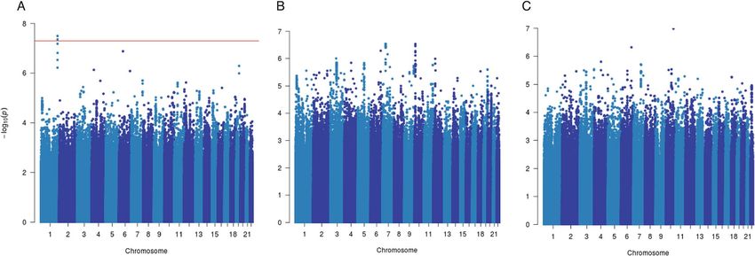
Figure 1. Manhattan plots of the meta-analysis of OncoArray and iCOGS datasets for the association of
common germline variants and breast cancer-specific survival for patients with metastases at primary breast
cancer diagnosis for (A) all breast tumors, (B) ER-positive tumors, and (C) ER-negative tumors. The y axis
shows the − log10 P values of each variant analyzed, and the x axis shows their chromosome position. The red
horizontal line represents P = 5 × 10⁻⁸.
array used and number of patients included are given in Supplementary Table 1. We analyzed the genotypes
and clinico-pathological data of a total of 1062 breast cancer patients, 606 of whom died of breast cancer within
15 years of follow-up. Of these, 721 of the patients had estrogen receptor (ER)-positive disease (388 deaths)
and 227 had ER-negative disease (148 deaths). All patients were women of European descent. The patients were
diagnosed from 1979 to 2014 (median: 2004) and aged 26–92 (median: 60) years.
Manhattan plots showing the association between germline variants and breast cancer-specific survival of all,
ER-positive and ER-negative metastasized breast cancers are shown in Fig. 1. We identified two genome-wide
significant (P < 5 × 10⁻⁸) variants (SNPs: rs138569520 and rs146023652) on chromosome 1 associated with breast
cancer-specific survival for all metastasized breast cancers (Table 1, Supplementary Table 2). The two variants
were part of a set of six highly correlated SNPs (Table 1, r2 > 0.88) based on European subjects in phase 3 of the
1000 Genomes Project14. No variant reached genome-wide significance for ER-positive or for ER-negative breast
cancer tumors alone (Supplementary Tables 3 and 4).
The variant with the strongest association was the SNP rs138569520 (HR = 3.67, 95% CI 1.86–7.23 and
P = 3.19 × 10⁻⁸). The HR estimates for rs138569520 in the ER-positive (HR = 3.38, 95% CI 1.48–7.70 and
P = 4.37 × 10⁻⁴) and ER-negative (HR = 2.76, 95% CI 1.16–6.64 and P = 8.70 × 10⁻³) were similar (P = 0.97 for
difference).
Several genes (SDE2, LEFTY2, PYCR2 and H3F3A) were located within 100 kb of the most significant SNP
rs138569520. We interrogated functional genomic data including annotations of enhancers, promoters and
transcription factor binding sites and found evidence consistent with gene regulation in the regions contain-
ing the associated variants (Fig. 2). Hi-C analysis in HMEC c ells15 showed that the lead variant rs138569520 is
located in a genomic region interacting with the promoter region of H3F3A. SNPs rs146023652 and rs114512448
overlapped with transcription factor (TF) binding sites which might reflect the active transcription of SDE2.
ChIP-seq signals from primary breast sub-populations16 also showed potential regulatory regions containing
rs114512448. ChIA-PET analysis in MCF-7 cells from E NCODE17, detected an interaction between rs114512448
and the PYCR2 gene. Finally, ChIA-PET also detected an interaction between rs72757046 and SDE2 and H3F3A.
Using KMplotter (kmplot.com/analysis)18, we tested the association of the mRNA tumor expression of SDE2
and H3F3A, the genes in closest proximity to rs138569520, with overall survival in grade 3 breast tumors (to
select the most aggressive subtype; selection for stage 4 was not available). Low mRNA expression levels of SDE2
gene were significantly associated (P = 0.01) with poorer breast cancer survival (Fig. 3a), while, in contrast, high
expression of H3F3A was associated with lower survival (P = 6.7 × 10⁻⁵) (Fig. 3b). These associations were not
statistically significant, neither for grade 1 or for grade 2 disease (P > 0.21).
Lastly, we aimed to evaluate the significance of the two genome-wide significant SNPs using an independent
set of 293 breast cancer patients with metastatic primary breast cancer at diagnosis from the SNPs to Risk of
Metastasis (StoRM) s tudy19. All patients were diagnosed in France from March 2012 to May 2014, aged 18 years
or older (median: 59 years) and followed up to July 2017. A total of 293 patients were available for the validation
study, 239 of whom had events, defined as progression and/or death occurring during follow-up. Both SNPs had
good imputation quality ( r2 ~ 0.7) and similar MAFs to those in the BCAC dataset (~ 2%). However, neither of
the two SNPs replicated in the survival analysis with the StoRM dataset (Table 2): rs138569520 (HR = 1.49, 95%
CI 0.60–3.71, P = 0.34) and rs146023652 (HR = 1.25, 95% CI 0.46–3.37, P = 0.66). Although the HR estimates
in the StoRM validation dataset were smaller than those from the BCAC analyses (HR = 3.67 and 3.64), the
confidence limits overlapped.
Because the BCAC dataset also included prevalent cases (n = 466), we repeated the analysis with incident
cases (n = 596) to match the study design in StoRM more closely. The HR estimates were similar to those for
Scientific Reports | (2021) 11:19787 | https://doi.org/10.1038/s41598-021-99409-3 4
Vol:.(1234567890)www.nature.com/scientificreports/
SNP Chr Position Ref Alt EAF r2 HR LCL UCL P value
rs138569520 1 226193175 T C 0.02 0.87 3.67 1.86 7.23 3.19 × 10⁻⁸
rs146023652 1 226158826 C T 0.02 0.86 3.64 1.84 7.19 4.42 × 10⁻⁸
rs114512448 1 226173980 G A 0.02 0.86 3.53 1.78 6.95 6.57 × 10⁻⁸
rs143653255 1 226157179 T C 0.02 0.86 3.26 1.68 6.34 1.53 × 10⁻⁷
rs115086585 1 226154721 C T 0.02 0.85 3.21 1.64 6.25 2.93 × 10⁻⁷
rs72757046 1 226235714 G C 0.02 0.84 3.62 1.78 7.37 6.02 × 10⁻⁷
Table 1. Results for the six correlated variants associated with breast cancer-specific survival for patients
with metastatic primary breast cancer at diagnosis. Genomic positions are based on the hg19 genome build.
ALT alternate, REF reference, EAF effect allele frequency, HR hazard rate, LCL lower control limit, UCL upper
control limit, r2 imputation quality.
Chr1 hg19 pos 226,120,000 226,140,000 226,160,000 226,180,000 226,200,000 226,220,000 226,240,000 226,280,000
rs115086585 rs138569520 rs72757046
SNPs rs143653255
rs146023652
Chromatin rs114512448
interactions
(ChIA-PET
and Hi-C)
TF binding
sites
SDE2
LEFTY2 H3F3A
Genes PYCR2
Basal
H3K27ac
Stromal
Luminal
H3K4me1 Basal
Stromal
Luminal
H3K27me3 Basal
Stromal
Luminal
Figure 2. Functional annotation of the six highly correlated SNPs: rs138569520, rs146023652, rs114512448,
rs143653255, rs115086585 and rs72757046. TF transcription factor.
the overall analysis (rs138569520: HR = 3.77, 95% CI 1.71–8.30, P = 3.12 × 10⁻⁵ and rs146023652: HR = 3.75,
95% CI 1.70–8.29, P = 3.60 × 10⁻⁵). Finally, since the maximum follow-up in the StoRM dataset was shorter
(5 years, compared with a maximum of 15 years in the BCAC dataset), we repeated the main analysis in BCAC
using a follow-up of 5 years (n = 1031, 476 deaths). The associations for the two SNPs were slightly less signifi-
cant (rs138569520: HR = 3.43, 95% CI 1.74–6.80, P = 1.83 × 10⁻⁷ and rs146023652: HR = 3.41, 95% CI 1.72–6.76,
P = 2.55 × 10⁻⁷) but the HR estimates were similar to those from the main analysis.
Discussion
In this analysis of breast cancer patients with metastatic primary breast cancer at diagnosis, involving 1062
patients with 606 breast cancer-specific deaths, we identified two variants on chromosome 1 (rs138569520 and
rs146023652) associated with survival, at genome-wide levels of statistical significance. The most significant
association was for the SNP rs138569520 (P = 3.19 × 10⁻⁸). The HR estimates were similar in patients with ER-
positive and ER-negative disease.
Two genes, SDE2 and H3F3A, were in closest proximity of rs138569520. Both genes have been previously
associated with oncogenic processes relevant for metastatic progression: the SDE2 gene (“silencing defective 2”)
is known to be involved in DNA replication, telomere maintenance and cell cycle c ontrol20,21. The functional
roles of SDE2 have been studied in a proteome dynamics analysis in prostate cancer cells; the results suggested
that alterations of the gene might diminish the error-prone DNA repair pathway activation and promote mis-
sense mutations22. The gene H3F3A encodes for histone H3.3, and mutations in this protein have been linked to
multiple cancer processes23, including breast invasive ductal carcinoma24. Additionally, the differential expression
of these two genes was significantly associated with survival in grade 3 tumors based on KMplotter. Previous
studies have also linked the expression of these genes to oncogenic processes. For example, downregulation of
SDE2 was associated with mutation disease phenotype as well as poorer mortality o utcomes22. Likewise, over-
expression of H3F3A was associated with lung cancer progression and promotion of lung cancer cell migration
by activation of metastasis-related g enes25. Unfortunately, in KMplotter it was not possible to specifically select
stage 4 tumors, which limits the interpretation of our findings. Future studies are needed in order to corroborate
the association of SDE2 and H3F3A expression with survival in this group of patients.
Additionally, there was predicted genomic activity in the locus based on the intersection of multiple genomic
regulatory features in breast tissue. Although the SNPs appeared to cluster around SDE2, there was also in-silico
evidence for two other potential target genes at this locus (H3F3A and PYCR2). PYCR2 encodes for a mito-
chondrial protein involved in proline biosynthesis. While little is known about this proline form, studies for
Scientific Reports | (2021) 11:19787 | https://doi.org/10.1038/s41598-021-99409-3 5
Vol.:(0123456789)www.nature.com/scientificreports/
A B
SDE2 (244103_at) H3F3A (200080_s_at)
1.0
1.0
HR = 0.51 (0.3 − 0.86) HR = 2.23 (1.49 − 3.34)
logrank P = 0.01 logrank P = 6.7e−05
0.8
0.8
0.6
0.6
Probability
Probability
0.4
0.4
0.2
0.2
Expression Expression
low low
high high
0.0
0.0
0 50 100 150 0 50 100 150
Time (months) Time (months)
Number at risk Number at risk
low 53 26 1 0 low 162 125 47 22
high 151 75 16 3 high 341 196 44 22
Figure 3. Kaplan–Meier overall survival plot for high versus low expression level of the genes (A) SDE2
(n = 204) and (B) H3F3A (n = 503) restricted to patients with a grade 3 tumor and 15 years of follow-up. The
differential expression analysis was performed in KMplotter.
SNP Chr Position Ref Alt EAF r2 HR LCL UCL P value
rs138569520 1 226193175 T C 0.02 0.69 1.49 0.60 3.71 0.34
rs146023652 1 226158826 C T 0.02 0.79 1.25 0.46 3.37 0.66
Table 2. Results for the validation of the two genome-wide significant variants in an independent dataset of
breast cancer patients with metastatic primary breast cancer at diagnosis. ALT alternate, REF reference, EAF
effect allele frequency, HR hazard rate, LCL lower control limit, UCL upper control limit, r2 imputation quality.
the close family member PYCR1 have found that higher levels of mRNA were associated with reduced survival
from breast cancer p atients26. To support further our hypothesis that the two genome-wide significant SNPs
(rs138569520 and rs146023652) were specific for survival in patients with metastatic disease, we confirmed that
there were no associations (HR = 1.04, P = 0.58, MAF = 0.02 and HR = 1.03, P = 0.60, MAF = 0.02 respectively)
with breast cancer-specific survival in the most recent BCAC dataset for all invasive early (stages I–III) breast
cancers (OncoArray and iCOGS, n = 86,627)27.
On the other hand, the two genome-wide significant variants, rs138569520 and rs146023652, were not repli-
cated (P = 0.34 and P = 0.66, respectively) using an independent dataset of patients with metastatic primary breast
cancer diagnosis (n = 293). The imputation quality and the minor allele frequency of the SNPs in the replication
cohort were comparable to those in the BCAC analyses (MAF = 2% and r2 > 7%), therefore the negative result
could not be attributed to those factors. Age of the patients could also not explain the difference since both
datasets had comparable median ages at diagnosis, 60 years for BCAC and 59 years for StoRM. On the other
hand, it is important to state that there were several factors that varied between the datasets. First, the sample
size differed considerably between BCAC (n = 1062) and the StoRM study (n = 293), the latter having a relatively
small sample size which limits the power to detect associations. Total follow-up time also varied: for the BCAC
dataset, patients were followed for a maximum of 15 years, while for the StoRM study the follow-up ended at
5 years. However, the results from the complementary analysis using the BCAC dataset and 5-year follow-up
were comparable to the initial 15 years follow-up results. This finding suggests that the disparity in estimates
between the two analyses is not due to shorter follow-up. There were several other differences between the main
BCAC dataset and the StoRM cohort used for validation. For example, the BCAC dataset included multiple
studies from several countries while the StoRM cohort included solely patients from France. Moreover, StoRM
was a recent cohort with the earliest reported diagnosis starting in 2012. On the other hand, in BCAC, the year
of patients’ diagnosis ranged between 1979 and 2014 and included prevalent cases. While the analysis in BCAC
using exclusively incident cases gave comparable estimates to the main analysis, the difference in the years of
diagnosis could be related to differences in treatment strategies that were not considered in the current analysis.
The lack of information about detailed treatment is a potential weakness of the current analysis and validation.
Treatment strategy, together with characteristics of the tumor, will also influence the final prognosis of metastatic
Scientific Reports | (2021) 11:19787 | https://doi.org/10.1038/s41598-021-99409-3 6
Vol:.(1234567890)www.nature.com/scientificreports/
breast cancer28. It is important to note that the associations observed in the BCAC study may be false positives,
and that further large replication studies will be required to confirm or refute the associations.
In conclusion, this analysis of BCAC patients with metastatic primary breast cancer at diagnosis from the
BCAC dataset identified a new region in chromosome 1 associated with breast cancer-specific survival. The
region includes six highly correlated SNPs that are predicted to be in an active region of the genome based
on in-silico evidence from breast cancer tissues and that are located in close proximity to genes involved in
oncogenic processes. However, we were unable to validate the association using a smaller, independent set of
patients. Overall, the role of germline variants in metastasis and progression remains unclear. Further analyses
with larger datasets including treatment information and functional analysis are needed to better understand
the underlying biological processes and the links between this locus and the nearby genes. Prior validation of
the reported associations is needed before these findings can be used in clinical-decision making. Therefore, a
next step is to study these SNPs in a, preferably, prospective large series of metastasized breast cancer patients.
Ultimately, germline variants could help identifying tailored treatments for patients with metastatic disease or
better strategies for risk management stratification of aggressive forms of breast cancer.
Methods
Breast cancer samples and genotype data: Breast Cancer Association Consortium (BCAC). We
used genotype and clinico-pathological data (database version 12) data from the Breast Cancer Association
Consortium (BCAC). The dataset included 1062 breast cancer patients with metastatic primary breast cancer at
diagnosis that were genotyped using one of the two different genotyping platforms: iCOGS12 and OncoArray13,
providing genome-wide coverage of common variants. The main analyses were based on imputed variants using
the Haplotype Reference Consortium29 as reference panel. All patients were women of European ancestry, aged
26–92 years (median: 60) years with metastasized breast cancer at diagnosis. Women were diagnosed between
1979 and 2014, with a median follow-up was three and a half years. Additional details about the genotype data
and sample quality control have been described previously7,27,30. We only analyzed variants that had a minor
allele frequency (MAF) > 0.01 and an imputation quality r 2 > 0.7 for at least one of the two genotyping platforms
(iCOGS or OncoArray). Details about the individual studies included in the analyses, including the array used,
associated country and number of patients with metastatic primary breast cancer at diagnosis are given in Sup-
plementary Table 1. The secondary use of data for the study was approved by the Data Access Committee of
the BCAC, under the legal provisions of the Memorandum of Understanding and Data Transfer Agreements of
Cambridge University which all the contributing institutions, which includes that all contributing institutions
provided the data with the appropriate approval of their institutional review boards and informed consent of the
participants of the individual studies.
Statistical and bioinformatic methods. We estimated the association of the germline variants with
breast-cancer specific survival using Cox proportional hazards regression. We analyzed separately the OncoAr-
ray and iCOGS datasets and combined the estimates using fixed-effect meta-analyses. Follow-up was right cen-
sored on the date of death, last date known alive if death did not occur, or at 15 years after diagnosis, whichever
came first27. Time at risk was calculated from the date of diagnosis with left truncation for prevalent cases. The
models were stratified by country and included the first two ancestry informative principal components12. We
performed the analysis for all breast cancers and for ER-positive and ER-negative tumors separately. To identify
evidence of potential cis-regulatory activity, we intersected germline variants with numerous sources of genomic
annotation information from primary breast cells (e.g., chromosome conformation, enhancer–promoter cor-
relations, transcription factor and histone modification ChIP-seq). To assess the effect of gene expression on
survival we used the Kaplan–Meier plotter on breast tissue data, grade 3 tumors and 15 years of follow-up
(180 months)18.
Validation dataset: SNPs to risk of metastasis (StoRM). To attempt to validate our results we used
data from the SNPs to Risk of Metastasis (StoRM) study. StoRM is a multicentric, prospective, cohort study of
metastatic breast cancer patients in France that was originally designed to identify genetic and other factors
associated with metastatic relapse and s urvival19. Patients aged 18 years or older, with a histologically proven
breast cancer that was metastatic for less than 1 year were included. All patients that had another coexisting
cancer or another cancer diagnosed within the last 5 years, were excluded from the study. Patients were followed
from March 2012 to July 2017. Time to progression on the first metastatic treatment was recorded and patients
were followed until death, every 6 months for 3 years, and then annually until July 2017. A total of 293 patients
were available for the validation. The median follow-up was of 3.2 years. Because of the short total follow-up time
(5 years) and the advanced disease stage of the patients in the cohort, both a recorded progression and/or death
were considered as an event in the survival analyses. Of the whole set of 293 patients, 239 had a progression and/
or died during the follow-up period.
Ethical approval. The study was performed in accordance with the Declaration of Helsinki. All individual
studies, from which data was used, were approved by the appropriate medical ethical committees and/or institu-
tional review boards. All study participants provided informed consent.
Scientific Reports | (2021) 11:19787 | https://doi.org/10.1038/s41598-021-99409-3 7
Vol.:(0123456789)www.nature.com/scientificreports/
Received: 28 March 2021; Accepted: 7 September 2021
References
1. Torre, L. A., Islami, F., Siegel, R. L., Ward, E. M. & Jemal, A. Global cancer in women: Burden and trends. Cancer Epidemiol.
Biomark. Prev. 26, 444–457 (2017).
2. Narod, S. A., Iqbal, J. & Miller, A. B. Why have breast cancer mortality rates declined?. J. Cancer Policy 5, 8–17 (2015).
3. Sledge, G. W. Curing metastatic breast cancer. J. Oncol. Pract. 12, 6–10 (2016).
4. Nguyen, D. X. & Massagué, J. Genetic determinants of cancer metastasis. Nat. Rev. Genet. 8, 341–352 (2007).
5. Weigelt, B., Peterse, J. L. & van’t Veer, L. J. Breast cancer metastasis: Markers and models. Nat. Rev. Cancer 5, 591–602 (2005).
6. Pharoah, P. D. P. et al. Polygenic susceptibility to breast cancer and implications for prevention. Nat. Genet. 31, 33–36 (2002).
7. Escala-Garcia, M. et al. Genome-wide association study of germline variants and breast cancer-specific mortality. Br. J. Cancer
120, 647–657 (2019).
8. Priestley, P. et al. Pan-cancer whole-genome analyses of metastatic solid tumours. Nature 575, 210–216 (2019).
9. Crawford, N. P. S. et al. Germline polymorphisms in SIPA1 are associated with metastasis and other indicators of poor prognosis
in breast cancer. Breast Cancer Res. 8, R16 (2006).
10. Crawford, N. P. S. et al. Rrp1b, a new candidate susceptibility gene for breast cancer progression and metastasis. PLoS Genet. 3,
e214 (2007).
11. O’Donnell, P. H. & Ratain, M. J. Germline pharmacogenomics in oncology: Decoding the patient for targeting therapy. Mol. Oncol.
6, 251–259 (2012).
12. Michailidou, K. et al. Association analysis identifies 65 new breast cancer risk loci. Nature 551, 92–94 (2017).
13. Amos, C. I. et al. The OncoArray Consortium: A network for understanding the genetic architecture of common cancers. Cancer
Epidemiol. Biomark. Prev. 26, 126–135 (2017).
14. Auton, A. et al. A global reference for human genetic variation. Nature 526, 68–74 (2015).
15. Rao, S. S. P. et al. A 3D map of the human genome at kilobase resolution reveals principles of chromatin looping. Cell 159,
1665–1680 (2014).
16. Pellacani, D. et al. Analysis of normal human mammary epigenomes reveals cell-specific active enhancer states and associated
transcription factor networks. Cell Rep. 17, 2060–2074 (2016).
17. Dunham, I. et al. An integrated encyclopedia of DNA elements in the human genome. Nature 489, 57–74 (2012).
18. Györffy, B. et al. An online survival analysis tool to rapidly assess the effect of 22,277 genes on breast cancer prognosis using
microarray data of 1809 patients. Breast Cancer Res. Treat. 123, 725–731 (2010).
19. Delrieu, L. et al. Analysis of the StoRM cohort reveals physical activity to be associated with survival in metastatic breast cancer.
Sci. Rep. 10, 10757 (2020).
20. Jo, U. et al. PCNA-dependent cleavage and degradation of SDE2 regulates response to replication stress. PLOS Genet. 12, e1006465
(2016).
21. Rageul, J. et al. SDE2 integrates into the TIMELESS-TIPIN complex to protect stalled replication forks. Nat. Commun. 11, 5495
(2020).
22. Luo, A., Gong, Y., Kim, H. & Chen, Y. Proteome dynamics analysis identifies functional roles of SDE2 and hypoxia in DNA damage
response in prostate cancer cells. NAR Cancer 2, zcaa010 (2020).
23. Yuen, B. T. K. & Knoepfler, P. S. Histone H3.3 mutations: A variant path to cancer. Cancer Cell 24, 567–574 (2013).
24. Sweeney, S. M. et al. AACR project genie: Powering precision medicine through an international consortium. Cancer Discov. 7,
818–831 (2017).
25. Park, S.-M. et al. Histone variant H3F3A promotes lung cancer cell migration through intronic regulation. Nat. Commun. 7, 12914
(2016).
26. Ding, J. et al. Human mitochondrial pyrroline-5-carboxylate reductase 1 promotes invasiveness and impacts survival in breast
cancers. Carcinogenesis 38, 519–531 (2017).
27. Escala-Garcia, M. et al. Breast cancer risk factors and their effects on survival: A Mendelian randomisation study. BMC Med. 18,
327 (2020).
28. Deluche, E. et al. Contemporary outcomes of metastatic breast cancer among 22,000 women from the multicentre ESME cohort
2008–2016. Eur. J. Cancer 129, 60–70 (2020).
29. Frye, F. L. & Cucuel, J. P. A reference panel of 64,976 haplotypes for genotype imputation. Nat. Genet. 48, 1279–1283 (2016).
30. Escala-Garcia, M. et al. A network analysis to identify mediators of germline-driven differences in breast cancer prognosis. Nat.
Commun. 11, 312 (2020).
Acknowledgements
BCAC: We thank all the individuals who took part in these studies and all the researchers, clinicians, techni-
cians and administrative staff who have enabled this work to be carried out. ABCFS: Maggie Angelakos, Judi
Maskiell, Gillian Dite. ABCS: Frans Hogervorst, Sten Cornelissen and Annegien Broeks. BBCS: Eileen Williams,
Elaine Ryder-Mills, Kara Sargus. BCINIS: Dr. K. Landsman, Dr. N. Gronich, Dr. A. Flugelman, Dr. W. Saliba,
Dr. E. Liani, Dr. I. Cohen, Dr. S. Kalet, Dr. V. Friedman, Dr. O. Barnet. BIGGS: Niall McInerney, Gabrielle Col-
leran, Andrew Rowan, Angela Jones. BREOGAN: Manuela Gago-Dominguez, Jose Esteban Castelao, Angel Car-
racedo, Victor Muñoz Garzón, Alejandro Novo Domínguez, Maria Elena Martinez, Sara Miranda Ponte, Carmen
Redondo Marey, Maite Peña Fernández, Manuel Enguix Castelo, Maria Torres, Manuel Calaza, José Antúnez,
Máximo Fraga; Joaquín González-Carreró and the Department of Pathology and Biobank of University Hospital
Complex of Vigo, Instituto de Investigacion Biomedica Galicia Sur, SERGAS. BSUCH: Peter Bugert, Medical
Faculty Mannheim. CCGP: Styliani Apostolaki, Anna Margiolaki, Georgios Nintos, Maria Perraki, Georgia Salou-
strou, Georgia Sevastaki, Konstantinos Pompodakis. CGPS: Dorthe Uldall Andersen, Maria Birna Arnadottir,
Anne Bank, Dorthe Kjeldgård Hansen and the Danish Cancer Biobank. CPS-II: Centers for Disease Control and
Prevention National Program of Cancer Registries. The National Cancer Institute Surveillance Epidemiology and
End Results program. Participants and the investigators of EPIC (European Prospective Investigation into Cancer
and Nutrition). ESTHER: Hartwig Ziegler, Sonja Wolf, Volker Hermann, Christa Stegmaier, Katja Butterbach.
GC-HBOC: Stefanie Engert, Heide Hellebrand, Sandra Kröber and LIFE. Markus Loeffler, Joachim Thiery, Mat-
thias Nüchter, Ronny Baber. GENICA: Christian Baisch, Hiltrud Brauch, Thomas Brüning, Hans-Peter Fischer,
UH, Volker Harth, RH, Yon-Dschun Ko, Wing-Yee Lo, Anne Lotz, Beate Pesch, Sylvia Rabstein. HABCS: Michael
Bremer, Natalia Bogdanova, Peter Schürmann, Johann H. Karstens, Peter Hillemanns. HEBCS: Carl Blomqvist,
Kristiina Aittomäki, Rainer Fagerholm, Kirsimari Aaltonen, Karl von Smitten, Irja Erkkilä. HUBCS: Darya
Scientific Reports | (2021) 11:19787 | https://doi.org/10.1038/s41598-021-99409-3 8
Vol:.(1234567890)www.nature.com/scientificreports/
Prokofyeva, Shamil Gantsev. KARMA and SASBAC: Swedish Medical Research Counsel. KBCP: Eija Myöhänen,
Helena Kemiläinen. LMBC: Gilian Peuteman, Thomas Van Brussel, EvyVanderheyden and Kathleen Corthouts.
MARIE: Petra Seibold, Nadia Obi, Sabine Behrens, Ursula Eilber and Muhabbet Celik. MBCSG: Siranoush
Manoukian, Bernard Peissel, Jacopo Azzollini, Erica Rosina, Daniela Zaffaroni, Bernardo Bonanni, Irene Feroce,
Mariarosaria Calvello, Aliana Guerrieri Gonzaga, Monica Marabelli, Davide Bondavalli and the personnel of the
Cogentech Cancer Genetic Test Laboratory. The following are NBCS Collaborators: Kristine K. Sahlberg (PhD),
Anne-Lise Børresen-Dale (Prof. Em.), Lars Ottestad (MD), Rolf Kåresen (Prof. Em.), Dr. Ellen Schlichting (MD),
Marit Muri Holmen (MD), Toril Sauer (MD), Vilde Haakensen (MD), Olav Engebråten (MD), Bjørn Naume
(MD), Alexander Fosså (MD), Cecile E. Kiserud (MD), Kristin V. Reinertsen (MD), Åslaug Helland (MD), Margit
Riis (MD), OSBREAC and Grethe I. Grenaker Alnæs (MSc). NHS/NHS2: the following state cancer registries:
AL, AZ, AR, CA, CO, CT, DE, FL, GA, ID, IL, IN, IA, KY, LA, ME, MD, MA, MI, NE, NH, NJ, NY, NC, ND, OH,
OK, OR, PA, RI, SC, TN, TX, VA, WA, WY. OBCS: Arja Jukkola-Vuorinen, Mervi Grip, Saila Kauppila, Meeri
Otsukka, Leena Keskitalo and Kari Mononen. ORIGO: E. Krol-Warmerdam, and J. Blom. PBCS: Louise Brinton,
Mark Sherman, Neonila Szeszenia-Dabrowska, Beata Peplonska, Witold Zatonski, Pei Chao, Michael Stagner.
The ethical approval for the PREFACE: Sonja Oeser and Silke Landrith. RBCS: Petra Bos, Jannet Blom, Ellen
Crepin, Elisabeth Huijskens, Anja Kromwijk-Nieuwlaat, Annette Heemskerk, the Erasmus MC Family Cancer
Clinic. SBCS: Sue Higham, Helen Cramp, Dan Connley, Ian Brock, Sabapathy Balasubramanian and Malcolm
W.R. Reed. We thank the SEARCH and EPIC teams. SKKDKFZS: Deutsches Krebsforschungszentrum (DKFZ),
Heidelberg, Germany [UH, MM], Staedtisches Klinikum Karlsruhe, Institute of Pathology [TR]. SZBCS: Ewa
Putresza. UCIBCS: Irene Masunaka. UKBGS: Breast Cancer Now and the Institute of Cancer Research and NHS
funding to the Royal Marsden/ICR NIHR Biomedical Research Centre.
Author contributions
M.K.S. and M.E.G. conceived the study. M.E.G. performed the main data analyses and drafted the initial manu-
script. M.K.S., S.C. and M.E.G. were involved in the interpretation of the data. R.K., Q.W., J.D. and M.K.B
provided database support. J.B. performed the functional analysis. M.K.S. and S.C. worked on revisions of the
manuscript. All authors contributed data from their own studies, helped revise the manuscript, and approved
the final version. All authors consented to this publication.
Funding
BCAC is funded the European Union’s Horizon 2020 Research and Innovation Programme (Grant numbers
634935 and 633784 for BRIDGES and B-CAST respectively), and PERSPECTIVE I&I, funded by the Govern-
ment of Canada through Genome Canada and the Canadian Institutes of Health Research, the Ministère de
l’Économie et de l’Innovation du Québec through Genome Québec, the Quebec Breast Cancer Foundation. The
EU Horizon 2020 Research and Innovation Programme funding source had no role in study design, data col-
lection, data analysis, data interpretation or writing of the report. Additional funding for BCAC is provided via
the Confluence project which is funded with intramural funds from the National Cancer Institute Intramural
Research Program, National Institutes of Health. Genotyping of the OncoArray was funded by the NIH Grant
U19 CA148065, and Cancer UK Grant C1287/A16563 and the PERSPECTIVE project supported by the Govern-
ment of Canada through Genome Canada and the Canadian Institutes of Health Research (Grant GPH-129344)
and, the Ministère de l’Économie, Science et Innovation du Québec through Genome Québec and the PSRSI-
IRI-701 grant, and the Quebec Breast Cancer Foundation. Funding for iCOGS came from: the European Com-
munity’s Seventh Framework Programme under Grant agreement n° 223175 (HEALTH-F2-2009-223175)
(COGS), Cancer Research UK (C1287/A10118, C1287/A10710, C12292/A11174, C1281/A12014, C5047/A8384,
C5047/A15007, C5047/A10692, C8197/A16565), the National Institutes of Health (CA128978) and Post-Cancer
GWAS initiative (1U19 CA148537, 1U19 CA148065 and 1U19 CA148112—the GAME-ON initiative), the
Department of Defence (W81XWH-10-1-0341), the Canadian Institutes of Health Research (CIHR) for the
CIHR Team in Familial Risks of Breast Cancer, and Komen Foundation for the Cure, the Breast Cancer Research
Foundation, and the Ovarian Cancer Research Fund. ABCFS was supported by Grant UM1 CA164920 from the
National Cancer Institute (USA). The content of this manuscript does not necessarily reflect the views or policies
of the National Cancer Institute or any of the collaborating centres in the in the Breast Cancer Family Registry
(BCFR), nor does mention of trade names, commercial products, or organizations imply endorsement by the
USA Government or the BCFR. The ABCFS was also supported by the National Health and Medical Research
Council of Australia, the New South Wales Cancer Council, the Victorian Health Promotion Foundation (Aus-
tralia) and the Victorian Breast Cancer Research Consortium. J.L.H. is a National Health and Medical Research
Council (NHMRC) Senior Principal Research Fellow. M.C.S. is a NHMRC Senior Research Fellow. The ABCS
study was supported by the Dutch Cancer Society [Grants NKI 2007-3839; 2009-4363; 2015-7632]. The work of
the BBCC was partly funded by ELAN-Fond of the University Hospital of Erlangen. The BBCS is funded by
Cancer Research UK and Breast Cancer Now and acknowledges NHS funding to the NIHR Biomedical Research
Centre, and the National Cancer Research Network (NCRN). For the BCFR-PA this work was supported by
Grant UM1 CA164920 from the National Cancer Institute. For BIGGS, ES is supported by NIHR Comprehensive
Biomedical Research Centre, Guy’s & St. Thomas’ NHS Foundation Trust in partnership with King’s College
London, United Kingdom. IT is supported by the Oxford Biomedical Research Centre. The BREOGAN is funded
by Acción Estratégica de Salud del Instituto de Salud Carlos III FIS PI12/02125/Cofinanciado FEDER; Acción
Estratégica de Salud del Instituto de Salud Carlos III FIS PI17/00918/Cofinanciado FEDER; Acción Estratégica
de Salud del Instituto de Salud Carlos III FIS Intrasalud (PI13/01136); Programa Grupos Emergentes, Cancer
Genetics Unit, Instituto de Investigacion Biomedica Galicia Sur. Xerencia de Xestion Integrada de Vigo-SERGAS,
Instituto de Salud Carlos III, Spain; Grant 10CSA012E, Consellería de Industria Programa Sectorial de Investi-
gación Aplicada, PEME I + D e I + D Suma del Plan Gallego de Investigación, Desarrollo e Innovación Tecnológica
Scientific Reports | (2021) 11:19787 | https://doi.org/10.1038/s41598-021-99409-3 9
Vol.:(0123456789)www.nature.com/scientificreports/
de la Consellería de Industria de la Xunta de Galicia, Spain; Grant EC11-192. Fomento de la Investigación Clínica
Independiente, Ministerio de Sanidad, Servicios Sociales e Igualdad, Spain; and Grant FEDER-Innterconecta.
Ministerio de Economia y Competitividad, Xunta de Galicia, Spain. The BSUCH study was supported by the
Dietmar-Hopp Foundation, the Helmholtz Society and the German Cancer Research Center (DKFZ). CCGP is
supported by funding from the University of Crete. The CGPS was supported by the Chief Physician Johan
Boserup and Lise Boserup Fund, the Danish Medical Research Council, and Herlev and Gentofte Hospital. The
American Cancer Society funds the creation, maintenance, and updating of the CPS-II cohort. The coordination
of EPIC is financially supported by the European Commission (DG-SANCO) and the International Agency for
Research on Cancer. The national cohorts are supported by: Ligue Contre le Cancer, Institut Gustave Roussy,
Mutuelle Générale de l’Education Nationale, Institut National de la Santé et de la Recherche Médicale (INSERM)
(France); German Cancer Aid, German Cancer Research Center (DKFZ), Federal Ministry of Education and
Research (BMBF) (Germany); the Hellenic Health Foundation, the Stavros Niarchos Foundation (Greece); Asso-
ciazione Italiana per la Ricerca sul Cancro-AIRC-Italy and National Research Council (Italy); Dutch Ministry
of Public Health, Welfare and Sports (VWS), Netherlands Cancer Registry (NKR), LK Research Funds, Dutch
Prevention Funds, Dutch ZON (Zorg Onderzoek Nederland), World Cancer Research Fund (WCRF), Statistics
Netherlands (The Netherlands); Health Research Fund (FIS), PI13/00061 to Granada, PI13/01162 to EPIC-
Murcia, Regional Governments of Andalucía, Asturias, Basque Country, Murcia and Navarra, ISCIII RETIC
(RD06/0020) (Spain); Cancer Research UK (14136 to EPIC-Norfolk; C570/A16491 and C8221/A19170 to EPIC-
Oxford), Medical Research Council (1000143 to EPIC-Norfolk, MR/M012190/1 to EPIC-Oxford) (United King-
dom). The ESTHER study was supported by a grant from the Baden Württemberg Ministry of Science, Research
and Arts. Additional cases were recruited in the context of the VERDI study, which was supported by a grant
from the German Cancer Aid (Deutsche Krebshilfe). The GC-HBOC is supported by the German Cancer Aid
(Grant no 110837, coordinator: Rita K. Schmutzler, Cologne). This work was also funded by the European
Regional Development Fund and Free State of Saxony, Germany (LIFE—Leipzig Research Centre for Civilization
Diseases, project numbers 713-241202, 14505/2470, 14575/2470). The GENICA was funded by the Federal
Ministry of Education and Research (BMBF) Germany Grants 01KW9975/5, 01KW9976/8, 01KW9977/0 and
01KW0114, the Robert Bosch Foundation, Stuttgart, Deutsches Krebsforschungszentrum (DKFZ), Heidelberg,
the Institute for Prevention and Occupational Medicine of the German Social Accident Insurance, Institute of
the Ruhr University Bochum (IPA), Bochum, as well as the Department of Internal Medicine, Evangelische
Kliniken Bonn gGmbH, Johanniter Krankenhaus, Bonn, Germany. The GESBC was supported by the Deutsche
Krebshilfe e. V. [70492] and the German Cancer Research Center (DKFZ). The HABCS study was supported by
the Claudia von Schilling Foundation for Breast Cancer Research. The HEBCS was financially supported by the
Helsinki University Hospital Research Fund, the Finnish Cancer Society, and the Sigrid Juselius Foundation.
The HUBCS was supported by the program for supporting the bioresource collections №007-030164/2, and the
study was performed as part of the assignment of the Ministry of Science and Higher Education of Russian
Federation (№2020-220-08-2197). Financial support for KARBAC was provided through the regional agreement
on medical training and clinical research (ALF) between Stockholm County Council and Karolinska Institutet,
the Swedish Cancer Society, The Gustav V Jubilee foundation and Bert von Kantzows foundation. The KARMA
study was supported by Märit and Hans Rausings Initiative Against Breast Cancer. The KBCP was financially
supported by the special Government Funding (EVO) of Kuopio University Hospital Grants, Cancer Fund of
North Savo, the Finnish Cancer Organizations, and by the strategic funding of the University of Eastern Finland.
LMBC is supported by the ’Stichting tegen Kanker’. The MARIE study was supported by the Deutsche Krebshilfe
e.V. [70-2892-BR I, 106332, 108253, 108419, 110826, 110828], the Hamburg Cancer Society, the German Cancer
Research Center (DKFZ) and the Federal Ministry of Education and Research (BMBF) Germany [01KH0402].
MBCSG is supported by grants from the Italian Association for Cancer Research (AIRC). The MCBCS was sup-
ported by the NIH Grants R36CA253187, CA192393, CA116167, CA176785 an NIH Specialized Program of
Research Excellence (SPORE) in Breast Cancer [CA116201], and the Breast Cancer Research Foundation. MCCS
cohort recruitment was funded by VicHealth and Cancer Council Victoria. The MCCS was further augmented
by Australian National Health and Medical Research Council Grants 209057, 396414 and 1074383 and by infra-
structure provided by Cancer Council Victoria. Cases and their vital status were ascertained through the Victo-
rian Cancer Registry and the Australian Institute of Health and Welfare, including the National Death Index and
the Australian Cancer Database. The MEC was supported by NIH Grants CA63464, CA54281, CA098758,
CA132839 and CA164973. The MISS study is supported by funding from ERC-2011-294576 Advanced grant,
Swedish Cancer Society, Swedish Research Council, Local hospital funds, Berta Kamprad Foundation, Gunnar
Nilsson. The MMHS study was supported by NIH Grants CA97396, CA128931, CA116201, CA140286 and
CA177150. The NBCS has received funding from the K.G. Jebsen Centre for Breast Cancer Research; the Research
Council of Norway Grant 193387/V50 (to A-L Børresen-Dale and V.N. Kristensen) and Grant 193387/H10 (to
A-L Børresen-Dale and V.N. Kristensen), South Eastern Norway Health Authority (Grant 39346 to A-L Børresen-
Dale) and the Norwegian Cancer Society (to A-L Børresen-Dale and V.N.Kristensen). The NC-BCFR was sup-
ported by Grant UM1 CA164920 from the National Cancer Institute (USA). The NCBCS was funded by Komen
Foundation, the National Cancer Institute (P50 CA058223, U54 CA156733, U01 CA179715), and the North
Carolina University Cancer Research Fund. The NHS was supported by NIH Grants P01 CA87969, UM1
CA186107, and U19 CA148065. The NHS2 was supported by NIH Grants UM1 CA176726 and U19 CA148065.
The OBCS was supported by research grants from the Finnish Cancer Foundation, the Academy of Finland
(Grant number 250083, 122715 and Center of Excellence Grant number 251314), the Finnish Cancer Founda-
tion, the Sigrid Juselius Foundation, the University of Oulu, the University of Oulu Support Foundation and the
special Governmental EVO funds for Oulu University Hospital-based research activities. The ORIGO study was
supported by the Dutch Cancer Society (RUL 1997-1505) and the Biobanking and Biomolecular Resources
Research Infrastructure (BBMRI-NL CP16). The PBCS was funded by Intramural Research Funds of the National
Scientific Reports | (2021) 11:19787 | https://doi.org/10.1038/s41598-021-99409-3 10
Vol:.(1234567890)You can also read