UC San Diego UC San Diego Previously Published Works
←
→
Page content transcription
If your browser does not render page correctly, please read the page content below
UC San Diego
UC San Diego Previously Published Works

Title
New bioactive peptides from the venom gland of a social hornet Vespa velutina.

Permalink
https://escholarship.org/uc/item/5775p1w5

Authors
Meng, Yi-Chuan
Mo, Xiang-Gui
He, Tian-Tian
et al.

Publication Date
2021-08-01

DOI
10.1016/j.toxicon.2021.06.002

Peer reviewed

 eScholarship.org                             Powered by the California Digital Library
                                                              University of California
Toxicon 199 (2021) 94–100
Contents lists available at ScienceDirect
Toxicon
journal homepage: www.elsevier.com/locate/toxicon
New bioactive peptides from the venom gland of social hornet
Vespa velutina
Yi-Chuan Meng a, b, Xiang-Gui Mo a, b, Tian-Tian He c, Xin-Xin Wen a, b, James-C Nieh d,
Xin-Wang Yang c, *, Ken Tan a, **
a
CAS Key Laboratory of Tropical Forest Ecology, Xishuangbanna Tropical Botanical Garden, Chinese Academy of Sciences, Kunming, Yunnan, 650000, China
b
University of Chinese Academy of Sciences, Beijing, 100049, China
c
Department of Anatomy and Histology & Embryology, Faculty of Basic Medical Science, Kunming Medical University, Kunming, Yunnan, 650500, China
d
Division of Biological Sciences, Section of Ecology, Behavior, and Evolution, University of California San Diego, La Jolla, CA, 92093, USA
A R T I C L E I N F O A B S T R A C T
Handling Editor: Dr. Raymond Norton Bacterial resistance to drugs is a global problem requiring the urgent development of new antibiotics. Antimi
crobial peptides (AMPs) are excellent candidates for the design of novel antibiotics to combat microbial resis
Keywords: tance. In this research, we identified four new peptides (U-VVTX-Vp1a, U-VVTX-Vp1b, U-VVTX-Vp2a, and U-
Vespa velutina VVTX-Vp2b, respectively) from the venom of Vespa velutina, and tested their antimicrobial, antioxidant, and
Venom gland
hemolytic effects. All four peptides showed scavenging ability against DPPH, ABTS+, and •OH free radicals. Of
Antimicrobial peptides
note, Vp1b strongly inhibited the growth of Staphylococcus aureus and Escherichia coli bacteria at concentrations
Antioxidative peptides
of 60 and 120 μM. Due to their low hemolytic activity, all four peptides could be utilized in the development of
new antioxidants and as candidates for the design of novel antimicrobial agents.
1. Introduction Therefore, AMPs may be a potential source of novel anti-infective agents
(Nijnik and Hancock, 2009). To date, thousands of AMPs have been
The excessive use of antibiotics and subsequent selection of micro identified in viruses, bacteria, fungi, fish, birds, insects, amphibians,
organisms that can develop resistance have resulted in the emergence of mollusks, and mammals (Wang et al., 2016). At present, a variety of
antibiotic-resistant bacteria and genes, which can have considerable AMPs have been approved for clinical application and food storage
impact on human health and environmental stability (Kennedy, 2013; (Jiang et al., 2021). For example, tigecycline (Wyeth, formerly
Pruden et al., 2006; Rysz and Alvarez, 2004). At present, more than GAR-936), licensed by the FAD in 2005, works against Gram-positive
700 000 people die each year from drug-resistant diseases and in and Gram-negative bacterial activity by inhibiting protein synthesis
fections, a figure that could increase to 10 million by 2050 if global (Livermore, 2005).
efforts to control antimicrobial resistance are not expanded (IACG, Given their lack of antibodies, thymus glands, and immune cells,
2019). As such, new antibiotics and antimicrobial agents are urgently insects rely on various innate immune defenses to resist microbial
needed. infection from environmental sources. In particular, the rapid synthesis
Antimicrobial peptides (AMPs), which are small active compounds of AMPs is one of the most important ways for insects to resist microbial
produced in bacteria, plants, insects, and vertebrates, can resist infection invasion (Gillespie et al., 1997; Kuhn-Nentwig, 2003; Otvos, 2000).
by pathogenic bacteria. These peptides range from 10 to 40 amino acids Wasp venom glands are highly specialized organs that produce venom
in size and show cationic and amphiphilic characteristics (Fjell et al., for protection against other insects and small vertebrates, even large
2012; Park et al., 2011). Cationic AMPs interact with negatively charged mammals under multiple sting attacks (Piek, 1986). In recent years,
bacterial outer membranes, thereby damaging the membrane structure wasp venom has attracted increasing research attention as a rich source
(Hancock and Annett, 2002). In addition, they exhibit strong selectivity, of pharmacologically active peptides (Baek and Lee, 2010).
killing speed, and antibacterial activity, without drug resistance. The Vespa velutina hornet, which belongs to the order Hymenoptera,
* Corresponding author.
** Corresponding author.
E-mail addresses: yangxinwang@kmmu.edu.cn (X.-W. Yang), kentan@xtbg.ac.cn (K. Tan).
https://doi.org/10.1016/j.toxicon.2021.06.002
Received 21 March 2021; Received in revised form 29 May 2021; Accepted 2 June 2021
Available online 12 June 2021
0041-0101/© 2021 Elsevier Ltd. All rights reserved.
Y.-C. Meng et al. Toxicon 199 (2021) 94–100
Table 1
Antibacterial activity of tested peptides against five kinds of bacteria. Data are mean ± standard deviation (SD) of three independent experiments (n = 9). Bacte
riostatic rates >0.50 and > 0.90 indicate strong and very strong antibacterial effects, respectively.
Peptide Peptide concentration (μM) Gram-positive bacteria Gram-negative bacteria
Staphylococcus aureus Bacillus subtilis Enterococcus faecalis Escherichia coli Klebsiella pneumoniae
Vp1a 120 0.410 ± 0.045 0.354 ± 0.006 0.269 ± 0.030 0.250 ± 0.039 0.313 ± 0.022
60 0.217 ± 0.034 0.340 ± 0.030 0.163 ± 0.056 0.239 ± 0.006 0.180 ± 0.038
30 0.131 ± 0.035 0.333 ± 0.023 0.134 ± 0.019 0.213 ± 0.015 0.097 ± 0.040
15 NA 0.304 ± 0.029 0.133 ± 0.050 0.176 ± 0.027 0.130 ± 0.042
7.5 NA 0.301 ± 0.080 NA 0.174 ± 0.006 0.078 ± 0.013
3.25 NA 0.301 ± 0.028 NA 0.117 ± 0.041 0.047 ± 0.012
Vp1b 120 0.998 ± 0.003 0.707 ± 0.001 0.106 ± 0.062 1.000 ± 0.001 0.317 ± 0.042
60 0.413 ± 0.016 0.533 ± 0.005 NA 0.244 ± 0.038 0.273 ± 0.049
30 0.173 ± 0.005 0.404 ± 0.033 NA 0.236 ± 0.019 0.194 ± 0.044
15 0.145 ± 0.032 0.347 ± 0.010 NA 0.216 ± 0.010 0.145 ± 0.034
7.5 NA 0.328 ± 0.014 NA 0.193 ± 0.038 0.121 ± 0.051
3.25 NA 0.325 ± 0.011 NA 0.136 ± 0.019 0.076 ± 0.014
Vp2a 120 0.566 ± 0.020 0.473 ± 0.030 0.358 ± 0.022 0.464 ± 0.013 0.685 ± 0.000
60 0.517 ± 0.040 0.444 ± 0.042 NA 0.373 ± 0.015 0.864 ± 0.036
30 0.449 ± 0.032 0.403 ± 0.005 NA 0.340 ± 0.042 0.783 ± 0.009
15 0.468 ± 0.019 0.397 ± 0.023 NA 0.345 ± 0.042 0.475 ± 0.019
7.5 0.375 ± 0.008 0.377 ± 0.041 NA 0.296 ± 0.007 0.323 ± 0.038
3.25 0.373 ± 0.033 0.371 ± 0.014 NA 0.189 ± 0.050 0.267 ± 0.022
Vp2b 120 0.237 ± 0.039 0.271 ± 0.007 0.351 ± 0.016 NA 0.533 ± 0.029
60 0.203 ± 0.033 0.261 ± 0.038 NA NA 0.400 ± 0.035
30 0.126 ± 0.042 0.256 ± 0.020 NA NA 0.342 ± 0.034
15 NA 0.254 ± 0.018 NA NA 0.209 ± 0.010
7.5 NA 0.272 ± 0.041 NA NA 0.118 ± 0.038
3.25 NA 0.285 ± 0.029 NA NA 0.122 ± 0.022
is primarily distributed in southern and eastern China, but also occurs in (5′ -ATGAAGTATATTAGTTTG-3′ ) were designed and used for
neighboring South Korea and Japan (Choi et al., 2011; Kishi and Goga, screening against known hornet AMPs found in the National Center
2017), as well as in France, Spain, and Portugal (Bertolino et al., 2016; for Biotechnology Information (NCBI) database. The PCR products
Budge et al., 2017; Grossosilva and Maia, 2012; López et al., 2011; were recovered using a Fast Pure Extraction Mini Kit (Vazyme,
Villemant et al., 2006), where it is considered a serious invasive pest. As Nanjing, China) and PMD19-T vector (TaKaRa, Dalian, China) and
such, most previous studies have focused on its biology and control, with then cloned into Escherichia coli DH5 cells and sequenced (Shuoyang
research on novel venom-derived peptides still limited. Technology Co., Ltd., Yunnan, China). Vector fragments and target
In this report, we identified potential AMPs from V. velutina venom gene fragments were extracted and identified, with peptides syn
using cDNA library screening, then tested their antibacterial, antioxi thesis based on gene fragments using solid phase synthesis by
dant, and hemolytic properties. The peptides identified in this research Shuoyang Technology Co., Ltd. (Yunnan, China). Four peptides (i.e.,
may be potential candidates for the design and development of new Vp1a, Vp1b, Vp2a, and Vp2b) were synthesized.
antimicrobial agents.
2.4. Detection of peptide activity
2. Materials and methods
2.4.1. Antimicrobial activity assays
2.1. Animal sample collection Table 1 lists the gram-positive and -negative bacteria used in the
antibacterial tests. The experimental data were the results of three in
V. velutina workers were collected whilst hawking outside the dependent experiments. All strains were obtained from the Kunming
entrance to several beehives located at the Yunnan Agricultural Uni Medical College (Yunnan, China). Bacteria were cultured to an optical
versity (Yunnan, China) and were immediately placed in liquid nitrogen. density of 1 (OD600 = 1) and diluted 3 000 times in Luria-Bertani (LB)
After removal from liquid nitrogen, the venom sacs of the wasps were medium. We added 50 μL of diluted bacterial solution and 50 μL of
removed, dissected, and placed in microcentrifuge tubes and stored in treatment solution to a 96-well plate, which was then cultured at 37 ◦ C
liquid nitrogen for later use. for 16–18 h. Optical density (OD600) was then measured to determine if
the various treatments inhibited bacterial growth. We tested different
2.2. RNA extraction concentrations (3.25, 7.5. 15, 30, 60, and 120 μM) of each peptide (MP-
2, MP-3, VP-2, and VP-3). DD water as the peptide solvent was used as a
RNA was extracted with a TransZol Up Plus RNA Kit (TransGen, blank control. The above concentrations were selected based on previ
Beijing, China) following the manufacturer’s protocols and then stored ous studies, which reported minimum inhibitory concentrations (MICs)
at − 80 ◦ C. of wasp AMPs against bacterial strains of 5–200 μM (Ji et al., 2011). The
following equation was used to measure the bacteriostatic rate:
2.3. Construction of cDNA library /
Bacteriostatic rate = (Ablank − Asample ) × 100 Ablank (1)
Construction of a cDNA library from the wasp venom was per
formed using previously reported methods (Ji et al., 2011; Loit et al., 2.4.2. Antioxidant assays
2008). cDNA was synthesized using a SMARTTM cDNA Construction We measured the antioxidant effects of the peptides by detecting
Kit (Clontech, Canada) following the manufacturer’s protocols. their ability to clear different free radicals. Each experiment required
cDNA synthesized by polymerase chain reaction (PCR) was three replicates of the independent experiment.
used as a template to screen for cDNA-encoding AMP precursors.
Primers S1 (5′ -CATCATGAAGAACACGAT-3′ ) and S2 (1) DPPH radical scavenging ability
95Y.-C. Meng et al. Toxicon 199 (2021) 94–100
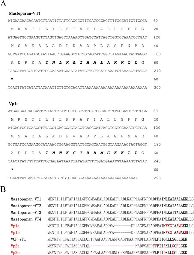
Fig. 1. Amino acid sequences of four new peptides identified in this study, together with other homologous peptides. (A) Nucleotide and amino acid sequences of
Mastoparan-VT1 and Vp1a. Stop codons of each sequence are indicated by asterisks. Mature AMP sequences were identified by sequence comparison with the
peptides of the same family through Clustal Omega (https://www.ebi.ac.uk/Tools/msa/clustalo/) and shown in bold type. (B) Comparison of four new peptides with
known peptides in same general family. Mature AMP sequences are shown in bold type.
We added 190 μL of DPPH solution (100 μM) to a 96-well plate,
followed by the addition of 10 μL of peptide solution (3, 6, 12, 30, 60, (2) ABTS + radical scavenging ability
and 120 μM, respectively) and incubation at 37 ◦ C for 30 min. Absor
bance was then measured at 517 nm. In addition, DPPH (alone), vitamin Methanol was used as a solvent to prepare the ABTS+ (7.4 mM) and
C (120 μM), and methanol solutions were used as the blank, positive, K2S2O8 (2.6 mM) reserve solutions. We mixed 5 mL of ABTS+ reserve
and negative controls, respectively. The following equation was applied solution and 88 μL of K2S2O8 reserve solution. After standing for 16 h,
to measure the DPPH radical scavenging rate: the ABTS+ working solution was prepared with 40 × dilution in meth
/ anol. Absorbance was determined at 734 nm after adding 200 μL of
DPPH radical scavenging rate = (Ablank − Asample ) × 100 Ablank
ABTS+ solution and 10 μL of peptide solution to each well of a 96-well
(2) plate for 6 min. In addition, ABTS+ (alone), vitamin C (120 μM), and
96Y.-C. Meng et al. Toxicon 199 (2021) 94–100
Fig. 2. DPPH radical scavenging rates of four V. velutina venom peptides tested at different concentrations in methanol. Vc is vitamin C (120 μM). Data are mean ±
SD of three independent experiments (n = 9). Statistical significance of differences was determined by t-test (*P < 0.05, **P < 0.01, and ***P < 0.001 indicate
significantly different from negative control, methanol was used as a negative control and compared with each group).
(2) Scavenging activity against ABTS+ radicals
methanol solutions were used as the blank, positive, and negative con centrifuged at 3 000 rpm for 3 min at 4 ◦ C. The absorbance of the
trols, respectively. The following equation was employed to measure the resulting supernatant was measured at 540 nm. We added 1 μL of Triton
ABTS+ radical scavenging rate: 100-x to the blood cells to determine the maximum hemolysis rate. The
/ experiment required three replicates of independent experiments.
ABTS+ radical scavenging rate = (Ablank − Asample ) × 100 Ablank
(3) 3. Results
(3) •OH radical scavenging ability 3.1. cDNA library
We added peptide solution (3, 6, 12, 30, 60, and 120 μM, respec We used DNAMAN v10 to analyze the sequencing data. The targeted
tively), 70 μL of distilled water, 10 μL of salicylic acid-methanol solution base sequences were translated into peptide sequences and then run
(9.1 mM), and 10 μL of FeSO4 solution (9 mM) to a 96-well plate, then through the NCBI database. Two V. velutina peptides corresponding to
added 10 μL of 30% H2O2 solution. Absorbance A1 was measured at 510 primer S1 were from the mastoparan (MP) family (Fig. 1), which we
nm. Absorbance A2 was measured with distilled water instead of FeSO4 named U-VPTX-Vp1a (Vp1a, GeneBank accession number: MZ147005):
solution. Absorbance A3 was measured with distilled water instead of INWKGIAAMGKKLL and U-VPTX-Vp1b (Vp1b, GeneBank accession
the sample. In addition, vitamin C (120 μM) and methanol solutions number: MZ147006): INWKGIAAMAKRLL. All G after the mature pep
were used as the positive and negative controls, respectively. The tide sequences are the amidation recognition sites (Fig. 1), these are
following equation was used to measure the •OH radical scavenging cleaved before yielding the mature peptides in the venom. The third
rate: peptide corresponding to primer S1 was previously reported in the
venom of Vespa xanthoptera (Hirai et al., 1979) and named
OH radical scavenging rate = [1 − (A1 − A2 ) / A3 ] × 100 Mastoparan-X (MP-X).
(4) Two peptides corresponding to primer S2 were in the vespid
chemotactic peptide (VCP) family (Fig. 1), which we named U-VPTX-
2.5. Hemolytic activity assays Vp2a (Vp2a, GeneBank accession number: MZ147007):
FFPIIAKLLGGFLGKK and U - VPTX-Vp2b (Vp2b, GeneBank accession
Red blood cells from mice were used to measure the hemolytic ac number: MZ147008): FLPIIDKLLGGLLGKK. The third peptide corre
tivity of each peptide (Bignami, 1993). In total, 1 mL of red blood cells sponding to primer S2 was previously isolated from the venom of Vespa
was washed twice with 500 μL of phosphate-buffered saline (PBS) and magnifica (Xu et al., 2006) and named VCP-5E.
then diluted with 5% PBS, followed by the addition of each peptide (120 An overall comparison with the same general family of peptides
μM) and incubation in a 37 ◦ C water bath for 30 min. Samples were then (VT1, VT2, VT3, and VT4) identified by Yang et al. (2013) showed that
97Y.-C. Meng et al. Toxicon 199 (2021) 94–100
Fig. 3. ABTS + radical scavenging rates of four V. velutina venom peptides tested at different concentrations in methanol. Vc is vitamin C (120 μM). Data are mean ±
SD of three independent experiments (n = 9). Statistical significance of differences was determined by t-test (*P < 0.05, **P < 0.01, and ***P < 0.001 indicate
significantly different from negative control, methanol was used as a negative control and compared with each group).
(3) Scavenging activity against •OH radicals
the four new peptides (Vp1a, Vp1b, Vp2a, and Vp2b) differed from other significant antioxidant properties against ABTS+ radicals (Fig. 3).
known peptides by two to four amino acids (Fig. 1B). Based upon their At concentrations ranging from 30 to 120 μM, all four peptides
amino acid sequences, Vp1a, Vp1b, Vp2a, and Vp2b were then synthe showed significant antioxidant abilities against •OH radicals (Fig. 4).
sized, resulting in four 20-mg samples with purities of 98% for the Notably, Vp1b showed significant antioxidant ability, even at the rela
biological activity tests. tively low concentration of 12 μM.
3.2. Detection of peptide activity 3.3. Hemolytic activity
3.2.1. Antimicrobial activity The potential toxicity of the peptides was assayed by measuring their
The four peptides differed in their antibacterial effects (Table 1). hemolytic activity against mouse red blood cells. At the highest con
Neither Vp1a nor Vp2b exhibited any strong inhibitory effects on bac centration of 120 μM, the hemolysis rates of Vp1a, Vp1b, Vp2a, and
terial strains at concentrations of 120 μM and below. However, Vp1b Vp2b were 2.37%, 4.08%, 3.46% and 2.12%, respectively. The absor
exhibited very strong inhibitory effects on Staphylococcus aureus and bance of the four peptides at 540 nm was not significantly different from
Escherichia coli bacteria at 120 μM, and inhibited Bacillus subtilis at 60 that of the control. As expected, the positive control, Triton-100x, had a
and 120 μM Vp2a inhibited S. aureus at concentrations of 60 and 120 μM highly significant hemolytic effect (P < 0.001, n = 9, t-test). At 120 μM,
and Klebsiella pneumoniae at 15, 60, and 120 μM. However, Vp1a showed none of the tested peptides exhibited hemolytic activity against mouse
no strong inhibitory effects on any of the tested strains at any of the blood cells, with activities all well below 5%.
tested concentrations.
4. Discussion
3.2.2. Antioxidant assays
Small peptides with molecular weights of 1.4–7 kDa account for
(1) Scavenging activity against DPPH radicals ~70% of the main components of wasp venom (Baek, 2013; Monsalve
and Lu, 1999; Yoon et al., 2015), and include mastoparan, kinin, and
Both Vp1b and Vp2a exhibited excellent antioxidant abilities against chemotactic peptides (Habermann, 1972). In the past few decades, an
DPPH radicals, with highly significant (P < 0.001) scavenging rates at increasing number of small peptides have been found in wasp species
concentrations of 3–120 μM (Fig. 2). Vp2b exhibited relatively good (Argiolas and Pisano, 1984; Bignami, 1993; Chen et al., 2012; Hirai
scavenging ability against DPPH radicals at concentrations of 6–120 μM et al., 1979; Wu et al., 2018; Vaara, 2009); however, reports on small
Vp1a showed significant scavenging ability at concentrations of 12, 60, peptides from V. velutina remain scarce.
and 120 μM, but not at 30 μM. In the current study, we identified four new peptides in the venom of
At concentrations of 6 to 120 μM, all four peptides exhibited V. velutina. Based on peptide sequence similarities with known AMPs, we
98Y.-C. Meng et al. Toxicon 199 (2021) 94–100
Fig. 4. •OH radical scavenging rates of four V. velutina venom peptides tested at different concentrations in methanol. Vc is vitamin C (120 μM). Data are mean ± SD
of three independent experiments (n = 9). Statistical significance of differences was determined by t-test (*P < 0.05, **P < 0.01, and ***P < 0.001 indicate
significantly different from negative control, methanol was used as a negative control and compared with each group).
investigated their antimicrobial properties. Results showed that Vp1b although all scavenging rates were lower than those achieved by the
and Vp2a significantly inhibited bacterial growth at concentrations positive control (vitamin C). In addition, the scavenging rates of free
above 60 μM, with the precise effect dependent on the bacterial species. radicals at 120 μM did not exceed 30%, and thus the antioxidant prop
Compared with other hornet venom AMPs (Xu et al., 2006; Yang et al., erties of our tested compounds were not high in comparison to other
2013), Vp1a, Vp2a, and Vp2b did not exhibit strong antibacterial effects, natural antioxidant peptides (Cao et al., 2018). However, our peptides
even at relatively high concentrations. Vp1b demonstrated good inhib may exhibit synergistic antioxidant activity when combined with other
itory effects against several experimental strains and therefore shows compounds, a potential avenue for future research. Moreover, as Vp1a,
promise as a new natural antibiotic agent. However, Vp1b showed good Vp1b, Vp2a, and Vp2b showed low hemolytic activity, they may be
antibacterial effect against bacteria such as Staphylococcus aureus at 120 useful as safe antioxidant drugs. The combined activity of these com
μM, while VT1-7 (Yang et al., 2013), which are also members of the MP pounds as antimicrobials and antioxidants warrants further study,
family, can achieve the same effect at concentration of about 3–60 μM. particularly their synergic activity with other compounds and against
High concentration is a potential challenge for future clinical use of antibiotic-resistant biofilms and microbes.
Vp1b. Additionally, the four new peptides had almost no inhibitory ef Due to the long-term overuse of antibiotics, bacterial resistance to
fect on Enterococcus faecalis, probably due to its good aggregation and drugs has become an urgent problem requiring the discovery and
fluidity, which made the external bacteria contact AMP first and die, development of new antimicrobial agents (Kennedy, 2013). Given their
while the internal bacteria were protected (Butler et al., 2010). As a good antimicrobial properties and low drug resistance, AMPs are
common infection-causing bacterium, Enterococcus faecalis may not be excellent candidates for drug development (Nijnik and Hancock, 2009).
suitable for AMP. Here, we identified four peptides from two families from the venom
Our study adds to the growing list of AMPs identified from wasp glands of V. velutina. Antimicrobial experiments indicated that several of
venom that show good anti-microbial activity. For example, the VCP these peptides showed good antibacterial effects, thus providing po
peptides, i.e., OdVP1, OdVP2, EpVP1, EpVP2a, and EpVP2b, from tential candidates for the design of effective antimicrobial agents.
Orancistrocerus drewseni and Eumenes pomiformis venom (Baek and Lee,
2010; Baek et al., 2013), mastoparan AMPs from Vespula lewisii venom
Declaration of competing interest
(Ji et al., 2011), and VesP-VB1 and MP-VB1 peptides from Vespa bicolor
venom (Chen et al., 2008) all exhibit good anti-bacterial activity against
The authors declare that they have no known competing financial
various standard and drug-resistant strains at MIC of 5–200 μM.
interests or personal relationships that could have appeared to influence
There is growing interest in the antioxidant abilities of natural
the work reported in this paper.
peptides, and therefore we also studied this property in the identified
peptides. All four peptides demonstrated significant antioxidant effects
Acknowledgments
at concentrations between 30 and 120 μM against the three different free
radicals. All peptides showed good scavenging ability against ABTS+,
This work was supported by grants from the National Natural Science
99Y.-C. Meng et al. Toxicon 199 (2021) 94–100
Foundation of China (81760648 and 31670776) and Key Laboratory of Hancock, R.E.W., Annett, R., 2002. Role of membranes in the activities of antimicrobial
cationic peptides[J]. FEMS Microbiol. Lett. (2), 143–149. https://doi.org/10.1016/
Tropical Forest Ecology, Xishuangbanna Tropical Botanical Garden,
S0378-1097(01)00480-3.
Chinese Academy of Sciences. Hirai, Y., Kuwada, M., Yasuhara, T., et al., 1979. A new mast cell degranulating peptide
homologous to mastoparan in the venom of Japanese hornet (Vespa xanthoptera) [J].
Appendix A. Supplementary data Chem. Pharmaceut. Bull. 27 (8), 1945–1946. https://doi.org/10.1248/cpb.27.1945.
IACG Interagency Coordination Group on Antimicrobial Resistance, 2019. No time to
wait: securing the future from drug-resistant infection. Report to the Secretary-
Supplementary data to this article can be found online at https://doi. General of the United Nations[R]. WHO, Switzerland. https://www.who.int/anti
org/10.1016/j.toxicon.2021.06.002. microbial-resistance/interagency-coordination-group/final-report/en/.
Ji, Hyeong, Baek, et al., 2011. Venom peptides from solitary hunting wasps induce
feeding disorder in lepidopteran larvae[J]. Peptides 32 (3), 568–572. https://doi.
Author contribution org/10.1016/j.peptides.2010.12.007.
Jiang, Y., Chen, Y., Song, Z., et al., 2021. Recent advances in design of antimicrobial
peptides and polypeptides toward clinical translation[J]. Adv. Drug Deliv. Rev. 170.
Yichuan Meng: Investigation, Writing – original draft; Xianggui Mo: https://doi.org/10.1016/j.addr.2020.12.016.
Investigation; Tiantian He: Investigation; Xinxin Wen: Software; James Kennedy, D., 2013. Time to deal with antibiotics[J]. Science 342 (6160), 777. https://
C Nieh: Writing – review & editing; Xinwang Yang: Supervision; Ken doi.org/10.1126/science.1248056.
Kishi, S., Goka, K., 2017. Review of the invasive yellowlegged hornet, Vespa velutina
Tan: Resources. nigrithorax, (Hymenoptera: Vespidae), in Japan and its possible chemical control[J].
Appl. Entomol. Zool. 52 (3), 361. https://doi.org/10.1007/s13355-017-0506-z.
Ethical statement Kuhn-Nentwig, L., 2003. Antimicrobial and cytolytic peptides of venomous arthropods
[J]. Cell. Mol. Life Sci. 60, 2651–2668. https://doi.org/10.1007/s00018-003-3106-
8.
The animals are cared for and treated in accordance with the re Livermore, D.M., 2005. Tigecycline: what is it, and where should it be used?[J].
quirements of the Ethics Committee of Xishuangbanna Tropical Botan J. Antimicrob. Chemother. (4), 611–614. https://doi.org/10.1093/jac/dki291.
ical Garden, Chinese Academy of Sciences. Loit, E., Wu, K., Cheng, X., et al., 2008. Functional whole-colony screening method to
identify antimicrobial peptides[J]. J. Microbiol. Methods 75 (3), 425–431. https://
doi.org/10.1016/j.mimet.2008.07.023.
References López, S., González, M., Goldarazena, A., 2011. Vespa velutina Lepeletier, 1836
(Hymenoptera: Vespidae): first records in Iberian Peninsula[J]. EPPO Bull. 41 (3),
Argiolas, A., Pisano, J.J., 1984. Isolation and characterization of two new peptides, 439. https://doi.org/10.1111/j.1365-2338.2011.02513.x.
mastoparan C and crabrolin, from the venom of the European hornet, Vespa crabro Monsalve, R.I., Lu, G., 1999. Expressions of recombinant venom allergen, antigen 5 of
[J]. J. Biol. Chem. 259 (16), 10106–10111. https://doi.org/10.1111/j.1467- yellowjacket (Vespula vulgaris) and paper wasp (Polistes annularis), in bacteria or
6494.2006.00408.x. yeast[J]. Protein Expr. Purif. 16, 410–416. https://doi.org/10.1006/
Baek, J.H., Lee, S.H., 2010. Isolation and molecular cloning of venom peptides from prep.1999.1082.
Orancistrocerus drewseni (Hymenoptera: Eumenidae) [J]. Toxicon 55 (4), 711–718. Nijnik, A., Hancock, R., 2009. Host defence peptides: antimicrobial and
https://doi.org/10.1016/j.toxicon.2009.10.023. immunomodulatory activity and potential applications for tackling antibiotic-
Baek, J.H., Oh, J.H., Kim, Y.H., Lee, S.H., 2013. Comparative transcriptome analysis of resistant infections[J]. Emerg. Health Threats J. 2, 1–7. https://doi.org/10.3134/
the venom sac and gland of social wasp Vespa tropica and solitary wasp Rhynchium ehtj.09.001.
brunneum[J]. J. Asia Pac. Entomol. 16, 497–502. https://doi.org/10.1016/j. Otvos, L., 2000. Antibacterial peptides isolated from insects[J]. J. Pept. Sci. 6, 497–511.
aspen.2013.08.003. https://doi.org/10.1002/1099-1387(200010)6:103.0.CO;2-W.
Bertolino, S., Lioy, S., Laurino, D., et al., 2016. Spread of the invasive yellow-legged Park, S.C., Park, Y., Hahm, K.S., 2011. The role of antimicrobial peptides in preventing
hornet Vespa velutina, (Hymenoptera: Vespidae) in Italy[J]. Appl. Entomol. Zool. 51 multidrug-resistant bacterial infections and biofilm formation[J]. Int. J. Mol. Sci. 12
(4), 589. https://doi.org/10.1007/s13355-016-0435-2. (9), 5971–5992. https://doi.org/10.3390/ijms12095971.
Bignami, G.S., 1993. A rapid and sensitive hemolysis neutralization assay for palytoxin Piek, T., 1986. Venoms of the Hymenoptera: biochemical, pharmacological and
[J]. Toxicon 31 (6), 817–820. https://doi.org/10.1016/0041-0101(93)90389-Z. behavioural aspects[J]. Q. Rev. Biol. 62 (3) https://doi.org/10.2307/2829032.
Budge, G.E., Hodgetts, J., Jones, E.P., et al., 2017. The invasion, provenance and Pruden, A., Pei, R., Storteboom, H., et al., 2006. Antibiotic resistance genes as emerging
diversity of Vespa velutina Lepeletier (Hymenoptera: Vespidae) in great Britain[J]. contaminants: studies in northern Colorado[J]. Environ. Sci. Technol. 40 (23), 7445.
PloS One 12 (9), e0185172. https://doi.org/10.1371/journal.pone.0185172. https://doi.org/10.1021/es060413l.
Butler, M.T., Wang, Q., Harshey, R.M., 2010. Cell density and mobility protect swarming Rysz, M., Alvarez, P.J.J., 2004. Amplification and attenuation of tetracycline resistance
bacteria against antibiotics[J]. Proc. Natl. Acad. Sci. U. S. A. 107 (8), 3776–3781. in soil bacteria: aquifer column experiments[J]. Water Res. 38 (17), 3705–3712.
https://doi.org/10.1073/pnas.0910934107. https://doi.org/10.1016/j.watres.2004.06.015.
Cao, X., Wang, Y., Wu, C., et al., 2018. Cathelicidin-OA1, a novel antioxidant peptide Vaara, M., 2009. New approaches in peptide antibiotics[J]. Curr. Opin. Pharmacol. 9,
identified from an amphibian, accelerates skin wound healing[J]. Sci. Rep. UK 8 (1), 571–576. https://doi.org/10.1016/j.coph.2009.08.002.
943. https://doi.org/10.1038/s41598-018-19486-9. Villemant, C., Haxaire, J., Streito, J.C., 2006. La découverte du Frelon asiatique Vespa
Chen, W., Yang, X., Yang, X., et al., 2008. Haining, Antimicrobial peptides from the velutina, en France[J]. Insectes 143 (4), 3.
venoms of Vespa bicolor fabricius[J]. Peptides 29, 1887–1892. https://doi.org/ Wang, G., Li, X., Wang, Z., 2016. APD3: the antimicrobial peptide database as a tool for
10.1016/j.peptides.2008.07.018. research and education[J]. Nucleic Acids Res. 44 (D1), 1087–1093. https://doi.org/
Chen, Z., Yang, X., Liu, Z., Zeng, L., Lee, W., Zhang, Y., 2012. Two novel families of 10.1093/nar/gkv1278.
antimicrobial peptides from skin secretions of the Chinese torrent frog, Amolops Wu, R., Li, D., Tang, Q., Wang, W., Xie, G., Dou, P., 2018. A novel peptide from Vespa
jingdongensis[J]. Biochimie 94 (2). https://doi.org/10.1016/j.biochi.2011.07.021, 0- ducalis induces apoptosis in osteosarcoma cells by activating the p38 MAPK and JNK
334. signaling pathways[J]. Biol. Pharm. Bull. 41 (4), 458–464. https://doi.org/10.1248/
Choi, M.B., Martin, S.J., Lee, J.W., 2011. Distribution, spread, and impact of the invasive bpb.b17-00449.
hornet Vespa velutina, in South Korea[J]. Entomol. Res. 41 (6), 276. https://doi.org/ Xu, X., Li, J., Lu, Q., et al., 2006. Two families of antimicrobial peptides from wasp
10.1111/j.1748-5967.2011.00370.x. (Vespa magnifica) venom[J]. Toxicon 47 (2), 249–253. https://doi.org/10.1016/j.
Fjell, C.D., Hiss, J.A., Hancock, R.E.W., et al., 2012. Designing antimicrobial peptides: toxicon.2005.10.015.
form follows function[J]. Nat. Rev. Drug Discov. 11 (1), 37–51. https://doi.org/ Yang, X., Wang, Y., Lee, W.H., Zhang, Y., 2013. Antimicrobial peptides from the venom
10.1038/nrd3591. gland of the social wasp Vespa tropica[J]. Toxicon 74, 151–157. https://doi.org/
Gillespie, J.P., Kanost, M.R., Trenczek, T., 1997. Biological mediators of insect immunity 10.1016/j.toxicon.2013.08.056.
[J]. Annu. Rev. Entomol. 42, 611–643. https://doi.org/10.1146/annurev. Yoon, K.A., Kim, K., Nguyen, P., 2015. Comparative functional venomics of social
ento.42.1.611. hornets Vespa crabro and Vespa analis[J]. J. Asia Pac. Entomol. 18, 815–823. https://
Grossosilva, J.M., Maia, M., 2012. Vespa velutina Lepeletier, 1836 (Hymenoptera: doi.org/10.1016/j.aspen.2015.10.005.
Vespidae), new species for Portugal[J]. Arquivos Entomolóxicos 6, 53.
100You can also read