Ultra-strong bio-glue from genetically engineered polypeptides
←
→
Page content transcription
If your browser does not render page correctly, please read the page content below
ARTICLE
                  https://doi.org/10.1038/s41467-021-23117-9                 OPEN

                  Ultra-strong bio-glue from genetically engineered
                  polypeptides
                  Chao Ma 1,2,3,12, Jing Sun3,4,12, Bo Li4,12, Yang Feng4, Yao Sun4, Li Xiang5, Baiheng Wu6, Lingling Xiao4,
                  Baimei Liu4, Vladislav S. Petrovskii7,8, Bin Liu4, Jinrui Zhang4, Zili Wang4, Hongyan Li2,9, Lei Zhang4, Jingjing Li4,
                  Fan Wang4, Robert Gӧstl 9, Igor I. Potemkin7,9,10, Dong Chen 6, Hongbo Zeng 5, Hongjie Zhang1,4,
                  Kai Liu 1,4 ✉ & Andreas Herrmann 2,9,11 ✉
1234567890():,;

                  The development of biomedical glues is an important, yet challenging task as seemingly
                  mutually exclusive properties need to be combined in one material, i.e. strong adhesion and
                  adaption to remodeling processes in healing tissue. Here, we report a biocompatible and
                  biodegradable protein-based adhesive with high adhesion strengths. The maximum strength
                  reaches 16.5 ± 2.2 MPa on hard substrates, which is comparable to that of commercial
                  cyanoacrylate superglue and higher than other protein-based adhesives by at least one order
                  of magnitude. Moreover, the strong adhesion on soft tissues qualifies the adhesive as bio-
                  medical glue outperforming some commercial products. Robust mechanical properties are
                  realized without covalent bond formation during the adhesion process. A complex consisting
                  of cationic supercharged polypeptides and anionic aromatic surfactants with lysine to sur-
                  factant molar ratio of 1:0.9 is driven by multiple supramolecular interactions enabling such
                  strong adhesion. We demonstrate the glue’s robust performance in vitro and in vivo for
                  cosmetic and hemostasis applications and accelerated wound healing by comparison to
                  surgical wound closures.

                  1 Department of Chemistry, Tsinghua University, Beijing, China. 2 Zernike Institute for Advanced Materials, University of Groningen, Groningen, The

                  Netherlands. 3 School of Engineering and Applied Sciences, Harvard University, Cambridge, MA, USA. 4 State Key Laboratory of Rare Earth Resource
                  Utilization, Changchun Institute of Applied Chemistry, Chinese Academy of Sciences, Changchun, China. 5 Department of Chemical and Materials
                  Engineering, University of Alberta, Edmonton, Alberta, Canada. 6 Institute of Process Equipment, College of energy engineering, Zhejiang University,
                  Hangzhou, China. 7 Physics Department, Lomonosov Moscow State University, Moscow, Russian Federation. 8 N. N. Semenov Institute of Chemical Physics,
                  Russian Academy of Sciences, Moscow, Russian Federation. 9 DWI - Leibniz Institute for Interactive Materials, Aachen, Germany. 10 National Research South
                  Ural State University, Chelyabinsk, Russian Federation. 11 Institute of Technical and Macromolecular Chemistry, RWTH Aachen University, Aachen, Germany.
                  12
                    These authors contributed equally: Chao Ma, Jing Sun, Bo Li. ✉email: kailiu@tsinghua.edu.cn; herrmann@dwi.rwth-aachen.de

                  NATURE COMMUNICATIONS | (2021)12:3613 | https://doi.org/10.1038/s41467-021-23117-9 | www.nature.com/naturecommunications                               1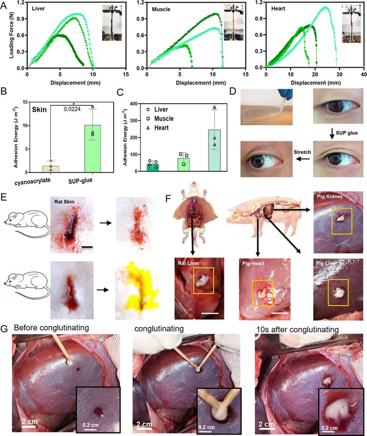
ARTICLE                                                                   NATURE COMMUNICATIONS | https://doi.org/10.1038/s41467-021-23117-9

S
       trong adhesives in both dry and wet conditions play an                 centrifugation a protein- and surfactant-rich liquid was obtained
       important role in many technical1–3 and clinical                       at the bottom of the tube (Fig. 1C). This behavior is termed
       applications4–6. Traditionally, polymer adhesives develop              complex coacervation and represents a liquid-liquid phase-
high adhesion strengths through coating asperities and retarding              separation process, which widely exists in nature, e.g. in mussel or
the fracture of adhesive joints. This is achieved by in situ poly-            sandcastle worm protein adhesives and in artificial biopolymeric
merization or crosslinking of reactive monomers that form per-                adhesion systems2,17,24. Specifically, the coacervate here can be
manent, non-adaptive covalent bonds or networks7,8. Recently,                 regarded as proteinaceous polyelectrolytes (SUPs) in an aqueous
systems based on supramolecular interfacial bond formation,                   solution combined with counterions (SDBS), yielding phase-
such as catechol or host-guest motifs, were introduced9–11.                   separated aggregates. The coacervate formation of the system was
However, they failed to deliver strong adhesion strengths under               further investigated by determining its phase diagrams in regard
ambient conditions and moreover often require hard-to-prepare                 to the concentration of the components, polypeptide to surfactant
or irritating components.                                                     ratio, pH, and ionic strength (Supplementary Fig. 4). It suggests
   Especially the latter should be avoided in glues employed in               that the occurrence of a coacervate in the system is a character-
biomedicine. In addition, biodegradability needs to be considered             istic phase-separation phenomenon. Particularly under the con-
for practical applications. Biodegradability can be implemented               dition of ~150 mM physiological saline, the complex indeed does
into glues by the utilization of biomacromolecules as adhesive                not swell.
threads since they are degraded by biochemical processes on                      After separation of the supernatant, the SUP-SDBS coacervates
reasonable timescales. One example for this is wound healing                  were viscous but plastically deformable (Fig. 1C–E). It is worth
where proteases are upregulated in the matrix microenvironment                noting that SDBS molecules are present as micelles during the
and actively degrade both exogenous entities and native                       process of complexation (Fig. 1B and Supplementary Fig. 5). A
components12. Several elastin-based adhesives, blood-derived                  representative and quantitative component determination of the
fibrin sealants, and other naturally derived adhesive matrices                 SUP-SDBS complexes was carried out by proton nuclear mag-
have been developed, but require laborious and time-consuming                 netic resonance (1H-NMR) spectroscopy. For the K18-SDBS
pre-treatment by heat or UV light irradiation to prime covalent               complex, a stoichiometry of K18:SDBS of 1:16 was measured,
bond formation risking secondary damage to the traumatized                    equaling a ca. 90% occupation of the positive lysine residues by
tissues13–16. Moreover, the existing bio-glue solutions, such as              surfactant molecules (Supplementary Fig. 5). Thermogravimetric
protein- or polypeptide-based structures, adhere only insuffi-                 analyses (TGA) showed that the SUP-SDBS complexes exhibited
ciently to substrates (i.e., soft tissue) and/or modestly promote             a water content of ~42% (w/w) (Supplementary Fig. 7). Besides
wound closure and natural healing processes17–26. Eventually, an              pristine SUP chains, fusions of SUPs with proteins of different
ideal adhesive for regenerative medicine should combine bio-                  absorption and emission colors, i.e., GFP and mCherry, were
compatibility and -degradability with high adhesive strength, yet             converted into adhesives by complexation with SDBS, analo-
still being adaptive and flexible to respond to remodeling tissues             gously to the pristine peptide chains (Fig. 1E).
where motile cells dynamically change their positional and                       After fabrication, the bulk adhesion strengths of the SUP glues
structural order27,28.                                                        were investigated by lap shear testing (Fig. 2A and Supplementary
   To accommodate these challenges, we here present the design                Fig. 10). K72-SDBS and commercial cyanoacrylate glue (as a
of a family of supercharged polypeptide-based adhesives for                   comparison) were applied on various substrates including glass,
in vivo tissue engineering applications. These glues were formed              steel, aluminum, polyethylene (PE), and polyvinyl chloride
by electrostatic complexation of cationic polypeptides and anionic            (PVC). The K72-SDBS glue adhered strongly on high-energy
aromatic surfactants avoiding covalent bond formation during                  surfaces (glass or metal) and exhibited fracture strengths in the
the gluing process. A well-balanced combination of non-covalent               range of 11.0–14.0 MPa, comparable to cyanoacrylate glue
bonds gives rise to ultra-high fracture strengths surpassing known            (Fig. 2A, Supplementary Figs. 10 and 11). This behavior indicates
protein-based adhesives by one order of magnitude. As a result,               that rough, high-energy surfaces adhere to SUP glue strongly.
external skin wounds and internal organ defects were sealed fast              This can be explained by the high potential of both chemical
and at the same time wound healing was accelerated.                           interactions and mechanical interlocking between the glue and
                                                                              the surface32. On low-energy surfaces (PVC or PE), the adhesion
                                                                              of SUP glue was reduced, but still as strong as cyanoacrylate, one
Results and discussion                                                        of the strongest commercially available adhesives. Notably, the
Cationic supercharged polypeptides (SUPs) are inspired by nat-                adhesive performance of SUP glue between two glass substrates
ural elastin and were recombinantly expressed in Escherichia                  was so robust that the substrate fractured before the glue failed as
coli29,30. The high net charge of SUPs is encoded in the penta-               visualized by intact adherent regions. Furthermore, lap shear
peptide repeat unit (VPGKG)n in which the fourth-position                     investigations involving K18-SDBS, K36-SDBS, K72-SDBS, K108-
valine is substituted with a lysine residue (K) (Fig. 1A) that is             SDBS, and K144-SDBS glues indicated that the adhesion strength
protonated under physiological conditions. A series of SUPs with              increased with increasing molar mass of the SUP (Fig. 2B and
different numbers of repeating units, and thus chain lengths,                 Supplementary Fig. 12) and by this the adhesion strengths could
including K18, K36, K72, K108, and K144, were produced. The                   be tuned between 3.0 and 16.5 MPa.
digit denotes the number of positive charges along the polypep-                  Importantly, there was no difference in adhesion properties
tide backbone (Supplementary Fig. 1–3 and Tables 1–2). In                     between the pristine K-SDBS groups and the fluorescent protein
addition, green and red fluorescent proteins (GFP and mCherry)                 fusion variants indicating that the adhesive behavior can be
were fused to the unfolded cationic SUPs to demonstrate their                 maintained even when exogenous functional protein entities are
easy functionalization with folded proteins and for facile tracing.           introduced. Notably, the fracture strength of 16.5 ± 2.2 MPa of
   Subsequently, the anionic surfactant sodium dodecylbenzene                 K144-SDBS was higher than any other reported protein-based
sulfonate (SDBS), which is an FDA-approved surfactant for                     adhesive and surpassed those by at least one order of
cosmetics31, was complexed with the cationic SUPs to form the                 magnitude15–20. The adhesion energies of SUP glue on different
adhesive. Therefore, SUPs and SDBS micelles were mixed in                     surfaces are summarized in Table S3. Comparison with chemi-
aqueous solution in a 1:1 molar ratio of lysine unit to surfactant            cally synthesized adhesives and genetically engineered protein-
(Fig. 1B). As a result, the solution became turbid and after                  based adhesives (Supplementary Table 4) shows that SUP glue

2                 NATURE COMMUNICATIONS | (2021)12:3613 | https://doi.org/10.1038/s41467-021-23117-9 | www.nature.com/naturecommunications
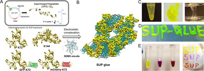
NATURE COMMUNICATIONS | https://doi.org/10.1038/s41467-021-23117-9                                                                      ARTICLE

Fig. 1 Fabrication and investigation of the SUP-SDBS glue system. A Schematic illustration of cationic supercharged polypeptide (SUP) expression
through genetically engineered E. coli. The polypeptide backbones are shown in a random coil conformation. Lysine is presented in a space-filling model in
yellow and the charged amino-group in gray. A series of SUPs with different chain lengths (K18, K36, K72, K108, and K144) and SUP fusions with GFP and
mCherry were produced. B The glue was prepared via electrostatic complexation of SUP and SDBS micelles. An all-atomistic simulated K18-SDBS complex
is shown. In this model, K18 is shown in yellow, SDBS in cyan, and some of SDBS micelle domains are encircled in red. C For the fabrication, a solution of
SUP (here GFP-K72) was mixed with a solution of SDBS micelles leading to liquid-liquid phase separation and the formation of a coacervate. After
centrifugation, the supernatant was removed (left) and the coacervate consisting of SUP-SDBS complex was collected by a pipette and applied to a glass
surface (middle and right). D Fluorescent SUP glues show robust adhesion behavior when applied on glass surfaces and are deformable. E GFP-SUP,
mCherry-SUP, and a mixture of both with different emission colors were used to prepare adhesives.

exhibits ultra-strong adhesion performance in both dry and wet                   Complexing SUPs with a surfactant lacking the phenyl moiety (by
conditions. To demonstrate the broad validity of the paradigm of                 using sodium dodecyl sulfate, SDS) does not result in any adhe-
the SUP glue system, the small molecule SDBS anionic surfactant                  sive properties. These experiments might emphasize the major
component was replaced by salmon sperm DNA (~2000 bp) and                        contribution of cation-π interactions between the free lysine
the new adhesive consisting exclusively of biomacromolecules                     residues and the phenyl ring of SDBS to the adhesive properties of
was termed SUP-DNA glue. The SUP-DNA showed similar                              the bio-based glue. The cation-π interaction is known to govern
adhesion properties as the SUP-SDBS system (Supplementary                        adhesive systems in nature, e.g., in mussel plaque10, and hence
Fig. 14).                                                                        might play a pivotal role in the cohesive properties of SUP-SDBS
   Adhesion not only involves rough but also wet surfaces. Hence,                complexes as well.
we investigated underwater adhesive strengths of SUP glues on                       To investigate the contributions of molecular interactions
two types of substrates (steel and glass) (Fig. 2C, Supplementary                involved in the complex to realize superb adhesion behavior,
Fig. 14, and Supplementary Table 3). The K72-SDBS glue                           surface force apparatus (SFA) measurements were carried out to
exhibited strong adhesion with fracture strengths of 490 and 330                 directly quantify the interaction forces between K72 and SDBS
kPa on steel and glass, respectively. These values are comparable                (Fig. 2D). It was found that when increasing NaCl concentration
to or higher than other proteinaceous underwater adhesives                       from 1 to 100 mM, the adhesion decreased to ~16.5 mN m−1 at
reported to date19–26. The underwater adhesion energy of SUP                     short contact (Fig. 2E). The reduced adhesion most likely stems
glue on glass and steel was 20 J m−2 and 51 J m−2, respectively,                 from the screening of the electrical double layer under high
which is higher than for recently reported chemically synthesized                salinity conditions, shortening the Debye length from ~10 nm in
adhesives (Supplementary Table 4)2,6.                                            1 mM NaCl to ~1 nm in 100 mM NaCl. To evaluate the con-
   Next, the effect of the molar ratio of SUP over SDBS to the                   tribution of cation-π interaction in the SUP-SDBS complex, we
adhesion properties was investigated. Since we showed that ~10%                  introduced interfering cation NMe4+ to the aqueous media and
of lysine moieties within the SUP are not complexed by surfactant                then measured the adhesion between SUP and SDBS layers33. We
molecules, we hypothesized that these free lysine residues may                   compared the adhesion (Fad/R ~29.6 mN m−1) between SUP and
contribute to the adhesion properties of the complexes. Therefore,               SDBS layers in 100 mM NMe4Cl with that (Fad/R ~39.5 mN m−1)
we prepared SUP-SDBS complexes with different stoichiometry.                     in 100 mM NaCl within 20 min contact (Fig. 2F, Supplementary
When a lysine:surfactant molar ratio of 1:5 was chosen, for each                 Fig. 18B, C). The weakened adhesion in 100 mM NMe4Cl can be
lysine on average 3.3 SDBS molecules were detected by 1H-NMR                     attributed to the presence of NMe4+ ions that form robust
measurements (Supplementary Fig. 16). As a result, the corre-                    NMe4+-π pairs with SDBS, thereby reducing the cation-π inter-
sponding fracture strength decreased by about one order of                       action between the NH3R+ on the lysine residue in the SUP and
magnitude suggesting that the non-complexed free lysine con-                     the phenyl group in SDBS. To further estimate the contribution of
tributes significantly to the overall adhesion performance (Sup-                  cation-π interaction in SUP-SDBS complex, we compared the
plementary Fig. 17A). In addition, a lysine:surfactant molar ratio               adhesion (Fad/R ~39.5 mN m−1) between SUP and SDBS layers in
of 1:0.5 was adjusted (Supplementary Fig. 17B) showing similarly                 100 mM NaCl with that (Fad/R ~27.8 mN m−1) of SUP and
low adhesion as the 1:5 complex. This suggests that the ratio of                 sodium dodecyl sulfate (SDS) under the same solution condition
lysine to SDBS needs to be properly adjusted to achieve optimal                  within 20 min contact (Fig. 2G and Supplementary Fig. 18B, E).
bulk adhesion properties. In addition, a systematic investigation                The adhesion for the SDS group was significantly reduced, which
of adhesion performance of SUP glues along with the initial ratios               can be explained by the absence of cation-π interactions between
from 1:0.5 to 1:2 with 0.1 intervals was performed, showing peak                 SUP and SDS due to the lack of phenyl group in SDS. Therefore,
strength in the subgroup of 1:1.0 (Supplementary Fig. 38).                       the above results demonstrate that cation-π interactions (i.e.,

NATURE COMMUNICATIONS | (2021)12:3613 | https://doi.org/10.1038/s41467-021-23117-9 | www.nature.com/naturecommunications                                3
ARTICLE                                                                     NATURE COMMUNICATIONS | https://doi.org/10.1038/s41467-021-23117-9

Fig. 2 Bulk adhesion behavior of the SUP glues. A Illustration of lap shear testing for the K72-SDBS glue and fracture strengths in comparison to
commercial cyanoacrylate-based glue on glass, steel, aluminum, polyethylene (PE), and polyvinyl chloride (PVC). ns, no significant difference. **p < 0.01. B
Correlation of bulk adhesion strength measured on steel substrate with SUP molar masses starting from K18 and ranging to K144. **p < 0.01; ***p < 0.001.
C SUP glue lap shear testing in an aqueous environment. Two sets of measurements were performed on steel and glass, respectively. D Schematic of
surface force apparatus (SFA) measurement. Two opposing curved mica surfaces with radius R in crossed-cylinder configuration coated with a K72 layer
and an SDBS (or a sodium dodecyl sulfate (SDS)) layer, respectively, were first approached to each other until they come into contact, and were then
separated to measure the adhesion force (Fad). E Impact of aqueous conditions on the adhesion between K72 and SDBS layers as revealed by SFA
measurements. 1 mM NaCl vs. 100 mM NaCl (pH 5.6 and short contact); F 100 mM NaCl vs. 100 mM NMe4Cl (pH 5.6 and 20 min contact); G Adhesion
comparison between K72 and SDBS layers, and between K72 and SDS layers in 100 mM NaCl at pH 5.6. *p < 0.05. H pH 5.6 vs. pH 12 (100 mM NaCl and
20 min contact). The short contact refers to a contact time of 1 min. I Proposed molecular mechanism for the strongly adhesive SUP-SDBS complexes. The
simulated structure of the K18-SDBS complex is presented in yellow and cyan, respectively. Different interactions govern the adhesive and cohesive
strength including electrostatic interactions (E.I.), cation-π interactions, van der Waals forces, hydrogen bonds, hydrophobic interactions, and other
aromatic interactions. All presented data are mean values ± SD from the mean from N = 3 independent measurements on independent samples. All
p-values were calculated using two-sided Student’s t-test.

NH3R+-π pairs) between SUP and SDBS play an important role                      reducing the adhesion. Interestingly, with increasing the contact
in the adhesion process, estimated ~9.9 mN m−1 (i.e., 39.5–29.6                 time to 20 min, the SUP-SDBS adhesion was greatly enhanced to
mN m−1), in addition to the electrostatic attraction. We further                Fad/R ~11.9 mN m−1. It is likely that due to the molecular rear-
investigated the impact of pH on the adhesion, and measured the                 rangement under contact, the initially hidden hydrophobic moi-
adhesion between SUP and SDBS layers in 100 mM NaCl at pH 12                    eties on one surface would expose to and interact with the
(Fig. 2H and Supplementary Fig. 18D). It was found that the                     opposing hydrophobic moieties on the other surface34–36. In view
adhesion was significantly weakened to Fad/R ~5.5 and ~11.9 mN                   of this, the adhesion contributed from hydrophobic interactions
m−1 at pH 12 under 1 min and 20 min contact, respectively, as                   could be roughly estimated to ~6.4 mN m−1. Given that the
compared to Fad/R ~16.5 and ~39.5 mN m−1 at pH 5.6. As the pI                   adhesion originating from electrostatic interaction and cation-π
of K72 is 11.87, the amino groups on lysine residues would                      interaction was estimated to ~20.9 mN m−1 (in 1 mM NaCl, pH
deprotonate at pH 12, which could largely weaken the electro-                   5.6) and ~9.9 mN m−1, respectively, the contribution ratio of
static and cation-π interactions between SUP and SDBS, thus                     electrostatic interaction, cation-π interaction, hydrophobic

4                   NATURE COMMUNICATIONS | (2021)12:3613 | https://doi.org/10.1038/s41467-021-23117-9 | www.nature.com/naturecommunications
NATURE COMMUNICATIONS | https://doi.org/10.1038/s41467-021-23117-9                                                                    ARTICLE

interaction, and other interactions (hydrogen bonding, van der                   combination with complex, irregular geometries and surfaces. We
Waals, and other aromatic forces) to the formation of SUP-SDBS                   used SUP-SDBS complexes to glue two pieces of porcine heart,
complex is approximately estimated as 4:2:1:1. Moreover, all-                    muscle, liver, and skin (Fig. 3A–C). With these samples, uniaxial
atomistic computer simulations showed the formation of dis-                      extension testing was performed by recording the corresponding
ordered nanodomains of isolated SUPs bound to each other by                      force-extension curves (Supplementary Movie 2). A peak force of
surfactant micelles (Fig. 2F) and demonstrate the importance of                  ~1.1 N with F w−1 = 200 N m−1 and adhesion energy of 260 ±
cation-π and other supramolecular interactions for the strong                    110 J m−2 on porcine heart tissues was achieved, which is higher
adhesive properties of the complex (Supplementary Movie 1, and                   than covalently crosslinked adhesives on soft tissues reported
Supplementary Fig. 19 and 20). Furthermore, by systematically                    earlier6. Adhesion performance of SUP glue on muscle, liver, and
investigating the water contents in K72-SDBS complex, we con-                    skin tissues was further characterized showing adhesion energies
clude that the presence of a certain amount of water in the                      of 80 ± 26 J m−2, 45 ± 15 J m−2, and 6 ± 2 J m−2, respectively
complex can strengthen and maintain intermolecular interactions                  (Fig. 3A–C and Supplementary Fig. 8). For wet tissue, no specific
in the system, allowing the lysine residues to be positively charged             adhesion strength was recorded due to the sliding nature of the
and available for electrostatic attraction and cation-π interaction              soft adhesive joints when applying with stretching stress. There-
with SDBS, as well as for maintaining the micellar structures of                 fore, the quantitative descriptions of parameters, including F w−1
SDBS (Supplementary Fig. 13).                                                    and adhesion energy, are employed using the width of the joint
   Therefore, the robust cohesion during SUP-SDBS complexa-                      area. Compared to the cyanoacrylate control on the skin, the SUP
tion is mainly realized through the synergy of multiple interac-                 glue shows significantly higher adhesion energy on this substrate
tions including electrostatic interactions, cation-π interactions,               (Fig. 3B). Proof-of-concept adhesive applications for cosmetics
hydrophobic interactions, hydrogen bonding, and van der Waals                    and skin healthcare were carried out on human skin and eyelids
interactions. When applying this SUP-SDBS glue to different                      (Fig. 3D and S29).
substrate surfaces, the flowability of the SUP-SDBS coacervate                       Subsequently, the hemostatic properties of SUP glue were
enables the intimate contact between the SUP glue and substrates;                investigated in vivo (Fig. 3E–G). Firstly, an incision of l × h × w =
meanwhile, the surface molecules and functional groups of the                    2 × 1 × 0.5 cm was performed on the back of a rat. Hereafter, K72-
coacervate possess good flexibility to rearrange and interact with                SDBS and GFP-K72-SDBS glues were applied to those hemor-
opposing substrate surfaces via a synergy of multiple interactions               rhaging skin defects as sealants. The wounds were sealed in 10 s
such as electrostatic interactions, cation-π interactions, hydro-                after the glues were applied, confirming the hemostatic properties
phobic interactions, hydrogen bonds and van der Waals inter-                     of the SUP glues (Fig. 3E). Beyond functioning in the context of
actions (depending on the substrate surface properties), thus                    external skin defects, SUP glues exhibited tissue adhering and
leading to the strong glue-substrate adhesion as we have mea-                    hemostatic properties internally on bleeding models of rat liver,
sured in bulk adhesion tests. Such strong cohesion and wet                       pig liver, pig kidney, and pig heart (Supplementary Movie 3–8,
adhesion mechanisms of the SUP glue are somewhat similar to                      Fig. 3F, G, and Supplementary Fig. 31). The results indicate that
marine mussel adhesives formed via the coacervation of the                       the in vivo wounded tissues can be fully sealed with SUP glue and
mussel adhesive proteins37,38.                                                   the hemostasis effect was realized within 5 s. Moreover, com-
   The non-covalent nature of the adhesive system endows the                     mercial adhesive Histoacryl® containing cyanoacrylate as an
SUP-SDBS complex with additional attractive features. Firstly,                   effective component was applied as a control, which solidified fast
glues are biodegradable. We showed that after enzymatic treat-                   on the pig organs. However, the bleeding still continued when the
ment with elastase, the SUP component of the glue was digested                   wound was brought in contact with Histoacryl®, indicating the
as confirmed by gel electrophoresis (Supplementary Figs. 21 and                   inappropriateness of this product when applied on the liver
22). Moreover, SUP glue applied on substrates could to a large                   (Movie S5, Supplementary Fig. 31). Quantification of adhesion
extent be detached from the wet substrates under sufficiently high                performance on organ surfaces was established in terms of
external shear or peeling forces, which is mainly attributed to the              hemostasis time (Supplementary Fig. 31C). It was shown that the
strong and synergistic non-covalent interactions between SUP                     SUP glue has superior performance on liver wounds and com-
and wet substrates. Afterwards, the polypeptide material was                     parable hemostasis effects on heart and kidney wounds compared
recovered and adhesive properties were regained demonstrating                    to Histoacryl®. These experiments demonstrate the flexibility and
the recyclability of the glue. It was found that the regenerated                 conformability of the SUP glue and indicate the potential of this
SUP-SDBS complex exhibited the same adhesion strength as the                     material for tissue hemostatic applications.
original, non-recycled batch (Supplementary Figs. 23 and 24).                       In addition, a systematic in vivo evaluation of wound healing
   Furthermore, the biocompatibility of the SUP glues was                        was performed employing a rat model with customized linear
investigated. We cultured HeLa cells in the presence of different                incisions and round openings (Fig. 4A and Supplementary
concentrations of K72-SDBS. Without significant deviation from                    Fig. 33). Four different animal groups for wound treatment were
the control group, the cell viability remained higher than 90%                   applied including a blank without treatment, suture closure,
after 24 h in all experiments (Supplementary Figs. 25–27).                       commercial medical adhesive COMPONT®, and SUP glue. In the
Moreover, mice mesenchymal stem cells (D1 cells), bone                           group treated with SUP glue, the wound was tightly sealed after
marrow-derived stem cells, human dermal fibroblasts, as well as                   the SUP glue was adjusted to the incision. The healing progress
HeLa cells were embedded into the SUP glue and SUP-DNA glue                      was evaluated quantitatively over 8 d (Fig. 4B). After 5 d, a sig-
matrices for 3D culturing (Supplementary Fig. 27). The viability                 nificant increase of repaired wound area was detected for the SUP
of encapsulated cells was quantified as well, showing good bio-                   glue compared to the other groups, demonstrating the capacity of
compatibility of the SUP glue groups (Supplementary Figs. 25–                    the SUP glue for regenerating skin. On day 8, the scar was almost
27). Compared with the alginate matrix, there is no significant                   invisible for the rats treated with SUP glue (4% wound area left),
difference between the SUP and alginate groups in terms of cell                  outperforming the commercial medical adhesive. A similar trend
viability.                                                                       was observed in a study involving injury dressing of round
   Motivated by the robust performance and non-toxic nature, we                  wounds (Supplementary Fig. 33). From these experiments, one
investigated the SUP glue’s suitability for biomedical applications              can conclude that the SUP glue is actively facilitating hemostasis
ex vivo and in vivo. The quantitative evaluation of adhesion on                  and accelerating the healing of wounds of different geometries. In
skin or organs is intrinsically challenging due to wet conditions in             stark contrast to suture closure and commercial chemical

NATURE COMMUNICATIONS | (2021)12:3613 | https://doi.org/10.1038/s41467-021-23117-9 | www.nature.com/naturecommunications                            5
ARTICLE                                                                  NATURE COMMUNICATIONS | https://doi.org/10.1038/s41467-021-23117-9

adhesives, the SUP glue with its biodegradable nature and                    Supplementary Fig. 34). Masson’s trichrome staining revealed
supramolecular bonding might accustom well to dynamics of                    that there was more collagen deposition when treated with SUP
matrix and tissue, which might explain accelerated healing and               glue than for the other treatment groups (Fig. 4D and Supple-
regeneration of skin defects27,28.                                           mentary Fig. 35). The H&E and Masson’s trichrome staining
   Histological analyses applying hematoxylin and eosin (H&E) as             results of the four groups were judged by double-blind scoring in
well as Masson’s trichrome staining were utilized to analyze the             regard to the healing performance on the 8th day according to the
regeneration of healed skin tissue. H&E staining showed the                  degree of skin repair, inflammatory cell infiltration, newborn
formation of new blood vessels and abundant follicle and                     capillary formation, and collagen levels around the wound. The
sebaceous glands in the group treated with SUP glue while the                evaluation indicates that the SUP-glue showed superior perfor-
recovery of control group tissues was inferior (Fig. 4C and                  mance among all test groups (Supplementary Table 6 and

6                NATURE COMMUNICATIONS | (2021)12:3613 | https://doi.org/10.1038/s41467-021-23117-9 | www.nature.com/naturecommunications
NATURE COMMUNICATIONS | https://doi.org/10.1038/s41467-021-23117-9 ARTICLE
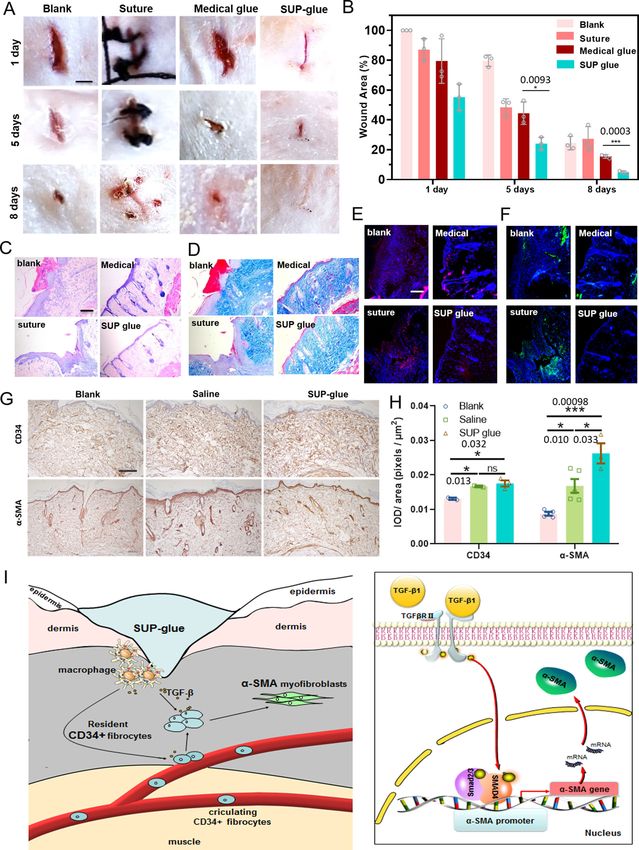
Fig. 3 Quantification and applications of SUP glues dealing with wet tissue adhesion and wound hemostasis. A Quantification of pre-clinical ex vivo
adhesion model with SUP-glue on porcine liver, muscle, and heart. The measurements were carried out via lap shear devices. Insets show images of the
adhesion joints. Three individual curves were collected for each group of specimens. B Adhesion energy comparison of cyanoacrylate and SUP glue (here
K72-SDBS) on pig skin. *p < 0.05. C Overview of SUP-glue performance in terms of adhesion energy on pig heart, liver, skin, and muscle. D SUP glue on
parafilm pasted on the skin of the arm exhibiting strong adhesion and good conformability. SUP glue applied on a single eyelid to achieve a double eyelid
effect, and reversal of the effect after stretching. Male volunteer with age of 35. E Bleeding wounds on rat skin treated with SUP glue. The skin wound was
sealed inducing hemostasis. The second skin wound was treated with fluorescent GFP-SUP glue for tracing the glue during wound healing over nine days. F
SUP glue worked as sealant on pig heart, kidney, liver as well as rat liver. The yellow boxes indicate the pasting position on organs. Scale bars: 10 mm
(black) and 30 mm (white). G The demonstration of in vivo adhesion and hemostasis model on pig kidney. The bleeding wound was ceased in 10 s when
SUP glue was applied. All presented data are mean values ± SD from the mean from N = 3 independent measurements on independent samples. All p-
values were calculated using a two-sided Student’s t-test.
Supplementary Fig. 32). On top of blood leakage, inflammation is aromatic surfactants endows the SUP glue with ultra-strong
another fatal consequence of severe wounds. Therefore, the effi- adhesive and biodegradable properties. This unique set of features
cacy of SUP glue to prevent injury-associated inflammation was renders the material perfectly suited for cosmetic skincare
assessed via immunofluorescence analysis. In this regard, pro- applications and as an in vivo bio-glue enabling tissue regen-
inflammatory cytokines including interleukin-6 (IL-6) and tumor eration after surgical interventions. The robust in vivo perfor-
necrosis factor-α (TNF-α) were measured. IL-6 accelerates scar mance was highlighted by wound-healing studies characterized
formation via stimulating keratinocyte migration and prolifera- by fast hemostasis, avoidance of an inflammatory response, and
tion, and mediating fibroblast proliferation by platelet-derived accelerated healing.
growth factor39,40. In our study, IL-6 in the wound tissue was
suppressed after administration of SUP glue, compared with
controls, indicating the potential function of SUP glue, i.e., pre- Methods
Materials. SDBS and other chemicals were obtained from Sigma-Aldrich
vention of keloid and hypertrophic scar occurrence (Fig. 4E and (Netherlands and China). Illustration figures of chemical structures in the
Supplementary Fig. 36). Moreover, for the same control groups, manuscript were plotted via ChemDraw software (version 11). Double-stranded
green fluorescence was recorded suggesting high levels of secreted salmon sperm DNA was purchased from Thermo Fisher (Waltham, MA). The
TNF-α (Fig. 4F and Supplementary Fig. 37). In stark contrast, the water used in this research (typically 18.2 MΩ cm at 25 °C) was from a Milli-Q
ultrapure water system (Merck, Germany). All biochemicals for cloning and SUP
wounds treated with SUP glue did not show these signs of expression, such as LB medium, salts, antibiotics as well as inducer compounds,
inflammation. Interestingly, when GFP-SUP glue was used for the were used as received (from Sigma-Aldrich) without any further purification. The
experiments, no GFP signal was detected in wound areas, which is pUC19 cloning vector, restriction enzymes, and GeneJET Plasmid Miniprep kit
a strong indication for the in vivo biodegradability of the glue were purchased from Thermo Fisher Scientific (Waltham, MA). Digested DNA
(Supplementary Fig. 37). Therefore, the degradation of the SUP fragments were purified using QIAquick spin miniprep kits from QIAGEN
(Valencia, CA). E. coli XL1-Blue competent cells for plasmid amplification were
backbone into body’s own amino acid building blocks might be purchased from Stratagene (La Jolla, CA). Oligonucleotides for sequencing were
important for the comparatively low immunogenicity and ordered from Sigma-Aldrich (St. Louis, MO). Alpha-cyano-4-hydroxycinnamic
remodeling of matrix and tissue. This is an attractive feature of acid and sinapinic acid was used as matrix during MALDI mass spectrometry and
the SUP glue for accelerating wound healing and especially pro- was purchased from Thermo Scientific (Waltham, MA). Mouse Anti-αSMA
(Abcam, ab7817, lot#GR31757-1), used at 1:1500 from 1 mg mL−1 stock solution.
mising for future surgical applications. Furthermore, we investi- Rabbit Anti-CD34 (Abcam, ab81289, lot#GR45485-1), used at 1:1500 from 1
gated whether the contribution of SUP glue on the process of mg mL−1 stock solution. Animal experiments and human skin adhesion experi-
wound healing involves certain signaling pathways. In the TGF-β/ ments are in agreement with the guidelines of the Regional Ethics Committee for
Smad pathway, resident CD34 positive (cluster of differentiation Animal and Clinical Experiments of Jilin University Institutional Animal Care and
Use and the Second Hospital of Jilin University, respectively. Informed, written
34, CD34+) fibrocytes are phenotypically metamorphosed to consent was given by human volunteers. Other solvents used in the work were of
SMA positive (α-smooth muscle actin, SMA+) myofibroblasts analytical grade.
under the stimulation of TGF-β1. Then SMA assists to remodel
the extracellular matrix (ECM) and promotes the contraction and
healing of wound tissue40,41. The immunohistochemistry staining Preparation of recombinant SUP proteins
Cloning/gene oligomerization. The building blocks of the SUP genes were ordered
tests showed that the level of CD34 around the wound was sig- from Integrated DNA Technologies (Iowa, USA). Gene and respective amino acid
nificantly upregulated in the SUP glue group particularly com- sequences of the monomer (K9) are shown in Supplementary Fig. 1. The SUP gene
pared with the control group after eight-days healing implying an was excised from the pCloneJET vector by restriction digestion and run on a 1%
increased influx of circulating CD34+ fibrocytes to wound areas agarose gel in TAE buffer (per 1 L, 108 g Tris base, 57.1 mL glacial acetic acid, 0.05
M EDTA, pH 8.0). The band containing the SUP gene was excised from the gel and
and intensive production of α-SMA (Fig. 4G, H). Thus, we sug-
purified using the QIAGEN spin-column purification kit. pUC19 was digested with
gest that the SUP glue might interact with macrophages or other EcoRI and HinDIII and dephosphorylated. The vector was purified by agarose gel
antigen-presenting cells, promoting the upregulation of inflam- extraction after gel electrophoresis. The linearized pUC19 vector and the SUP-
matory cytokines and chemokines around wound tissue and thus encoding gene were ligated and transformed into chemically competent DH5α cells
facilitating the wound healing process (Fig. 4I). (Stratagene, Texas, USA) according to the manufacturer’s protocol. Cells were
plated and colonies were picked and grown overnight in LB medium supplemented
To summarize, we here demonstrated that robust adhesive with 100 µg mL−1 ampicillin, and plasmids were isolated using the GenElute
strength, comparable to cyanoacrylate superglue, can be achieved Plasmid Miniprep Kit (Sigma-Aldrich, Missouri, USA). Positive clones were ver-
in the absence of polymerization or crosslinking processes ified by plasmid digestion with PflMI and BglI and subsequent gel electrophoresis.
involving the formation of covalent bonds. Instead, an intricate The sequences of inserts were further verified by DNA sequencing (GATC, Kon-
stanz, Germany). Gene oligomerization, known as recursive directional ligation
set of non-covalent interactions establishes strong adhesion and (RDL), was performed as described by Chilkoti and co-workers42. In brief,
cohesion in the dry and wet states. Adhesive threads were realized monomer K9 was digested using PflMI and BglI from the parent vector as one
on various hard substrates and soft tissues. The adhesive per- insert. A second parent vector with K9 was cut with PflMI only, dephosphorylated
formance is more than ten times higher than for all other bio- and afterwards applied as a host plasmid. Ligation between the insert fragment and
the host vector was performed in the presence of T4 ligase at 22 °C for 1 h. Positive
inspired protein-based adhesives reported to date. The supra- clones were verified by plasmids miniprepand gel electrophoresis. Consequently,
molecular buildup of cationic SUPs complexed electrostatically to doubled SUP fragments (i.e., K18) were obtained. For dimerizing K18–K36, K36–
NATURE COMMUNICATIONS | (2021)12:3613 | https://doi.org/10.1038/s41467-021-23117-9 | www.nature.com/naturecommunications 7ARTICLE NATURE COMMUNICATIONS | https://doi.org/10.1038/s41467-021-23117-9
K72, and K72–K144, a similar protocol was applied. The same holds true for the obtained via restriction enzyme digest using PflMI and BglI from cloning vector
fabrication of K108. Therefore, K36 and K72 gene fragments were combined. and ligated into the expression vector pET-SfiI.
For GFP-K72 and mCherry-K72 fusion proteins, the pET-SfiI was further
Expression vector construction. The expression vector pET 25b(+) was modified by digested with XbaI and NdeI, dephosphorylated and purified using a
cassette mutagenesis, for incorporation of a unique SfiI recognition site and an microcentrifuge spin column kit. The GFP or mCherry genes including the
affinity tag consisting of six histidine residues at the C-terminus (hence in the ribosomal binding site were excised from the pGFP and pmCherry vectors,
following sections called pET-SfiI), as described before43. SUP fragments were respectively (both vectorsare kind gifts from Prof. D. Hilvert, Federal Institute of
8 NATURE COMMUNICATIONS | (2021)12:3613 | https://doi.org/10.1038/s41467-021-23117-9 | www.nature.com/naturecommunicationsNATURE COMMUNICATIONS | https://doi.org/10.1038/s41467-021-23117-9 ARTICLE
Fig. 4 SUP glue facilitates wound sealing and tissue regeneration in vivo characterized by histological investigation of rat skin tissue in an 8 d post-
wounding process. A Different treatments for wound healing, including blank-no treatment, suture closure, commercial medical adhesive COMPONT®, and
SUP glue. Three times each experiment was repeated independently with similar results. Scale bar: 10 mm. B Quantitative analysis of SUP glue treatment
over time monitoring the wound closure area. Three trials were performed for each treatment. C Histological investigation: H&E staining to investigate
tissue regeneration. D Masson’s trichrome staining to show collagen recovery in the wound area. E Red immunofluorescent staining as an indicator of the
level of IL-6. F Green immunofluorescent staining revealing the level of TNF-α. C–E Independent evaluation by clinical pathologists of histological results is
presented in Supplementary Table 5 and Supplementary Fig. 30. C–F Three times each experiment was repeated independently with similar results. Scale
bar: 100 µm. G Immunohistochemistry analysis on the levels of CD34 and α-SMA in the wounded areas after eight-day treatment. Anti-CD34 and anti-α-
SMA antibodies were employed in these experiments, respectively. Scale bar: 200 μm. H Quantification of CD34 and α-SMA in the wounded areas (*p <
0.05; **p < 0.01; ***p < 0.001). I Proposed mechanism of the wound healing process applying SUP glue. The SUP glue accelerates the healing process by
activating TGF-β/Smad pathway. All presented data are mean values ± SD from the mean from N = 3 independent measurements on independent samples.
All p-values were calculated using a two-sided Student’s t-test.
Technology, Zurich, Switzerland) by digestion with XbaI and SacI, and the excised (https://www.ams.usda.gov/sites/default/files/media/SDBSTR052617.pdf), which is
gene fragments were purified by DNA extraction from agarose gel after far below the initial SDBS concentration in our prepared solution (~15 g L−1).
electrophoresis. A linker sequence that connects GFP or mCherry gene and the SfiI Thus, the SDBS adopts micelles during complexation with the polypeptide
restriction site was constructed. Thus, pET-SfiI, the insert containing GFP or component.
mCherry and the linker were ligated, yielding pET-GFP-SfiI or pET-mCherry-SfiI. As a result of mixing, the transparent solution became cloudy because the SUP-
To introduce the K72 gene, pET-gfp-SfiI was linearized with SfiI, dephosphorylated SDBS complex segregated from the aqueous phase. After centrifugation, the SUP-
and purified using a microcentrifuge spin column kit. The K72 gene was excised SDBS complex sediments at the bottom of the vial as a coacervate and was
from the pUC19 vector by digestion with PflMI and BglI. The excised K72 gene and separated from the aqueous supernatant. The supernatant was removed by a
the linearized GFP vector were ligated, transformed into XL1-Blue cells, afterwards pipette and the SUP-SDBS glue material was collected.
screened for containing the insert and verified by DNA sequencing. The When the original solution after complex formation was shaken heavily
construction of the vector containing the mCherry-K72 fusion was performed in an overnight, the solution became cloudy as the complex could not be re-dissolved in
analogous manner. the aqueous medium. Due to this fact, smaller droplets formed that scatter light.
This indicates that the occurrence of a coacervate in the system is a characteristic
Protein expression and purification. E. coli BLR (DE3) cells (Novagen) were phase-separation phenomenon.
transformed with the pET-SfiI expression vectors containing the respective SUP
genes. For protein production, Terrific Broth medium (for 1 L, 12 g tryptone, and Characterization of the SUP glues
24 g yeast extract) enriched with phosphate buffer (for 1 L, 2.31 g potassium Nuclear magnetic resonance spectroscopy. Proton nuclear magnetic resonance (1H-
phosphate monobasic and 12.54 g potassium phosphate dibasic) and glycerol (4 mL NMR) spectroscopy was employed to determine the optimal molar ratio of the two
per 1 L TB) and supplemented with 100 µg mL−1 ampicillin, was inoculated with components in the SUP-SDBS system. The experiment was carried out by taking
an overnight starter culture to an initial optical density at 600 nm (OD600) of 0.1 the short K18-SDBS complex as an example. The specific primary structure of the
and incubated at 37 °C with orbital agitation at 250 rpm until OD600 reached 0.7. SUP, i.e., its repeating amino acid sequence (VPGKG)n, renders a quantitative
Protein production was induced by a temperature shift to 30 °C. Cultures were then evaluation of the 1H-NMR spectra via the integration of valine’s CH3 groups
continued for an additional 16 h post-induction. Cells were subsequently harvested possible. In the K18-SDBS sample, both the solutions of SUP and SDBS were mixed
by centrifugation (7000 × g, 20 min, 4 °C), resuspended in lysis buffer (50 mM in an aqueous solution with a molar ratio of lysine to surfactant of 1:1.
sodium phosphate buffer, pH 8.0, 300 mM NaCl, 20 mM imidazole) to an OD600 of The analysis of the stoichiometry of the K18-SDBS complex by 1H-NMR (400
100 and disrupted with a constant cell disrupter (Constant Systems Ltd., North- MHz) was performed in D2O/CD3OD (delay time: 10 s). The signal of aromatic
ands, UK). Cell debris was removed by centrifugation (40,000 × g, 90 min, 4 °C). ring protons (marked by a), methylene protons (marked by b) in SDBS and
Proteins were purified from the supernatant under native conditions by Ni- dimethyl group of Valine (marked by c) in the SUP were utilized to quantify the
sepharose chromatography. Product-containing fractions were pooled and dialyzed molar ratio of SUP and SDBS. If one SUP molecule can be combined with n SDBS
against ultrapure water and then purified by anion exchange chromatography using molecules (SUP: n∙SDBS), then after complexation, the total number of protons
a Q HP column. Protein-containing fractions were dialyzed extensively against (marked b + c) in K18-SDBS can be expressed as SUP (having 22 valine units, each
ultrapure water. Purified proteins were frozen in liquid N2, lyophilized, and stored valine carrying 2 CH3) × 6 + SDBS (having 3 CH2) × n. According to the
at −20 °C until further use. integration of the protons of SDBS surfactant and SUP-SDBS in their 1H-NMR
spectra as shown above, we have: 22 ´ 6 þ 5:78n ¼ 13:66n, where n can be
determined to be 16.7. Therefore, SUP (K18): SDBS = 1:16.7 and the stoichiometric
Characterization of SUPs. The concentrations of the purified polypeptides were ratio of SDBS and lysine moiety is roughly 0.9:1, indicating that ~10% of lysine
determined by measuring absorbance at 280 nm using a spectrophotometer due to moieties are not complexed with a surfactant molecule.
the presence of a Trp residue at the C-terminus of the SUP backbone (Spectra Max To further confirm the NMR experiments, we have employed inductive coupled
M2, Molecular Devices, Sunnyvale, USA). Product purity was determined by plasma mass spectrometry (ICP-MS) of sulfur atoms in the liquid supernatant
sodium dodecyl sulfate-polyacrylamide gel electrophoresis (SDS-PAGE) on a 10% during SUP glue complex formation. In this way, residual SDBS can be quantified.
polyacrylamide gel. Afterwards, gels were stained with Coomassie staining solution Three individual experiments were performed and the results showed that there is
(40% methanol, 10% glacial acetic acid, 1 g L−1 Brilliant Blue R250). Photographs ~20% SDBS present in the supernatant compared to the SDBS solution before
of the gels after staining were taken with an LAS-3000 Image Reader (Fuji Photo mixing with the peptide solution. The analysis by UV spectroscopy of the
Film GmbH, Düsseldorf, Germany). The resulting stained gel is shown in Sup- supernatant at 280 nm showed that there is ~10% SUP left compared to the starting
plementary Fig. 2. The SUPs exhibit different electrophoretic mobility according to solution, implying that not all components were incorporated into SUP-SDBS
their charge and molar mass (Supplementary Table 1). complexes during the coacervate formation. Since during the glue preparation the
Mass spectrometric analysis was performed using a 4800 MALDI-TOF lysines within the SUP and SDBS are combined in equimolar ratio the above
Analyzer in linear positive mode. The protein samples were mixed 1:1 v/v with analysis suggests that 90% of the lysine residues in the coacervate (i.e., the glue) are
sinapinic acid matrix (SIGMA) or alpha-cyano-4-hydroxycinnamic acid (100 mg involved in complexation with SDBS.
mL−1 in 70% MeCN and 0.1% TFA). Mass spectra were analyzed with the Data
Explorer software (version 4.9) and the data were plotted with Origin Pro 9.1.
Values determined by mass spectrometry were in good agreement with the masses Thermogravimetric analysis. TGA was carried out using a TA Instruments
that were calculated (shown in Supplementary Fig. 3 and Supplementary Table 2) Q1000 system in an N2 atmosphere and with a heating/cooling rate of 10 °C min−1.
based on the amino acid sequence. The TGA test is used to evaluate water content of the freshly prepared SUP-SDBS
glue (here taking K72-SDBS as an example), collected from an Eppendorf vial
(Supplementary Fig. 7).
Preparation of the SUP glue. An aqueous solution of the SUP with a con-
centration of ~220 μM (K18, K36, K72, K108, K144, GFP-K72, and mCherry-K72) Structure determination of the SUP glue. Polarized optical microscopy (POM) was
was obtained by dissolving the lyophilized SUP in milliQwater. In a second solu- conducted on a Zeiss Axiophot. Small-angle X-ray scattering (SAXS) was per-
tion made from ultrapure water, the concentration of SDBS lipid was adjusted formed by employing a conventional X-ray source with radiation wavelength of λ
to 10–20 mM at room temperature. Both solutions were combined in a 1:1 molar = 1.54 Å and a Bruker Nano/microstar machine was used to obtain small-angle
ratio so that ~1 mol of surfactant equals 1 mol of lysine residues within the scattering profiles, where the sample-to-detector distance was 24 cm. The sample
SUP. Important to note, the critical micelle concentration of SDBS is 0.1 g L−1 holder is a metal plate with a small hole (diameter ~0.25 cm, thickness ~0.15 cm),
NATURE COMMUNICATIONS | (2021)12:3613 | https://doi.org/10.1038/s41467-021-23117-9 | www.nature.com/naturecommunications 9ARTICLE NATURE COMMUNICATIONS | https://doi.org/10.1038/s41467-021-23117-9
where the X-ray beam passes through. The SUP-SDBS complex was loaded into the dimethyl group of valine (marked by c) in SUP were utilized to quantify the
hole by a pipette and was then sealed by kapton. The scattering vector q is defined product molar ratio of SUP and SDBS. It was assumed that one SUP molecule can
as q = 4π sinθ λ−1 with 2θ being the scattering angle and the x-ray signal is further combine with n SDBS molecules (SUP : n∙SDBS). After complexation, the total
processed in FIT2D (version 18) software. number of protons (marked b + c) in K18-SDBS can be expressed as SUP (having
The absence of birefringence in the K72-SDBS sample indicated its disordered 22 valine units à 2 CH3) × 6 + SDBS (having 3 CH2) × n. According to the
molecular packing (Supplementary Fig. 8). SAXS was used to investigate the integration of the protons of SDBS surfactant and SUP-SBDS in the 1H-NMR
average packing distance of the SUP-SDBS complexes. Based on a rough estimation spectra, as shown above, we obtain: 22 ´ 6 þ 6:3n ¼ 8:5n, where n equals 60. Thus,
of volumes and a comparison between TGA and SAXS experimental data, we the stoichiometric ratio of SDBS and lysine moieties within K18 is roughly 3.3:1,
estimate that the complex is composed of hydrated SUP units of ~2.2 nm thickness indicating an excess of surfactant molecules being present within the complex.
separated by SDBS surfactant domains of ~1.8 nm thickness.
Characterization of the adhesion of the SUP glue prepared with molar ratioof lysine
to surfactant molecule of 1:5 and of 1:0.5. To investigate the underlying mechanism
Mechanical characterization of the SUP glue of the SUP-SDBS glue, control experiments involving K72-SDBS complexes with
Evaluation of SUP glue with TGA. The freshly prepared SUP-SDBS complex was different molar ratios of lysine to surfactant were carried out. The K72-SDBS
briefly freeze dried for 3–5 min prior to adhesion investigation. The water content complexes were prepared with a starting ratio of lysine to surfactant of 1:5 and
of SUP glue before lap shear testing was characterized with TGA (Supplementary 1:0.5. For lap shear measurements with 12 h curing the same method as described
Fig. 9). above was used. For the lysine:SDBS ratio 1:5, experiments were performed on steel
substrates while for the ratio 1:0.5 steel and PVC were employed.
Evaluation of SUP glue with lap shear measurements. All lap shear measurements A bilayer adhesion test was used to measure the adhesion energy. The adhesives
were carried out on different substrates including steel, glass, aluminum, PE, and tested with this method include SUP glues and cyanoacrylate. After adding the glue
PVC. Steel/aluminum substrates (10 × 0.5 × 0.2 cm) were sanded with 120 grit onto one substrate, a second piece of substrate was placed atop the first one to
sandpaper, washed with soapy water, and rinsed with ethanol prior to testing. create a lap shear joint with an overlap area of 5 × 5 mm. The substrates were then
Glass, PE, and PVC substrates (10 × 0.5 × 0.2 cm) were cleaned with soap water, allowed to cure for 12 h at room temperature. Office clamps were used to hold the
rinsed with deionized water, and dried overnight in air. After adding the glue onto substrates together during the curing period. The measurements were carried out
one substrate, a second piece of substrate was then placed atop the first one to with an INSTRON universal tensile tests system (model 5565) equipped with a
create a lap shear joint with an overlap area of 5 × 5 mm. The substrates were then 1000 N load cell, at a rate of 10 mm min−1. At the same time stress-strain curves
allowed to cure 12 h at room temperature. Office clamps were used to hold the were recorded. In our system, the adhesion energy E can be calculated by using a
substrates together during the curing period. simplified equation according to previous reports44: E = F(λ−1), where F and λ are
Lap shear measurements were carried out with an INSTRON universal material the breaking force per unit width (the force in the current state divided by the
testing system (model 5565) equipped with a 1000 N load cell, at a rate of 10 mm width of the sample in the pristine state) and the breaking extensibility after
min−1 or 40 mm min−1. The bonding strength for each trial was obtained by stretching, respectively.
dividing the maximum load (kN) observed at bond failure by the area of the adhesive
overlap (m2), giving the bonding strength in Mega Pascals (MPa = 1000 kN m−2).
Each sample was tested a minimum of three times and gave the average value. Molecular force measurements using surface forces apparatus (SFA). The
normal force-distance profiles and adhesion forces between two components
(namely cationic polypeptide and anionic surfactant) that form the bio-glue were
Testing of commercial adhesive (cyanoacrylate). Lap shear measurements for com- determined using an SFA. The typical experimental setup and working principle of
mercial cyanoacrylate glue as control experiments were conducted using the same SFA have been reported previously45–48. Briefly, thin back-silvered mica sheets
method. For bulk dry lap shear measurements, all samples were cured for 12 h. (thickness 1–5 μm) were first glued onto cylindrical silica disks (radius R = 2 cm).
Both polypeptide and surfactant coatings were prepared by drop coating method.
Investigation of adhesion performance of K72-SDBS complex with varied water For polypeptide coatings, 100 μL polypeptide aqueous solution (50 μg mL−1) was
ratios. To investigate the effects of the water content on adhesion performance of placed on mica substrates. After 20 min adsorption, the mica surfaces were thor-
the SUP-SDBS glue, control experiments involving K72-SDBS complex with dif- oughly rinsed by Milli-Q water in order to remove unbound or loosely bound
ferent water contents were carried out. In this regard, the K72-SDBS complexes polypeptides. For surfactant coatings, mica surfaces were first pretreated with (3-
were prepared with varying freeze-drying times (2–12 min) to yield a series of aminopropyl) triethoxysilane (APTES) ethanol solution (5% w/w) for 20 min to
samples with different water contents. Lap shear measurements on steel substrates form APTES-grafted mica substrates. The modified mica substrates were drop-
were conducted using the same method as described above. For bulk dry lap shear coated by 100 μL surfactant aqueous solution (50 μg mL−1) and incubated for 20
measurements, all samples were cured for 4 h. The weight ratio of water contents min, followed by a thorough rinsing with Milli-Q water. One polypeptide-coated
was calculated as following: Water content (%) = Wwater∙(Wwater + Wcomplex)−1. mica surface and one surfactant-coated mica surface were mounted into the SFA
chamber in a crossed-cylinder geometry, the interaction of which is equivalent to a
Preparation of the K72-DNA glue. The lyophilized K72 was dissolved in milliQ sphere of radius R approaching a flat surface when their separation D is much
water at a concentration of 2.14 mm (stock solution). Moreover, an aqueous smaller than R based on the ‘Derjaguin approximation’49. Desired testing aqueous
solution of DNA was obtained by dissolving salmon sperm DNA in milliQ water at solution was then injected between two surfaces and the system was allowed to
a concentration of 7.69 μm. For the production of the K72-DNA complex, the equilibrate for 30 min before force measurements. The normal interaction forces F
solution of K72 was combined with salmon DNA solution in a 1:6.7 volume ratio. between the two curved surfaces were measured as a function of surface separation
In this way, 1 mol of phosphate of the DNA is combined with 1 mol of lysine distance D. Adhesion was measured when the two attractive surfaces were sepa-
residues of K72. After mixing, a cloudy solutions were formed. The complex was rated and jumped apart from each other (so-called “jump out”). D could be
centrifuged at 14,500 × g for 5 min and separated from the supernatant. The K72- monitored in real time and in situ using an optical technique called multiple-beam
DNA complex was collected and freeze-dried for 10–15 min. Lap shear measure- interferometry (MBI) by employing the fringes of equal chromatic order (FECO)50.
ments for K72-DNA glue were then carried out on steel and glass substrates using The reference distance (D = 0) was defined as the contact of two bare mica surfaces
the same method as for the SUP-SDBS glue. in air. The coating thickness DT could be determined via the shift of FECO
wavelength before and after coating. F was determined according to the Hooke’s
Underwater lap shear measurements. To test underwater adhesion, steel substrates law50. During a typical force measurement, two surfaces were first brought to
were polished before performing the lap shear measurements. Glass substrates were approach each other (“approach”) and were kept in contact for a certain time
cleaned with soap water, rinsed with deionized water, and dried overnight in air. followed by separation (“separation”). The adhesion Fad measured during separa-
The SUP-SDBS complex was added atop of the two different substrates. After tion is related to the adhesion energy per unit area Wad for two flat surfaces of the
adding the complex onto one substrate, a second piece of the respective substrate same materials based on the Johnson−Kendall−Roberts (JKR) model Fad/R
was placed atop the first one to create a lap shear joint with an overlap area of 5 × 5 =1.5∙π∙Wad, which is generally applied for soft deformable surfaces with relatively
mm. In addition, clamps were used to hold the substrates together during the large curvature and strong adhesion 51–53.
curing period. Then the substrates were immersed into water for 60 min. Finally,
the bonding strength was measured as described above. Computer simulations of SUP-SDBS and SUP-SDS complexes. All-atomistic
simulations were conducted in NVT ensemble using GROMACS 2018 package54.
Proton nuclear magnetic resonance with SUP-SDBS complex in a molar ratio of 1:5. We used force field Optimized Parameters for Liquid Simulation - All Atomfor
To investigate the underlying adhesion mechanism of the SUP-SDBS glue, 1H- simulations of peptides, surfactants, ions, and TIP3P for water55–57. The integra-
NMR experiments were performed. For this, the SUP-SDBS complex was prepared tion of equations of motion was performed by using Verlet algorithm with time
from a starting ratio of lysine to surfactant of 1:5. The NMR measurements step 1 fs58. The shot-range electrostatic and Lennard-Jones interactions were cal-
revealed a stoichiometry of 3.3 SDBS surfactant molecules per lysine within the culated with a cutoff radius of 1.2 nm. The particle mesh Ewald technique was used
resulting SUP-SDBS product. for the long-range electrostatic interactions. All bonds involving hydrogen are
The analysis of the stoichiometry of the K18-SDBS complex by 1H-NMR (400 constrained using a LINCS algorithm59,60. The velocity rescale temperature cou-
MHz) was performed in D2O/CD3OD (delay time: 10 s). The signal of aromatic pling scheme was employed for NVT ensemble at 310 K and time constant 0.01 ps.
ring protons (marked by a), methylene protons (marked by b) in SDBS, and The cubic box size was varied from 8 to 25 nm depending on the number of SUPs
10 NATURE COMMUNICATIONS | (2021)12:3613 | https://doi.org/10.1038/s41467-021-23117-9 | www.nature.com/naturecommunicationsYou can also read