University of Bristol M.Sc. in Palaeobiology Research Projects 2018-2019 - Bristol Palaeobiology
←
→
Page content transcription
If your browser does not render page correctly, please read the page content below
University of Bristol M.Sc. in Palaeobiology Research Projects 2018-2019
Contents

Project 1: Ecomorphology, convergence and disparity in aquatic tetrapods           4
Project 2: Ontogenetic brain development in birds and crocodilians with
applications to non-avian dinosaurs                                                6
Project 3: External anatomy of Megalodon                                           8
Project 4: Morphological diversity of Strawberry Bank crocodiles                   9
Project 5: The Triassic fauna of the Ruthin fissure                               11
Project 6: The Eocene-Oligocene Transition and American Larger Benthic
foraminifera                                                                      12
Project 7: Morphological variation in reticulate Nummulites across the Eocene-
Oligocene Transition                                                           13
Project 8: Impact of oil spills on benthic foraminiferal growth and development 15
Project 9: The preservation potential of cell organelles and the implications for the
early eukaryote fossil record                                                      17
Project 10: The affinities of an exceptionally preserved larva from the early
Cambrian                                                                          18
Project 11: Biomechanics of the earliest vertebrate skeleton                      19
Project 12: Experimental taphonomy of bivalved crustaceans                        21
Project 13: Anatomy, affinity, and evolutionary significance of the earliest land
plants                                                                            23
Project 14: Evolution and development of early vertebrate skeletons               25
Project 15: Do crown-group ctenophores exist in the fossil record?                27
Project 16: Does the way we code the data affect the estimates of divergence times?
                                                                                 28
Project 17: Testing the phylogenetic placement and morphological evolution of
fossil cnidarians and comb jellies                                            29

Project 18: Merging morphology and molecules to reconstruct the starfish tree of
life                                                                           30
Project 19: The biomechanics of kangaroo feet – big and small                     32
Project 20: Digital reconstruction of form and function in the skeleton of the
“archosaur exemplar” Euparkeria                                                   34
Project 21: How does jaw shape relate to jaw function in small mammal jaws? 35

                                          2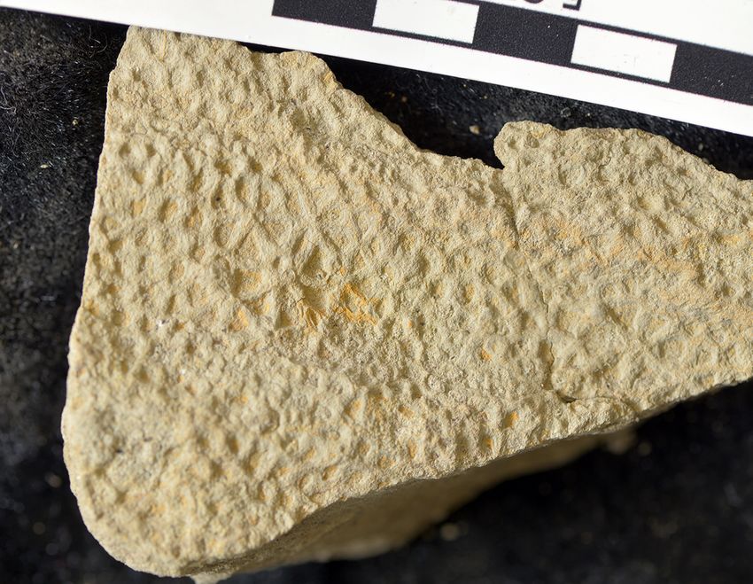
Project 22: Determinate growth and tooth replacement in the basal
mammaliaform Morganucodon                                                        37
Projects 23 and 24: How does the size of foraminifera respond to Milankovitch
Cycles?                                                                       38
Project 25: Morphological variation in response to changing ecology              39
Project 26: Biotic response to a breathless ocean                                40
Project 27: Southern Ocean diatoms and climate change: quantifying the relative
roles of diversity and plasticity in evolution                                42
Project 28: Feather taphonomy in terrestrial/fluvial settings                    44
Project 29: On the nature of rosettes in hadrosaur skin: sensory bristles or glands?
                                                                                  46
Project 30: Pathways to exceptional fossil preservation—the role of polymerization
                                                                                48
Project 31: Vulcanisation—does sulphur make more than just rubber?               49

Additional specialist projects:
Project 32: The impact of environmental stress on isotopic fractionation by marine
diatoms                                                                         50
Project 33: Changing Arctic Ocean microfossils: Radiocarbon ages of sedimentary
carbonate from the Barents Sea                                               52
Project 34: The evolution of eye regulatory control                              54

                                         3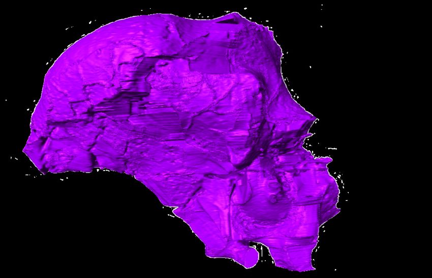
Project 1: Ecomorphology, convergence and disparity in aquatic tetrapods

Supervisors: Tom Stubbs, Susana Gutarra Diaz and Mike Benton

Many tetrapods have adapted to living in aquatic ecosystems, including cetaceans,
pinnipeds, sirenians, turtles, crocodiles and birds, and extinct marine reptiles such as
ichthyosaurs, mosasaurs and sauropterygians (Kelley and Pyenson 2015).
Understanding patterns and processes of morphological divergence and convergence is
a key objective for palaeontologists and evolutionary biologists. When organisms
explore novel ecospace, this can drive evolution, but shared environmental and
ecological pressures can give rise to the same adaptive traits (Stayton 2015). Although
the shared morphologies of many aquatic tetrapods are textbook examples of
evolutionary convergence, tests for convergence are rare.

Previous research has illustrated how
morphospaces can be effective tools for
examining ecomorphological trends.
Gingerich (2003) used postcranial
measurements to construct a morphospace
for aquatic mammals and explore the degree
of terrestrial versus aquatic specialization in
early whales. This study provided a
framework to explore locomotory trends in
fossil pinnipeds (Bebej, 2009).
Kelley et al. (2015) illustrated widespread
convergence in morphology linked to
feeding ecology in marine tetrapods and incorporated a phylogenetic framework.

Comparable studies of postcranial anatomy, with links to locomotory drivers, using
state-of-the-art methods are absent. There are also many unanswered questions about
comparative trends amongst aquatic tetrapods, do some groups have greater
morphospace occupation, are there clear
phylogenetic constraints, and do some groups
have faster evolutionary rates?

In this project, the student will explore skeletal
morphospace in modern aquatic tetrapods to test
trends of morphological divergence and convergence. The student will examine
specimens, and then assemble a database of key measurements from the postcranial
skeleton that have links to overall body plans and locomotory functions. Using this
database, the student will perform a series of computational analyses to generate
morphospaces, calculate disparity statistics to compare clades, and examine
convergence and evolutionary rates using comparative phylogenetic methods.

The project will provide broad training in vertebrate comparative anatomy and
numerical palaeobiology, including morphometrics and evolutionary modelling. The
student will have the opportunity to visit museums to examine specimens.

                                            4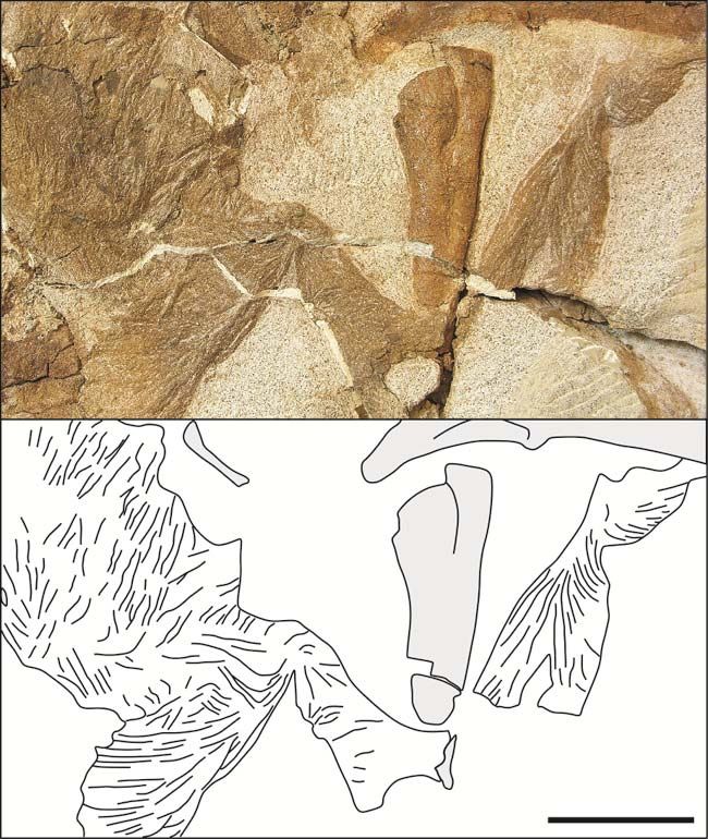
References
Bebej RM. 2009 Swimming mode inferred from skeletal proportions in the fossil
  pinnipeds Enaliarctos and Allodesmus (Mammalia, Carnivora). Journal of
  Mammalian Evolution 16: 77–97
Gingerich, P. D. 2003. Land-to-sea transition in early whales: evolution of Eocene
   Archaeoceti (Cetacea) in relation to skeletal proportions and locomotion of living
   semiaquatic mammals. Paleobiology 29: 429-454.
Kelley, N.P., & Pyenson, N. D. 2015. Evolutionary innovation and ecology in marine
   tetrapods from the Triassic to the Anthropocene. Science 348, aaa3716.
Kelley, N.P., & Motani, R. 2015. Trophic convergence drives morphological
   convergence in marine tetrapods. Biology Letters 11: 20140709.
Stayton, C.T. 2015. The definition, recognition, and interpretation of convergent
   evolution, and two new measures for quantifying and assessing the significance of
   convergence. Evolution 69: 2140-2153.

                                           5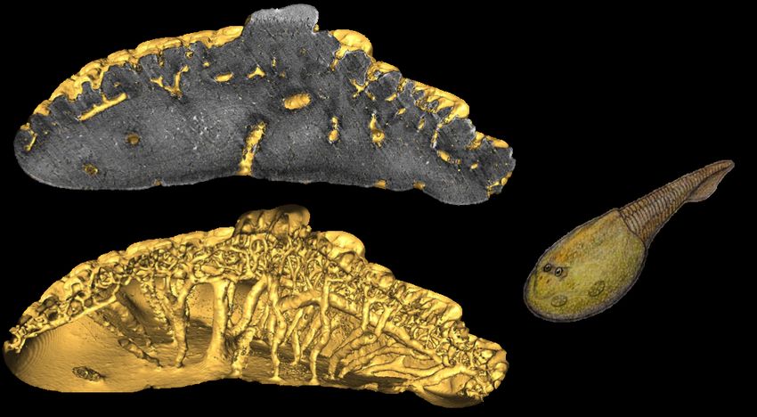
Project 2: Ontogenetic brain development in birds and crocodilians with
applications to non-avian dinosaurs

Supervisors: Logan King, Mike Benton, Emily Rayfield

Advancements in CT scanning and continued 3D discoveries of non-avian dinosaur
skulls from deposits in China and the United States have allowed for an unprecedented
look into the ontogenetic development of ceratopsian endocasts. However, the rate of
progression and innovation in palaeontology has, to a certain extent, outpaced our
understanding of model modern taxa. For instance, a shape analysis of the brains in
alligators and ostriches—common outgroups used to compare changes in dinosaurs—
has yet to be completed. Palaeoneurology is currently experiencing almost exponential
growth but still requires further research with modern taxa to make a baseline to
measure fossil endocasts against.

This project will build endocasts from ostriches (Struthio camelus) and alligators
(Alligator mississippiensis), measure the changes in flexure points in both genera, and
analyse them both via a 2D geometric morphometric generate two datasets that can be
compared to two ceratopsian dinosaurs, Psittacosaurus lujiatunensis and Triceratops
horridus.

The alligator endocasts to be used for this project have been premade and will be ready
to use for shape analysis; however, the ostrich CT data will need to be made into 3D
models before being analysed further. The student undertaking this project will utilize
Avizo and R (or TPS) to measure flexure angles and shape change along the x- and y-
axis, respectively. The end goals of the project seek to clarify four main points:

1) How does the overall shape of the Struthio camelus/Alligator mississippiensis
endocast vary throughout ontogeny?
2) How do cephalic and pontine flexure points significantly differ between the youngest
stage and adults?
3) When, ontogenetically speaking, do ostriches and alligators experience the most
endocranial change?
4) Are any trends in shape/flexure change noted in the modern taxa mirror any in non-
avian dinosaurs from recent literature?

This project is best suited for a student with an interest in 3D imaging and how it
benefits either dinosaur palaeobiology or endocranial development. The work will be
quantitative in nature and be intensive in statistical and morphometric analyses. At its
core, this project will identify trends in brain development that can be applied to an
ontogenetic series of non-avian dinosaurs. Future uses of this project will include
ceratopsian dinosaurs as well as modern crocodilians.

                                           6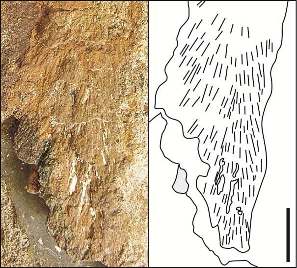
Figure 1: Examples of shape change that occurs between a 10-month-old (lower left)
and 2-year-old (upper right) Psittacosaurus lujiatunensis. The amount of shape change
needs to be cross referenced with modern taxa to understand if the developmental form
and rate of dinosaur endocasts are faster, slower, or unique. Bar = 1cm.

Recommended reading
Jirak, D., and J. Janacek. 2017. Volume of the crocodilian brain and endocast during
  ontogeny. PLOS ONE, 12: e0178491
Kawabe, S., S. Matsuda, N. Tsunekawa, and H. Endo. 2015. Ontogenetic Shape Change
  in the Chicken Brain: Implications for Paleontology. PLOS ONE, 10: e0129939
Racicot, R. 2016. Fossil Secrets Revealed: X-ray CT Scanning Applications in
  Paleontology. The Paleontological Society Papers, 22: 21–38.

                                         7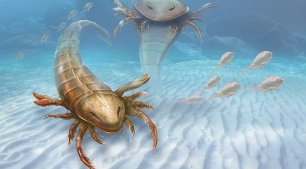
Project 3: External anatomy of Megalodon

Supervisors: Catalina Pimiento and Mike Benton

It has been long assumed that Megalodon (the biggest shark that has ever lived) was
simply a larger (and fatter) version of the great white shark. However, it has been
evidenced that the great white shark did not derive directly from Megalodon. This
suggests that these two species could have been anatomically, very different. This
project aims to infer the potential external anatomy of Megalodon by extrapolating
anatomical measurements from living species.

The student will collect anatomical measurements (e.g. tooth size, size of the head and
fins, total length) and morphological proportions (e.g. distance from head to the dorsal
fin, from the dorsal fin to the anal fin, etc.) from phylogenetically related and
ecologically similar species to Megalodon (e.g. great white shark, tiger shark, mako
shark, bull shark). This data will be collected from online platforms (e.g. Fishbase) and
from the literature (e.g. from main compendia like the FAO species catalogue and
scientific papers found through the Web of Science, GeoRef, Google Scholar and
Shark-References). This data will be then analysed to infer the potential relationships
between of Megalodon's body parts, which will be then scaled up to Megalodon's body
based on known dimensions (e.g. total length, tooth size, vertebral centra diameter). Dr
Pimiento will provide the student with 3D and CT scans of Megalodon’s teeth and
vertebral column that can be used as reference, as well as a large dataset of teeth
photographs from museums around the world.

Recommended reading
Gottfried MD, Compagno LJV, Bowman SC (1996) Size and skeletal anatomy of the
  giant megatooth shark Carcharodon megalodon. In: Klimley AP, Ainley DG, editors.
  Great white sharks: the biology of Carcharodon carcharias. San Diego: Academic
  Press. pp. 55–89.
Mollet, H. F., & Cailliet, G. M. (1996). Using allometry to predict body mass from
  linear measurements of the white shark. Great White Sharks: The Biology of, 81-89.
Pimiento, C., and Balk, M. (2015). Body size trends of the extinct giant
  shark Carcharocles megalodon: A deep-time perspective on marine apex predators.
  Paleobiology 41: 479-490.
Shimada, K. 2002. The relationship between the tooth size and total body length in the
  white shark. Journal of Fossil Research 35: 28-33.
Pimiento, C., Ehret, D.J., MacFadden, B.J., and Hubbell, G. 2010. Ancient nursery area
  for the extinct giant shark Megalodon from the Miocene of Panama. Plos One 5:
  e10552.

                                            8
Project 4: Morphological diversity of Strawberry Bank crocodiles

Supervisors: Benjamin Moon, Antonio Ballell, Mark Young (Edinburgh), Steve
Brusatte (Edinburgh), Matt Williams (BRLSI), Neil Gostling (Southampton), Mike
Benton, Emily Rayfield

The Toarcian Strawberry Bank lagerstätte preserves a large number of marine tetrapods
in a ‘nursery lagoon’ environment, including several thalattosuchian crocodiles referred
to the teleosaurid taxon Pelagosaurus typus (Pierce and Benton 2006; Caine and Benton
2011). These crocodiles have the specialised longirostry characteristic of piscivores,
however their ecological interactions with other marine tetrapods in Strawberry Bank
and broader Toarcian seas has been understudied (Benton and Taylor 1984; Godefroit
1994; Stubbs et al. 2013). So too with the morphological and functional changes that
occur during the growth of taxa, despite the presence of several ontogenetic stages.
Having several fully three-dimensional, articulated specimens presents an opportunity
to assess the morphological and ecological diversity of this restricted environment
(Williams, Benton, and Ross 2015). This project will ask the following questions:

1. How does the cranial morphology of Pelagosaurus typus change through
ontogeny? what effect does this have on sensory and ecological inferences?
2. Does the specimen BRLSI M1420 represent Pelagosaurus typus? how does this
specimen differ morphologically and functionally from the other Strawberry Bank
crocodiles?

The preservation of these specimens lends itself to the use of µCT scans. Segmented CT
scan data sets of three specimens of Pelagosaurus typus from three ontogenetic stages
will be used – an infant (BRLSI M1418 scanned at Southampton by NG), juvenile
(BRLSI M1413), and an adult (NHM specimen from SB and MY) – to form a sequence
from which manifest changes in skull, endocast, and functional morphology will be
measured. Modification of the morphology during ontogeny will be assessed using a
combination of anatomical observation – e.g. endocranium, jaw morphology – classic
morphometric regression, and 3D landmark geometric morphometrics.

An additional juvenile specimen – BRLSI M1420 – has been captured but not
segmented. The student will segment this data set, then use detailed anatomical
observation and comparisons to other Thalattosuchia from Strawberry Bank and in
museum collections (e.g. Johnson et al. 2015; Pierce et al. 2017) to assess whether this
represents a distinct taxon. This specimen may also be compared with the above to
further assess the diversity of form and function present in the Strawberry Bank
thalattosuchians.

The student will be given training in capturing and segmenting µCT data sets and
visualisation of results using Avizo; anatomical observation and description from hand
specimens and µCT data sets; geometric morphometric analysis using R package
geomorph; statistical analyses in R. This research is expected to lead to at least one
publication, including helping to name a new taxon if BRLSI M1420 is determined to
distinct.

                                            9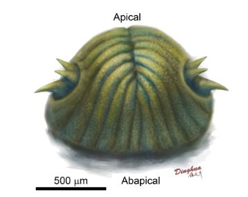
References
Benton, M. J., and Taylor, M. A. 1984. “Marine Reptiles from the Upper Lias (Lower
  Toarcian, Lower Jurassic) of the Yorkshire Coast.” Proceedings of the Yorkshire
  Geological Society 44 (4): 399–429.
Caine, H., and Benton, M. J. 2011. “Ichthyosauria from the Upper Lias of Strawberry
  Bank, England.” Palaeontology 54 (September): 1069–93.
  https://doi.org/10.1111/j.1475-4983.2011.01093.x.
Godefroit, P. 1994. “Les Reptiles Marins Du Toarcien (Jurassique Inférieur) Belgo-
  Luxembourgeois.” Mémoires Pour Servir à L’Explication Des Cartes Géologiques et
  Minières de La Belgique 39 (September): 1–98.
Johnson, M. M,, Young, M. T., Steel, L. and LePage, Y. 2015. “Steneosaurus Edwardsi
  (Thalattosuchia: Teleosauridae), the Largest Known Crocodylomorph of the Middle
  Jurassic.” Biological Journal of the Linnean Society 115 (4): 911–18.
  https://doi.org/10.1111/bij.12525.
Pierce, S. E., and Benton, M. J. 2006. “Pelagosaurus Typus Bronn, 1841
  (Mesoeucrocodylia: Thalattosuchia) from the Upper Lias (Toarcian, Lower Jurassic)
  of Somerset, England.” Journal of Vertebrate Paleontology 26 (3): 621–35.
  https://doi.org/10.1671/0272-4634(2006)26[621:PTBMTF]2.0.CO;2.
Pierce, S. E., Williams, M. and Benson, R. B. J. 2017. “Virtual Reconstruction of the
  Endocranial Anatomy of the Early Jurassic Marine Crocodylomorph Pelagosaurus
  Typus (Thalattosuchia).” PeerJ 5 (April): e3225. https://doi.org/10.7717/peerj.3225.
Stubbs, T. L., Pierce, S. E., Rayfield, E. J. and Anderson, P. S. L. 2013. “Morphological
  and Biomechanical Disparity of Crocodile-Line Archosaurs Following the End-
  Triassic Extinction.” Proceedings of the Royal Society B: Biological Sciences 280:
  20131940. https://doi.org/10.1098/rspb.2013.1940.
Williams, M., Benton, M. J. and Ross, A. 2015. “The Strawberry Bank Lagerstätte
  Reveals Insights into Early Jurassic Life.” Journal of the Geological Society, London
  172 (October): 683–92. https://doi.org/10.1144/jgs2014-144.

                                           10
Project 5: The Triassic fauna of the Ruthin fissure
Supervisors: Mike Benton and David Whiteside
There have been detailed studies of the Late Triassic and Early Jurassic faunas of the
fissure deposits of South Wales and near Bristol. These fissures have yielded globally
important terrestrial reptiles such as Thecodontosaurus, Kuehneosaurus, Clevosaurus,
Planocephalosaurus, Diphydontosaurus and Gephyrosaurus. Bristol students have
recently described new species of sphenodontians such as Clevosaurus sectumsemper
and C. cambrica. Many of the fissure deposit faunas including Cromhall, Durdham
Down, Tytherington, Pant-y-ffynnon and Holwell are documented in detail but the
fauna of Ruthin Quarry in South Wales is much less well known. That is despite Ruthin
being the type locality for the poorly known genus Tricuspisaurus.
Ruthin offers an opportunity to describe a range of reptiles including archosauromorphs,
lepidosaurs and procolophonids. There are many undescribed specimens from Ruthin in
museum collections in the National Museum of Wales and in the Natural History
Museum, London. Further, the Ruthin fissure can still be visited as it is accessible from
a public road, so the sedimentological features can be documented.
The study will investigate and document the fauna of the Ruthin Quarry Triassic fissure,
comparing it with other fissure localities, and applying appropriate computational
methods in ecology. There is also an opportunity to compare the sedimentological
settings associated with the tetrapod-bearing rocks to those from Tytherington,
Woodleaze and Cromhall. The student will compile species descriptions and provide a
quantitative analysis of the fauna. There is the opportunity to describe hand specimens
collected previously and in the field. It will then be possible to establish whether there
are distinctive sedimentological conditions related to individual or groups of localities.
The project involves detailed descriptions and analysis of the tetrapod fauna using
optical microscopy, acid preparation and possibly CT scanning, as well as appropriate
ecological analysis in R. The student will also describe the geomorphology of the
fissures, the nature of the sedimentary infills and have access to thin-cut microscope
slides and hand specimens. In addition, there is the possibility of using the XRD and/or
electron microscope probes and fieldwork. Training in identification and analytical
methods will be provided.
Recommended reading:
Whiteside, D. I. & Marshall, J. E. A. 2008. The age, fauna and palaeoenvironment of
the Late Triassic fissure deposits of Tytherington, South Gloucestershire, UK.
Geological Magazine 145: 105-147.
Whiteside DI, Duffin CJ, Gill PG, Marshall JEA, Benton MJ. 2016. The Late Triassic
and Early Jurassic fissure faunas from Bristol and South Wales: stratigraphy and
setting. Palaeontologia Polonica 67: 257–287.
Whiteside D.I., Duffin C.J, 2017. Late Triassic terrestrial microvertebrates from
Charles Moore’s ‘Microlestes’ quarry, Holwell, Somerset, U.K. Zoological Journal
of the Linnean Society 179: 677-705.
Wings, O. 2004. Authigenic minerals in fossil bones from the Mesozoic of England:
poor correlation with depositional environments Palaeogeography,
Palaeoclimatology, Palaeoecology 204: 15-32.
11Project 6: The Eocene-Oligocene Transition and American Larger Benthic
foraminifera
Supervisor: Laura Cotton
The Eocene–Oligocene transition (EOT) was a time of profound climatic and
oceanographic change associated with the first major continental-scale glaciation of
Antarctica (see Coxall and Pearson, 2007, for review). This environmental disruption
led to a global peak in biotic turnover, seen in both terrestrial and marine records and in
shallow water and deep-sea environments. This includes the global extinction of a
number of long-ranging and widespread larger benthic foraminifera (LBF), however the
mechanism for this extinction remain unclear (Cotton and Pearson, 2011). This LBF
event has been well studied in Europe, East Africa and Indonesia, but exceptionally
little work has been carried out recently in the Americas.
Shallow-marine carbonate deposits are well-known from the Eocene of the US Gulf
Coast and Caribbean (Frost and Langenheim, 1974; Robinson, 1993). These deposits
frequently contain abundant larger benthic foraminifera (LBF). The American LBF
assemblages, however, are distinctly different to those of Europe and the Indo-Pacific
(Adams, 1983; BouDagher- Fadel, 2008). Within the Eocene, they lack the huge
diversity of Nummulites seen elsewhere (e.g. Schaub, 1981), instead assemblages are
often dominated by lepidocyclinids, which do not occur in the rest of the world until at
least the upper part of the lower Oligocene (Serra-Kiel et al., 1998; BouDagher-Fadel,
2008). It is therefore essential that the American LBF bio-province is included instudies
of LBF evolution, migration and biodiversity, to understand these processes on a global
scale. However, this is currently not the case.
This project therefore involves the study of consists of two field sections from North
Florida spanning the Upper Eocene to Lower Oligocene, and including abundant larger
benthic foraminifera. The student will learn preparation and identification of a number
of larger benthic foraminiferal taxa, as both loose specimens and in petrological thin
section, and independent dating techniques. Larger benthic foraminiferal ranges and
biodiversity will be analysed and tied to global stratigraphy, allowing comparisons with
global sites.
References
Adams, C. G. 1967. Tertiary foraminifera in the Tethyan, American and Indo-Pacific
provinces. Aspects of Tethyan biogeography 7: 195–217.
BouDagher-Fadel, M. K. 2008. Evolution and geological significance of larger benthic
foraminifera, Elsevier, Amsterdam, the Netherlands.
Cotton, L. J. & Pearson, P. N. 2011. Extinction of larger benthic foraminifera at the
Eocene/Oligocene boundary. Palaeogeography, Palaeoclimatology, Palaeoecology
311: 281296.
Frost, S. and Langenheim, R.: Cenozoic reef biofacies, Tertiary larger foraminifera and
scleractinian corals from Chiapas, Mexico, Northern Illinois University Press,
DeKalb, IL, USA, 1974.
12Schaub, H. 1981. Nummulites et Assilines de la Tethys Paleogene. Taxonomie,
phylogenese et biostratigraphie. Schweizerische Palaeontologische Abhandlungen
104106: 1236.
Serra-Kiel, J., Hottinger, L., Caus, E., Drobne, K., Ferrandez, C.,Jauhri, A. K., Less, G.,
Pavlovec, R., Pignatti, J., Samso, J. M.,Schaub, H., Sirel, E., Strougo, A., Tambareau,
Y., Tosquella, J., and Zakrevskaya, E. 1998. Larger foraminiferal biostratigraphy of
theTethyan Paleocene and Eocene, B. Soc. Geol. Fr. 169: 281–299.
Robinson, E. 2003. Zoning the White Limestone Group of Jamaica using larger
foraminiferal genera: a review and proposal. Cainozoic Research 3: 39–75.
13Project 7: Morphological variation in reticulate Nummulites across the Eocene-
Oligocene Transition
Supervisor: Laura Cotton
The reticulate Nummulites are a widespread and abundant group of larger benthic
foraminifera (LBF) found throughout Europe, Asia and Africa, and are extensively used
within Eocene biostratigraphy. Models of their evolution have previously been very
simplistic, suggesting a single lineage with increasing proloculus size through time
(e.g. Schaub 1981; Less & Ozcan 2012). However, recent and on-going work has
shown their evolution to be far more complex with several lineages and potential
migration events, and morphological change linked to climatic events (Cotton and
Pearson, 2011; Cotton et al., 2015).
The Eocene-Oligocene transition (EOT) is one of the most dramatic climate shifts of the
Cenozoic, associated with widespread cooling and biological overturning, including
extinctions within the LBF (see Coxall and Pearson, 2007). The reticulate Nummulites
continue through this event, but a continuous high resolution record from Tanzania
shows changes in their morphology, coincide with the EOT (Cotton and Pearson, 2011).
Traditionally larger benthic foraminifera are studied in oriented thin sections, this is a
destructive process where only the equatorial slice is preserved and measured, meaning
a large amount of data is lost (e.g. see Renema and Cotton, 2015 for comparison of 3D
and 2D morphologies). Micro-CT allows for comparison of the entire foraminiferal test,
however, unlike thin sections there are no standard measurements used for this.
This project aims to examine and define the morphological change across the EOT
using both thin sections and 3D micro-CT scans. Students will learn micro-CT
techniques, preparation of foraminiferal specimens and application of multivariate
statistical clustering methods following Pearson and Ezard (2014).
References
Cotton, L. J. & Pearson, P. N. 2011. Extinction of larger benthic foraminifera at the
Eocene/Oligocene boundary. Palaeogeography, Palaeoclimatology, Palaeoecology 311:
281296.
Cotton, L. J., Pearson, P. N., Renema, W., 2015. A place for Nummulites ptukhiani? A new
lineage of reticulate Nummulites from Kilwa District, Tanzania. Journal of Systematic
Palaeontology http://dx.doi.org/10.1080/14772019.2015.1079562.
Coxall, H.K., Pearson, P.N., 2007. The Eocene–Oligocene transition. In: Williams, M.,
Haywood, A.M., Gregory, F.J., Schmidt, D.N. (Eds.), Deep-time Perspectives on Climate
Change: Marrying the Signal from Computer Models and Biological Proxies: The
Micropalaeontological Society, Special publications, London, pp. 351–387.
Less, G. & Ozcan, E. 2012. Bartonian Priabonian larger benthic foraminiferal events in the
Western Tethys. Austrian Journal of Earth Sciences, 105, 129140.
Pearson, P.N. and Ezard, T.H., 2014. Evolution and speciation in the Eocene planktonic
foraminifer Turborotalia. Paleobiology 40: 130-143.
Schaub, H. 1981. Nummulites et Assilines de la Tethys Paleogene. Taxonomie, phylogenese et
biostratigraphie. Schweizerische Palaeontologische Abhandlungen 104106: 1236.
14Project 8: Impact of oil spills on benthic foraminiferal growth and development
Supervisors: Laura Cotton and Patrick Schwing (University of South Florida)
Benthic foraminifera, which are single celled protists that primarily produce calcite
shells, have been commonly used as bioindicators of anthropogenic and natural
perturbations (Sen Gupta 1999; Morvan et al. 2004; Mojtahid et al. 2006; Nigam et al.
2006; Denoyelle et al. 2010; Brunner et al. 2013, Lei et al. 2015). Within the Gulf of
Mexico numerous surveys have been conducted prior to any major oil spill, which has
allowed for detailed studies of benthic foraminiferal response to the Deepwater Horizon
(DWH) in 2010 (Poag 2015 and references therein).
Using a series of short core records taken by the University of South Florida, Schwing
et al (2015; 2017) showed an 80-93% decrease in benthic foraminiferal abundance and a
30-40% decrease in benthic foraminiferal species richness and heterogeneity in the
northern Gulf of Mexico. However, the majority of these records were deep water, and
included only two relatively shallow water sites from 400-500m water depth.
This project therefore aims to increase the spatial coverage of the shallow water sites
with a third site close to the Mississippi outflow, and provide data on whether growth
and development of benthic foraminifera was affected by the DWH. Taxa such as
Uvigerina spp., Cibicidoides spp. and Bolivina spp., which are known to continue
through the oil spill, as well as examples of those that show local disappearances, will
be examined from 0 - 50 mm in five short cores, taken in the north of the Gulf of
Mexico ~85 km from the DWH wellhead from 2010 to 2017. The student will learn
sample preparation - washing and picking foraminifera and identification of a number
of benthic foraminiferal taxa. The specimens will then be measured and data collected
on their morphology (e.g. number of chambers) and whether abnormalities occur.
Statistical analysis will then be carried out to examine if the oil spill affected their
growth. In addition to this, representative specimens from various levels will be micro
CT-scanned and segmented using Avizo to test for three-dimensional growth variations.
References
Brunner CA, Yeager KM, Hatch R, Simpson S, Keim J, Briggs KB, Louchouarn P (2013).
Effects of Oil from the 2010 Macondo Well Blowout on Marsh Foraminifera of Mississippi
and Louisiana, USA. Environ. Sci. Technol. 47: 9115−9123. dx.doi.org/10.1021/es401943y
Denoyelle M, Jorissen FJ, Martin D, Galgani F, Mine J (2010). Comparison of benthic
foraminifera and macrofaunal indicators of the impact of oil-based drill mud
disposal. Marine Pollution Bulletin 60: 11:2007-
2021. dx.doi.org/10.1016/j.marpolbul.2010.07.024
Lei YL, Li TG, Bi H, Cui WL, Song WP, Li JY, Li CC (2015). Responses of benthic
foraminifera to the 2011 oil spill in the Bohai Sea, PR China. Marine Pollution Bulletin 96:
245–60. http://doi.org/10.1016/j.marpolbul.2015.05.020
Mojtahid M, Jorissen F, Durrieu J, Galgani F, Howa H, Redois F, Camps R (2006). Benthic
foraminifera as bio-indicators of drill cutting disposal in tropical east Atlantic outer shelf
environments. Marine Micropaleontology 61:58-75.
Morvan J, Le Cadre V, Jorissen FJ, Debenay JP (2004). Foraminifera as potential bio-indicators
of the “Erika” oil spill in the Bay of Bourgneuf: field and experimental studies. Aquatic
Living Resources 17: 317-322.
15Nigam R, Saraswat R, Panchang R (2006) Application of foraminifers in ecotox- icology:
retrospect, perspect and prospect. Environ. Int. 32: 273–283.
Poag WC (2015). Benthic Foraminifera of the Gulf of Mexico: Distribution, Ecology,
Paleoecology. Texas A&M University Press, College Station.
Schwing PT, Romero IC, Brooks GR, Hastings DW, Larson RA, Hollander DJ (2015). A
Decline in Deep-Sea Benthic Foraminifera Following the Deepwater Horizon Event in the
Northeastern Gulf of Mexico. PLOSone 10(3): e0120565. doi:10.1371/journal.pone.0120565
Schwing PT, O’Malley BJ, Romero IC, Martinez-Colon M, Hastings DW, Glabach MA,
Hladky EM, Greco A, Hollander DJ (2017). Characterizing the variability of benthic
foraminifera in the northeastern Gulf of Mexico following the Deepwater Horizon event
(2010-2012).
Sen Gupta BK (ed) (1999) Modern Foraminifera. Kluwer Academic Publishers, Great Britain.
16Project 9: The preservation potential of cell organelles and the implications for the
early eukaryote fossil record
Supervisors: John Cunningham and Phil Donoghue
A fossil record of cell organelles would yield important information that could allow
unequivocal identification of fossil eukaryotes and might help constrain the timing of
early eukaryote evolution. However, reports of fossilized organelles have been
controversial, particularly when they originate from Precambrian rocks. In the 1970s
experiments showed that collapse of the cell during the decay of prokaryotic organisms
can produce structures that resemble nuclei and have been termed ‘pseudonuclei (Knoll
and Barghoorn 1975). Since then many palaeontologists have considered that most, if
not all, reports of fossil organelles fail to reject the null hypothesis that they can be
interpreted as pseudonuclei.
In recent years, however, there have been a number of more convincing reports of fossil
nuclei that have generally been considered too regular to be interpreted as pseudonuclei
Bomfleur et al. 2014). Yet some authors (e.g. Xiao et al. 2012, Pang et al. 2013) remain
skeptical of these claims for three main reasons: (1) nuclei and other organelles are
expected to decay very rapidly; (2) nuclei and other organelles have never been
fossilized in laboratory experiments; and (3) if nuclei are preserved then one might
expect other organelles, thought to have higher preservation potential, to be preserved
alongside them. However, there is in fact very little evidence regarding the preservation
potential of organelles or the relative preservation potential of different organelles.
This project aims to address this by taking an experimental taphonomy approach to
examine the decay of organelles under controlled conditions in the laboratory. The
student will build on successful trials from a previous MSc project by combining this
approach with the use selective stains to identify organelles such as nuclei,
mitochondria and chloroplasts and track their decay over time. This will allow
important questions to be answered, such as whether these structures survive decay for a
time scale compatible with fossilization and which organelles have the highest relative
preservation potential. The findings will then be used to help interpret fossil structures,
such as the potential nuclei from the literature, which will be studied using Synchrotron
Radiation X-ray Tomographic Microscopy (SRXTM). The student will gain valuable
skills in experimental taphonomy, microscopy and 3-D analysis of fossils.
References:
Bomfleur B., McLoughlin S., Vajda V. 2014 Fossilized Nuclei and Chromosomes
Reveal 180 Million Years of Genomic Stasis in Royal Ferns. Science 343: 1376-1377.
Huldtgren, T. et al. 2011 Fossilized nuclei and germination structures identify Ediacaran
'animal embryos' as encysting protists. Science 334: 1696-1699.
Knoll A., Barghoorn E.S. 1975 Precambrian eukaryotic organisms: A reassessment of
the evidence. Science 190: 52-54.
Pang K. et al. 2013 The nature and origin of nucleus-like intracellular inclusions in
Paleoproterozoic eukaryote microfossils. Geobiology 11: 499-510.
Xiao, S. et al. 2012. Comment on “Fossilized nuclei and germination structures identify
Ediacaran ‘animal embryos’ as encysting protists”. Science, 335.
17Project 10: The affinities of an exceptionally preserved larva from the early
Cambrian
Supervisors: John Cunningham, Phil Donoghue and Michael Steiner (Frei
University, Berlin)
Invertebrate larvae tend to have an incredibly poor fossil record as they tend to decay
and disintegrate very rapidly after death (Gostling et al. 2009). Eolarva is a rare
exception that is preserved in exquisite detail in the early Cambrian Kuanchuanpu Biota
from South China (ca. 535 Ma). It is the oldest larva in the fossil record and has the
potential to provide vital information on the developmental evolution of early animals.
However, this potential can only be realised if the phylogenetic affinities of Eolarva can
be resolved, and this is the goal of the project.
Eolarva has been interpreted as cnidarian-grade
animal based on its overall external anatomy
(Zhang and Dong 2015). However, its internal
anatomy remains unknown and it has not been
subject to a rigorous phylogenetic analysis. The
student will firstly analyse tomographic data
gathered previously by the supervisors using
Synchrotron Radiation X-ray Tomographic
Microscopy (SRXTM) in order to resolve the
details of the internal anatomy. Next they will
make a detailed anatomical description of the
fossil specimens using SRXTM and Scanning
Electron Microscopy (SEM) data. Finally they
will carry out a phylogenetic analysis by
incorporating the new anatomical information
into an existing matrix (Duan et al. 2017).
This will allow the student to assess the
significance of Eolarva for understanding the
developmental evolution of early animals and
its role in the Cambrian radiation. The student
will receive full training in 3D reconstruction
and analysis, interpretation of exceptionally
preserved fossils and relevant phylogenetic methods. If completed successfully the
project should lead to a high profile publication.
References:
Duan, B., Dong, X. P., Porras, L., Vargas, K., Cunningham, J. A., and Donoghue, P. C.
J., 2017. The early Cambrian fossil embryo Pseudooides is a direct-developing
cnidarian, not an early ecdysozoan. Proc Roy Soc B 284: 20172188.
Gostling, N.J., Dong, X.P. & Donoghue, P.C.J. 2009. Ontogeny and taphonomy: an
experimental taphonomy study of the development of the brine shrimp Artemia salina.
Palaeontology 52: 169-186.
Zhang, H. and Dong, X. P. 2015. The oldest known larva and its implications for the
plesiomorphy of metazoan development. Science Bulletin 60: 1947-1953.
18Project 11: Biomechanics of the earliest vertebrate skeleton
Supervisors: Philip Donoghue, Emily Rayfield, Emma Randle (University of
Manchester)
Sharks and boney fishes are among the most primitive living vertebrates with a
mineralised skeleton and, consequently, they have strongly influenced hypotheses on
the evolution of the vertebrate skeleton, invariably envisaged to have emerged
primitively in a shark-like condition of cartilaginous endoskeletons and an external
dermal skeleton comprised of microscopic tooth-like scales. Nothing could be further
from the truth. The earliest skeletonizing vertebrates evolved much earlier, in meek
jawless deposit feeding fish (Keating et al. 2015). Indeed, it has been argued that the
thick dermal armour that these ‘ostracoderms’ exhibit, is an adaptation to ward off the
unrequited attentions of the top predators of the day, the eurypterids (Romer 1933).
However, while there has been some conjecture on the force that eurypterid claws could
exhibit, there has been no research into the biomechanical properties of body armour
possessed by the earliest skeletonising vertebrates, the heterostracans. This project aims
to test the hypothesis that the exoskeleton of the earliest vertebrates was an adaptation
to predation through analysing the functional performance of simplified models of early
vertebrate body armour using Finite Elements Analysis (FEA).
Heterostracans exhibit diversity of architectural styles to the gross histology of their
body armour, within which there are end members of (a) an anastomosing structure
dominated by coarse calibre canals, and (b) a vaulted honeycomb-like structure; both
architectures have evolved multiple times in heterostracans and their relatives (Keating
et al. 2015), and may relate to constraints imposed by scaling of vastly different sized
organisms (from tens of centimetres to many metres). Exploiting existing high
resolution synchrotron-based tomographic scans of representatives of these armour
types you will create simple models characterising their structure. The relative
performance of these models will be evaluated and compared to one another and
relative to scaling, under biologically realistic loads informed by existing work on
19arthropod limbs (Bicknell et al. 2018). For a successful and time-efficient student, there
is scope within the project to evaluate the skeleton of other early vertebrates, allowing
the student to generalise their conclusions and broaden their relevance and interest.
Training
The student will receive specialist training in histology, computed tomography, meshing
and finite element analysis. This skill set is recognised by morphologists and
palaeontologists alike and can be used to study the evolution of anatomy in both living
and fossil taxa. There will be an opportunity for the student to attend the Swiss
Synchrotron Light Source to collect their own data, if they can secure funds for travel
and subsistence – through the research project funding competition open to all MSc
Palaeobiology students.
References
Bicknell, R.D.C., Ledogar, J.A., Wroe, S., Gutzler, B.C., Watson, W.H., 3rd &
Paterson, J.R. 2018. Computational biomechanical analyses demonstrate similar shell-
crushing abilities in modern and ancient arthropods. Proc Biol Sci, 285, doi:
10.1098/rspb.2018.1935.
Keating, J.N., Marquart, C.L. & Donoghue, P.C.J. 2015. Histology of the heterostracan
dermal skeleton: Insight into the origin of the vertebrate mineralised skeleton. Journal
of Morphology, 276, 657–680, doi: 10.1002/jmor.20370.
Romer, A.S. 1933. Eurypterid influence on vertebrate history. Science, 78, 114-117.
20Project 12: Experimental taphonomy of bivalved crustaceans
Supervisors: Philip Donoghue, John Cunningham and David Horne (Queen Mary)
The known fossil record of early crustacean arthropods is dominated by microscopic,
perhaps meiofaunal, species that have a bivalved carapace, resembling (but not
representing) the living ostracods. They are surprisingly commonly preserved in three
dimensions as part of the global Orsten Fauna (Maas et al. 2006). While the Orsten
fauna is largely a Cambrian – early Ordovician phenomenon, this style of exceptional
preservation through calcium phosphate replication of cuticle (and of internal organs in
some instances) occurs in younger Lagerstatte including the Late Cretaceous Santana
Lagerstatten of Brasil where true ostracods are exceptionally preserved associated with
the guts and the environment around the carcases of exceptionally preserved fish (Smith
2000). While Orsten fossils have been described anatomically in exquisite detail,
surprisingly, there have been no explicit attempts to understand their preservation, either
from analysis of the fossils or through experimental taphonomy of living analogues.
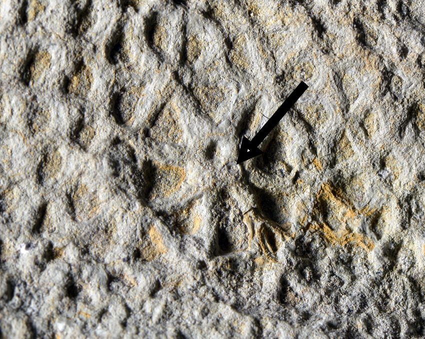
This project aims to overcome these shortcomings through (a)
analysis of the preservation of exceptionally preserved
phosphatocopids from the late Cambrian Wangcun
Lagestatten of Hunan, China (Dong et al. 2005), and
exceptionally preserved ostracods from the Cretaceous
Santana Lagerstatten of Brasil (Smith 2000); and (b) an
experimental taphonomy study of the decay and
(potentially, the fossilization) of living marine ostracods.
The analysis of fossil phosphatocopids and ostracods will
entail analysis of their diagenetic history through
synchrotron tomographic (SRXTM) characterization of
their fossilization history (this method reveals the
‘stratigraphy’ of mineral replication of soft tissues and
subsequent infilling and alteration) supplemented by scanning electron microscopy
(SEM) and electron microprobe analysis of polished sections through specimens. This
will uncover the process through which the original biological remains of the organisms
were replicated in geologically stable mineral phases (Cunningham et al. 2014). You
will also characterise the preserved anatomy of the fossils for comparison to the results
of taphonomy experiments in which you will decay marine ostracods (which we will
collect from the field) under a series of controlled conditions, attempting to determine
the key variables that promote the maintenance and replication of gross anatomy. The
results of the taphonomy experiments will be investigated using SEM and SRXTM,
aiding comparison to the fossils.
The outcomes of this integrated project, completed successfully, will provide a
framework for interpreting the fossil record of bivalved arthropods and, consequently,
our interpretation of the fossil record of early crustaceans.
Training
The student will receive specialist training in computed tomography, comparative
anatomy, and experimental taphonomy. This skill set is recognised by morphologists
21and palaeontologists alike and can be used to study the evolution of anatomy in both
living and fossil taxa. You will help to collect living marine ostracods from the Kent
coast. There will be an opportunity for the student to attend the Swiss Synchrotron
Light Source to collect their own data, if they can secure funds for travel and
subsistence – through the research project funding competition open to all MSc
Palaeobiology students.
References
Cunningham, J.A., Donoghue, P.C.J. & Bengtson, S. 2014. Distinguishing biology from
geology in soft tissue preservation. Paleontological Society Special Publication 20:
275-288.
Dong, X.-P., Donoghue, P.C.J., Liu, Z., Liu, J. & Peng, F. 2005. The fossils of Orsten-
type preservation from Middle and Upper Cambrian in Hunan, China. Chinese
Science Bulletin 50: 1352-1357.
Maas, A., Braun, A., Dong, X.-P., Donoghue, P.C.J., Muller, K.J., Olempska, E.,
Repetski, J.E., Siveter, D.J., Stein, M. & Waloszek, D. 2006. The `Orsten'--More than
a Cambrian Konservat-Lagerstatte yielding exceptional preservation. Palaeoworld 15:
266-282.
Smith, R.J. 2000. Morphology and ontogeny of Cretaceous ostracods with preserved
appendages from Brazil. Palaeobiology 43: 63-98.
22Project 13: Anatomy, affinity, and evolutionary significance of the earliest land
plants
Supervisors: Philip Donoghue and Dianne Edwards (Cardiff University)
Forget animals – plants transformed the planet through the terraforming of the
continents, creating habitats suitable for (ungrateful) animals, providing them with
sustenance, and irrevocably changing global biogeochemical cycles. When and how
they an originally unassuming lineage of pond scum achieved these profound
evolutionary feats remains uncertain, principally because we don’t fully understand the
anatomy of the earliest land plants. Consequently, we cannot determine their
evolutionary relationship – to other extinct species and to their living relatives. Without
knowing these phylogenetic relationships, there is no way to resolve the evolutionary
significance of these early Palaeozoic fossil remains. And they make for amazing
fossils. Microscopic, to be sure, but preserved to a cellular level through the action of
wildfires, converting living plant tissue to charcoal. The challenge lies in recovering
that preserved anatomy, routinely studied through SEM analysis from the outside,
sometimes peering at their innards through breakages. However, we can now
characterise the complete anatomy of these fossils using synchrotron tomography,
allowing the fossils to be dissected, virtually, in any manner and any number of times.
The project will entail analysis of many tens of existing synchrotron tomographic scans
of early plant fossils from a Lower
Devonian Lagerstatte in the Welsh
Borderlands – the principal fossil
archive of early plant evolution
(Edwards et al. 2014). The data
will be analysed using computed
tomography to uncover the internal
anatomy of key early land plant
species, specifically focussing on
early land plant bodyplan
innovations, such as conducting
strands, stomata, tracheids, etc.
The ensuing data will contribute to
a redescription of the species, but
also feed into a morphological
phylogenetic analysis of the fossil
species and their living bryophyte
and tracheophyte relatives (Puttick
et al. 2018).
Training
The student will receive specialist training in computed tomography, plant comparative
anatomy and phylogenetic inference. This skill set is recognised by morphologists and
palaeontologists alike and can be used to study the evolution of anatomy in both living
and fossil taxa. There will be an opportunity for the student to attend the Swiss
Synchrotron Light Source to collect their own data, if they can secure funds for travel
23and subsistence – through the research project funding competition open to all MSc
Palaeobiology students.
Figure: tomographic data from the early land plants Tortilicaulus (upper) and Rhynia
(lower) showing cellular level of preservation.
References
Edwards, D., Morris, J.L., Richardson, J.L. & Kenrick, P. 2014. Cryptospores and
cryptophytes reveal hidden diversity in early land floras. New Phytologist 202: 50-78.
Puttick, M.N., Morris, J.L., Williams, T.A., Cox, C.J., Edwards, D., Kenrick, P.,
Pressel, S., Wellman, C.H., Schneider, H., Pisani, D. & Donoghue, P.C.J. 2018. The
interrelationships of land plants and the nature of the ancestral embryophyte. Current
Biology 28: 1-13, doi: 10.1016/j.cub.2018.01.063.
24Project 14: Evolution and development of early vertebrate skeletons
Supervisor: Philip Donoghue
The origin of a complex mineralized skeleton, and of its canonical cell and tissue types,
represents perhaps the most formative episode in vertebrate evolution. Despite this, our
knowledge of this important interval is currently rudimentary. Living jawless
vertebrates (cyclostomes) possess only unmineralised cartilaginous rudiments of the
braincase, fin radials and axial skeleton. In contrast, living jawed vertebrates
(gnathostomes) possess mineralised axial, appendicular and dermal skeletons, a
neurocranium and a splanchnocranium. Thus, there is a lack of experimental models
representative of distinct grades in the evolutionary assembly of the vertebrate skeleton.
However, there is a rich fossil record of jawless vertebrates, characterized as the
‘ostracoderms’, that record this episode (Donoghue et al. 2014), revealing the gradual
assembly of mineralized skeletal systems manifest in living jawed vertebrates
(Donoghue and Sansom 2002).
A mineralized dermal skeleton is manifest first in vertebrate evolutionary history
(Donoghue and Sansom 2002) and it is in this skeletal system that the canonical skeletal
cell and tissue types are apparent from the first, with dermal bones comprising acellular
bone surmounted by tooth-like tubercles composed of enameloid, dentine and bone of
attachment (Keating and Donoghue 2016; Keating et al. 2015; Keating et al. 2018).
However, the early evolution of the dermal skeleton remains unclear, principally
because these numerous disparate studies have lacked coherence and synthesis, as well
as a phylogenetic framework in which to derive evolutionary interpretations from these
data. A major obstacle to achieving this objective is our rudimentary knowledge of the
diversity and evolution of the dermal skeleton in osteostracans. This is surprising, since
osteostracans constitute one of the most diverse ostracoderm groups and are perceived
as the sister lineage to all jawed vertebrates (Donoghue et al. 2014). Thus, though they
do not evidence the plesiomorphic nature of the skeleton in and of themselves, through
comparison to other skeletonizing vertebrates, and within an explicit phylogenetic
framework, it is possible to infer the ancestral skeleton shared by all jawed vertebrates.
To this end, the project aims to characterise the dermal skeletal histology of
representative taxa spanning osteostracan diversity and, with reference to previous
histological studies, infer the primitive skeletal histology for jawed vertebrates. Data
will be obtained principally through scanning electron microscopy. However there is
opportunity to expand the scope of the research to include some synchrotron radiation
X-ray tomographic microscopy (SRXTM) conducted at the Swiss Light Source
(Keating et al. 2018). The histological results will be integrated into an existing data to
infer the nature of the skeleton in the ancestral gnathostome using the latest
phylogenetic methods. The overall results of this study will provide the last piece of the
evolutionary puzzle that is the evolutionary assembly of the vertebrate skeleton and, as
such, a successful completion of the project will be eminently publishable.
Training
25The student will receive specialist training in histology, computed tomography,
comparative anatomy and phylogenetic inference. This skill set is recognised by
morphologists and palaeontologists alike and can be used to study the evolution of
anatomy in both living and fossil taxa. There will be an opportunity for the student to
attend the Swiss Synchrotron Light Source to collect their own data, if they can secure
funds for travel and subsistence – through the research project funding competition
open to all MSc Palaeobiology students.
References
Donoghue, P.C.J., Keating, J.N. & Smith, A. 2014. Early vertebrate evolution.
Palaeontology, 57, 879-893, doi: 10.1111/pala.12125.
Donoghue, P.C.J. & Sansom, I.J. 2002. Origin and early evolution of vertebrate
skeletonization. Microscopy Research and Technique, 59, 352-372, doi:
10.1002/jemt.10217.
Keating, J.N. & Donoghue, P.C.J. 2016. Histology and affinity of anaspids, and the
early evolution of the vertebrate dermal skeleton. Proceedings of the Royal Society B:
Biological Sciences, 283, 20152917, doi: 10.1098/rspb.2015.2917.
Keating, J.N., Marquart, C.L. & Donoghue, P.C.J. 2015. Histology of the heterostracan
dermal skeleton: Insight into the origin of the vertebrate mineralised skeleton. Journal
of Morphology, 276, 657–680, doi: 10.1002/jmor.20370.
Keating, J.N., Marquart, C.L., Marone, F. & Donoghue, P.C.J. 2018. The nature of
aspidin and the evolutionary origin of bone. Nat Ecol Evol, doi: 10.1038/s41559-018-
0624-1.
Fig. 1 (right). A zenaspid osteostracan from the Lochkovian (early Devonian) of
Herefordshire. Fig. 2 (left). SRXTM virtual model of a dermal tessera from a Baltic
thyestidian. Virtual section through the tessera (A), surface render applied to the same
section in order to visualise the internal vasculature (B).
26Project 15: Do crown-group ctenophores exist in the fossil record?
Supervisors: Davide Pisani, John Cunningham, Luke Parry, Gert Wörheide and
Mike Reich
The relationships at the root of the animal tree of life are highly debated. While it has
long been suggested that sponges are sister group of all the other animals, recent studies
suggested that ctenophores (comb jellies) might represent the sister of all the other
animals instead (Feuda et al. 2017, Whelan et al. 2017 and references therein).
A similar, but related problem is that of dating the evolutionary history of the comb
jellies. Putative stem group ctenophores are known from the Ediacaran (Tang et al.
2011), however only two fossil ctenophores have been suggested to represent crown
group ctenophores. These are Archaeocydippida hunsrueckiana and Paleoctenophora
brasseli (Stanley and Stürmer 1983)) from the Hunsrück Slate Lagerstätte. Only one
specimen exists for each of these species and, unfortunately, they are both too delicate
to be prepared. Accordingly, to date, these specimens have only been studied from their
X-ray images (Stanley and Stürmer 1987, Simion et al. 2017), from which, however,
ctenophoran apomorphies cannot be clearly identified. This has led to significant
uncertainty on the age of the ctenophoran crown group which has been suggested by
some authors to be as young as the K-Pg boundary (Simeon et al. 2017), and by others
to be ~350 to 400 Ma (Whelan et al. 2017).
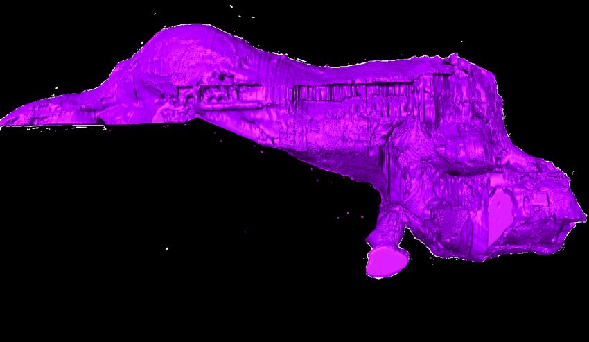
We CT-scanned Archaeocydippida hunsrueckiana, and this project proposes to use
AVIZO to reconstruct a 3D model for this species, use it to better understand its
morphology, and evaluate whether the apomorphies characterising the crown
ctenophores can be identified in this species. We shall further investigate the
phylogenetic relationship of Archaeocydippida hunsrueckiana by including it into the
morphological dataset assembled by the co-supervisor Luke Parry.
You will learn the use of AVIZO to reconstruct fossils from CT scanned data and
phylogenetic methods as you will be investigating the phylogenetic relationships of this
taxon using modern phylogenetic methodologies.
Feuda et al. (2017) Improved Modeling of Compositional Heterogeneity Supports
Sponges as Sister to All Other Animals. Current Biology.
Stanley, G. D. and Sturmer, W. 1983. The first fossil ctenophore from the lower
Devonian of West Germany. Nature.
Stanley, G.D. and Sturmer, W. 1987. A new fossil ctenophore discovered by X- rays.
Nature, 328: 61-63.
Simion, P. et al. 2017. A Large and Consistent Phylogenomic Dataset Supports
Sponges as the Sister Group to All Other Animals. Current Biology.
Tang, F. et al. 2011. Eoandromeda and the origin of Ctenophora. Evolution and
Development 13(5): 408-14.
Whelan et al. (2017). Ctenophore relationships and their placement as the sister group to
all other animals. Nat. Ecol. Evol. 1: 1737–1746.
27You can also read