Use of BERT (Bidirectional Encoder Representations from Transformers)-Based Deep Learning Method for Extracting Evidences in Chinese Radiology ...
←
→
Page content transcription
If your browser does not render page correctly, please read the page content below
JOURNAL OF MEDICAL INTERNET RESEARCH                                                                                                                 Liu et al

     Original Paper

     Use of BERT (Bidirectional Encoder Representations from
     Transformers)-Based Deep Learning Method for Extracting
     Evidences in Chinese Radiology Reports: Development of a
     Computer-Aided Liver Cancer Diagnosis Framework

     Honglei Liu1,2, PhD; Zhiqiang Zhang1,2, BS; Yan Xu3, MD; Ni Wang1,2, BS; Yanqun Huang1,2, BS; Zhenghan Yang3,
     MD; Rui Jiang4, PhD; Hui Chen1,2, PhD
     1
      School of Biomedical Engineering, Capital Medical University, Beijing, China
     2
      Beijing Key Laboratory of Fundamental Research on Biomechanics in Clinical Application, Capital Medical University, Beijing, China
     3
      Department of Radiology, Beijing Friendship Hospital, Capital Medical University, Beijing, China
     4
      Ministry of Education Key Laboratory of Bioinformatics, Research Department of Bioinformatics at the Beijing National Research Center for Information
     Science and Technology, Center for Synthetic and Systems Biology, Department of Automation, Tsinghua University, Beijing, China

     Corresponding Author:
     Hui Chen, PhD
     School of Biomedical Engineering, Capital Medical University
     No 10, Xitoutiao, Youanmen, Fengtai District
     Beijing
     China
     Phone: 86 010 83911545
     Email: chenhui@ccmu.edu.cn

     Abstract
     Background: Liver cancer is a substantial disease burden in China. As one of the primary diagnostic tools for detecting liver
     cancer, dynamic contrast-enhanced computed tomography provides detailed evidences for diagnosis that are recorded in free-text
     radiology reports.
     Objective: The aim of our study was to apply a deep learning model and rule-based natural language processing (NLP) method
     to identify evidences for liver cancer diagnosis automatically.
     Methods: We proposed a pretrained, fine-tuned BERT (Bidirectional Encoder Representations from Transformers)-based
     BiLSTM-CRF (Bidirectional Long Short-Term Memory-Conditional Random Field) model to recognize the phrases of APHE
     (hyperintense enhancement in the arterial phase) and PDPH (hypointense in the portal and delayed phases). To identify more
     essential diagnostic evidences, we used the traditional rule-based NLP methods for the extraction of radiological features. APHE,
     PDPH, and other extracted radiological features were used to design a computer-aided liver cancer diagnosis framework by
     random forest.
     Results: The BERT-BiLSTM-CRF predicted the phrases of APHE and PDPH with an F1 score of 98.40% and 90.67%,
     respectively. The prediction model using combined features had a higher performance (F1 score, 88.55%) than those using APHE
     and PDPH (84.88%) or other extracted radiological features (83.52%). APHE and PDPH were the top 2 essential features for
     liver cancer diagnosis.
     Conclusions: This work was a comprehensive NLP study, wherein we identified evidences for the diagnosis of liver cancer
     from Chinese radiology reports, considering both clinical knowledge and radiology findings. The BERT-based deep learning
     method for the extraction of diagnostic evidence achieved state-of-the-art performance. The high performance proves the feasibility
     of the BERT-BiLSTM-CRF model in information extraction from Chinese radiology reports. The findings of our study suggest
     that the deep learning–based method for automatically identifying evidences for diagnosis can be extended to other types of
     Chinese clinical texts.

     (J Med Internet Res 2021;23(1):e19689) doi: 10.2196/19689

     http://www.jmir.org/2021/1/e19689/                                                                      J Med Internet Res 2021 | vol. 23 | iss. 1 | e19689 | p. 1
                                                                                                                           (page number not for citation purposes)
XSL• FO
RenderX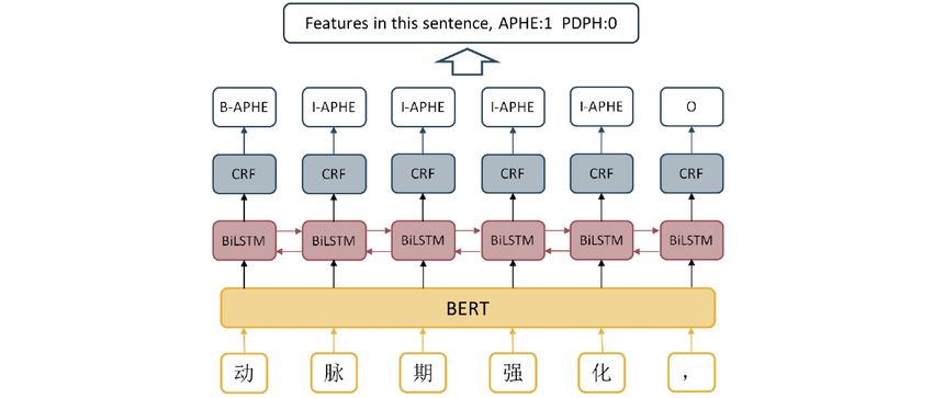
JOURNAL OF MEDICAL INTERNET RESEARCH                                                                                                       Liu et al

     KEYWORDS
     BiLSTM-CRF; natural language processing; radiology reports; information extraction; computer-aided diagnosis; BERT

                                                                          NER performance through multi-layer data representations. Of
     Introduction                                                         the popular deep learning methods, BiLSTM (bidirectional long
     In the past decades, electronic health records (EHRs) from           short-term memory) can capture long-range related information
     millions of patients have become massive sources of valuable         effectively. Furthermore, BiLSTM with CRF, known as
     clinical data. Machine learning–based algorithms, especially         BiLSTM-CRF, outperforms the traditional models with feature
     deep learning algorithms, have been applied effectively to           extraction and reduces the workload of feature selection [13].
     analyze patient data and they have shown promising results,          Due to the difference in the grammatical features from English
     thereby advancing medical research and better informing clinical     and the limitation of the EHR corpus, information extraction
     decision making for the secondary use of EHRs [1,2]. Owing           of Chinese EHRs using NLP remains challenging. In the medical
     to the high dimensionality, noise, heterogeneity, random errors,     field, researchers have developed information extraction
     and systematic biases, the data mining of EHRs remains               algorithms for varied implementations, including diagnostic
     challenging. Natural language processing (NLP) technologies          models for different diseases such as cancers [14] and childhood
     could extract meaningful information, thus facilitating the          diseases [15]. For Chinese NER tasks, BiLSTM-CRF is the
     application of clinical texts. There are 2 types of methods for      most common and practical approach [16,17]. BERT has also
     information extraction, namely, rule-based methods and machine       received extensive attention in Chinese EHRs. Zhang et al used
     learning methods [1]. The use of machine learning methods for        fine-tuning BERT for NER and relation extraction in several
     data mining of EHRs can derive previously unknown clinical           types of Chinese clinical documents. The comprehensive clinical
     insights and be applied powerfully in clinical decision-making       information related to breast cancer was extracted [14]. Wu et
     and computer-aided diagnosis of diseases [3,4]. Recently, deep       al developed an aided clinical diagnosis service on EHRs by
     learning methods have had a profound impact in various areas         using a deep learning model [3]. Liang et al applied an automatic
     of research because of their simplicity, efficient processing, and   NLP system and achieved a high diagnostic accuracy in
     state-of-the-art results [5,6]. In particular, recurrent neural      childhood diseases [15].
     networks and Word2Vec embedding are the most popular
     methods that are utilized in clinical NLP tasks [2]. Deep learning   The radiology report is a crucial component of EHRs, as it is
     methods have made improvements in various clinical                   the communication bridge between radiologists and physicians.
     applications, especially for text classification, named-entity       The accuracy and efficiency of diagnosis are limited since it is
     recognition (NER), relation extraction, and question answering       formulated based on subjective judgment, especially for
     [7,8]. With growing acceptance and increasing number of              inexperienced physicians. Hence, extracting useful radiological
     applications, deep learning methods have become a baseline in        information from radiology reports has considerable significance
     many clinical NLP tasks.                                             in advancing radiological research and clinical practice [18,19].
                                                                          NLP technologies have received great attention in the processing
     Word embedding is an essential step for sequencing labelling         of radiology reports and have been successfully applied in
     tasks. Learning word representations from massive unannotated        identifying     biomedical     concepts      [20],     extracting
     documents is a long-established method. The Word2Vec method          recommendations [21], determining the change level of clinical
     [9] is the first word embedding approach based on deep learning      findings [22], and so on.
     methods. The model derives the semantic and synthetic meaning
     of a word on account of its adjacent words by using                  With the development of machine learning methods in recent
     unsupervised learning. Global Vector word representation [10]        eras, computer-aided early diagnosis for cancer based on
     is another effective word embedding method, which constructs         massive clinical data becomes feasible. Many diseases have
     a global word-word co-occurrence matrix and utilizes matrix          been investigated to date, such as hepatocellular cancer [23]
     factorization to learn embeddings in a lower dimensional space.      and colorectal cancer [24]. In this study, we focused on the
     However, the word-level representations have a limitation that       computer-aided diagnosis of liver cancer, which remains to be
     only a single embedding is provided for a word with no thought       a substantial disease burden in China. For liver cancer diagnosis,
     for polysemy under different contexts. Unlike the traditional        dynamic contrast-enhanced computed tomography (CT) is one
     embedding methods, ELMo (Embeddings from Language                    of the primary diagnostic tests. Imaging findings of the key
     Models) [11] uses a bidirectional language model to embed the        enhanced scan phases such as the arterial phase, portal phase,
     context information into word representations. BERT                  and delayed phase are recorded in the radiology reports.
     (Bidirectional Encoder Representations from Transformers)            According to the guidelines of the Chinese Society of Clinical
     [12] is another prominent contextualized word representation         Oncology (CSCO), hyperintense enhancement in the arterial
     model, which uses a masked language model that predicts              phase (APHE) and hypointense enhancement in the portal and
     randomly masked words in a context sequence. Different from          delayed phases (PDPH) are significant diagnostic evidences for
     ELMo, BERT targets different training objectives and uses a          liver cancer [25].
     masked language model to learn bidirectional representations.        In this study, we designed deep learning–based methods to
     For clinical sequence labelling tasks such as NER, rule-based        identify evidences for liver cancer diagnosis automatically. We
     approach and conditional random fields (CRFs) have been used         recognized the phrases of APHE and PDPH by using a
     widely. Deep learning technologies substantially improve the         BERT-BiLSTM-CRF model by combining a pretrained,

     http://www.jmir.org/2021/1/e19689/                                                            J Med Internet Res 2021 | vol. 23 | iss. 1 | e19689 | p. 2
                                                                                                                 (page number not for citation purposes)
XSL• FO
RenderX
JOURNAL OF MEDICAL INTERNET RESEARCH Liu et al
fine-tuned BERT language model with BiLSTM-CRF. We also
applied the FENLP (feature extraction using the rule-based
Methods
NLP) method based on the content of radiology reports to extract Evidence Extraction for Diagnosis of Liver Cancer
the radiological features. Therefore, the evidences for diagnosis,
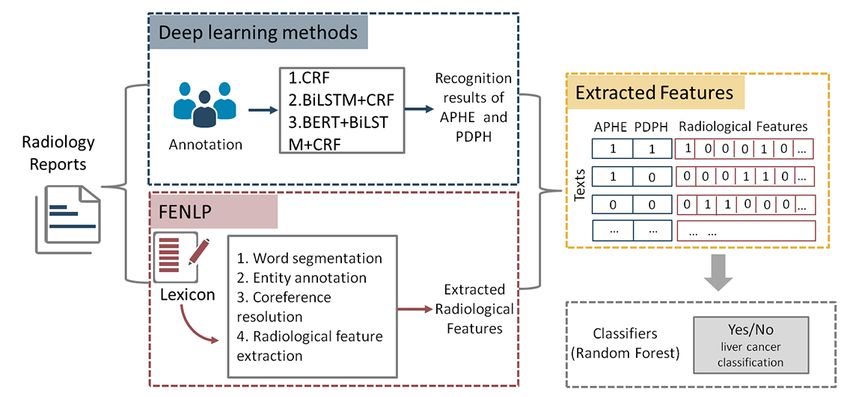
considering both clinical knowledge and radiology findings, Figure 1 shows the workflow of the evidence extraction for the
contained APHE, PDPH, and radiological features extracted by diagnosis of liver cancer. We implemented 2 feature extraction
FENLP [26]. With these evidences, we designed a methods based on clinical knowledge and the content of
computer-aided liver cancer diagnosis framework by using radiology reports to generate a radiological feature set. Then,
random forest. we built a random forest model to predict liver cancer by using
these features as inputs.
Figure 1. The workflow of this research. Labels 0/1 represent the absence/presence of a certain feature. BERT: Bidirectional Encoder Representations
from Transformers; BiLSTM: bidirectional long short-term memory; CRF: conditional random field; APHE: hyperintense enhancement in the arterial
phase; PDPH: hypointense in the portal and delayed phases.
Chinese character. In this study, BIO tags contained B-APHE,
Data Sets I-APHE, B-PDPH, I-PDPH, and O-Outside. We invited 2
We collected abdomen and pelvic CT radiology reports from a radiologists with more than 5 years of medical experience to
tertiary hospital in Beijing, China, between 2012 and 2019. To annotate all the data. If there was any inconsistency, another
protect patient privacy, we removed all the identifying experienced radiological expert was then asked to make the
information. An unstructured radiology report has different final annotation, to obtain the gold standard annotated data. To
sections, including Type of Examination, Clinical History, ensure the consistency of the annotation, radiologists were
Comparison, Technique, Imaging Findings, and Impressions. trained in advance. At the report level, APHE and PDPH were
The Impressions section summarizes crucial radiology findings not mutually exclusive, that is, 1 report could contain both
from the Findings section and contains a diagnosis indicated APHE and PDPH. Of all the reports, 602 had the phrase of
by a radiologist. In this study, the diagnosis of liver cancer was APHE and 330 had the phrase of PDPH. For the 480 reports
determined according to the Impression section and the diagnosed with liver cancer, the numbers of APHE and PDPH
annotation of experienced radiologists, resulting in 480 patients were 442 and 330, respectively.
with liver cancer. We randomly selected 609 patients without
liver cancer from our data set. Therefore, 480 and 609 radiology BiLSTM-CRF is commonly used in the sequence labeling task.
reports for patients with and without liver cancer, respectively, To further improve the recognition performance for the features
were used in this study. We then trained and evaluated an NER of APHE and PDPH, we performed the BERT-BiLSTM-CRF
model on the Imaging Findings section. The reports were model comprising a fine-tuned BERT language model for word
randomly divided into the training set and the test set in a ratio embedding and BiLSTM-CRF method for feature recognition.
of 8:2. CRF and BiLSTM-CRF model were applied as the baseline.
APHE and PDPH in Chinese radiology reports had a variety of
BERT-BiLSTM-CRF for Recognition of APHE and presentations such as detailed presentation, CT values of
PDPH different phases, and abbreviations (Table 1). The deep learning
We considered the recognition of APHE and PDPH as a model for the recognition of APHE and PDPH consisted of 3
sequence labelling task at the character level, where the goal layers, namely, the word embedding layer, BiLSTM layer, and
was to assign the BIO (Begin, Inside, Outside) tags to each CRF layer (Figure 2).
http://www.jmir.org/2021/1/e19689/ J Med Internet Res 2021 | vol. 23 | iss. 1 | e19689 | p. 3
(page number not for citation purposes)
XSL• FO
RenderXJOURNAL OF MEDICAL INTERNET RESEARCH Liu et al
Table 1. Some expressions of APHEa and PDPHb in Chinese.
Expressions of APHE and PDPH in Chinese Detailed descriptions
增强后动脉期明显不均匀强化 The arterial phase shows the heterogeneous density in the enhanced scan.
动脉期强化明显 Marked enhancement is shown in the arterial phase.
动脉期可见多发强化灶 Multiple enhancement areas are seen in the arterial phase.
门脉期相对低密度 The portal phase has relatively low density.
门脉期可见消退 PDPH occurs in the portal phase.
a
APHE: hyperintense enhancement in the arterial phase.
b
PDPH: hypointense in the portal and delayed phases.
Figure 2. The architecture of the BERT-BiLSTM-CRF model for the recognition of APHE and PDPH. BERT: Bidirectional Encoder Representations
from Transformers; BiLSTM: bidirectional long short-term memory; CRF: conditional random field; APHE: hyperintense enhancement in the arterial
phase; PDPH: hypointense in the portal and delayed phases.
network. Compared with LSTM, BiLSTM can learn forward
Word Embedding Layer and backward information of input words by splitting the
The word embedding layer could map and transform the discrete neurons into 2 directions of a text sequence. We set the number
characters into distributed representations. A word-level vector of hidden units in BiLSTM to 100 and the optimizer to Adam.
representation learned a real valued vector to represent a word
from a large amount of unannotated text. On most NLP tasks, CRF Layer
BERT could achieve state-of-the-art performance while For the sequence labelling step in our study, adjacent tags had
requiring minimal architectural modification [27]. In this study, dependencies. For example, an inside tag I must follow a begin
we applied Word2Vec and BERT to train the character vectors, tag B. We applied the sequential CRF to calculate optimal
followed by a comparison of the results. The Word2Vec was sequence combinations on top of the BiLSTM layer that could
used with a dimension size of 100 and a batch size of 120. The consider the dependencies of adjacent tags.
Word2Vec was pretrained on the Chinese Wikipedia data. The
sentence embedding had been pretrained and fine-tuned by
APHE and PDPH Labels at the Report Level
BERT on the original Google BERT GitHub repository [28]. Considering the characteristics of Chinese language and also
The maximum sequence length was set to 256 with a batch size avoiding the noise, we defined the following as APHE or PDPH
of 64. features at the report level: (1) 2 continuous characters that were
the abbreviations of APHE (ie, 快进) or PDPH (ie, 快出); (2)
BiLSTM Layer more than 3 continuous characters that were predicted as APHE
Recurrent neural networks is a family of neural networks, which or PDPH. Criterion (1) was checked first and was only based
is usually used for modelling sequential data. The LSTM (Long on the characters. If not met, criterion (2) was checked, which
Short-Term Memory Networks) is a variant of the recurrent was based on CRF results.
neural networks, and it can effectively capture high
dependencies and retrieve rich global information. LSTM solves
FENLP for Radiological Feature Extraction
the problem by using the gating mechanism. An LSTM unit We implemented the NLP pipeline in the Findings section to
consists of 3 gates (ie, Input Gate, Output Gate, and Forget extract useful features from the unstructured radiology reports
Gate), which can select semantic information in a neural to facilitate liver cancer diagnosis. As shown in Figure 1, the
http://www.jmir.org/2021/1/e19689/ J Med Internet Res 2021 | vol. 23 | iss. 1 | e19689 | p. 4
(page number not for citation purposes)
XSL• FO
RenderXJOURNAL OF MEDICAL INTERNET RESEARCH Liu et al
NLP pipeline consisted of 4 successive steps, that is, word
segmentation, entity annotation, coreference resolution, and
Results
relationship extraction, resulting in radiological features We extracted the features of APHE and PDPH by using 3
consisting of 1 or more terms. The detailed description of the different models, that is, CRF, BiLSTM-CRF, and
pipeline is provided in our previous study [26]. The whole BERT-BiLSTM-CRF. The recognition results were presented
pipeline was based on a lexicon that was constructed manually both at the report level and character level (Table 2). At the
according to Chinese grammatical characteristics. A small report level, the performance was computed depending on
number of reports were sampled randomly for generating the whether the radiology reports contained a feature of APHE or
lexicon by manual reading. The lexicon contained clinical terms PDPH. At the character level, the recognition results of BIO
and lists of synonyms. The lexicon was collected in the same tags for each Chinese character were counted. For the
hospital and clinical text type with this study. Five entity types character-level recognition results of APHE and PDPH, the
(Location, Morphology, Density, Enhancement, and Modifier) BERT-BiLSTM-CRF model obtained the best performance,
were recognized. After coreference resolution, according to the with F1 scores of 89.14% and 82.19%, respectively. At the
synonym list in the lexicon, we then used several patterns of report level, the BERT-BiLSTM-CRF model also achieved the
entity types as rules to obtain the final radiological features best performance (F1 scores of 98.40% for APHE and 90.67%
(Table S1 of Multimedia Appendix 1). Therefore, the for PDPH). For the other 2 baseline models, the BiLSTM-CRF
radiological features could be seen as a combination of several model outperformed the CRF model but underperformed the
entities such as 肝脏+低密度影 (liver + low density) and 肝脏 BERT-BiLSTM-CRF model. If a single character was
+增强扫描未见强化 (liver + enhancement scan showed no recognized as a feature, it would be regarded as noisy
enhancement). information, thereby leading to its exclusion from the
Prediction Models report-level results. As a result, the recognition performances
at the report level were higher than those at the character level
Using the radiological features obtained by
in all the models. We chose the recognition results of APHE
BERT-BILSTM-CRF and FENLP, we used a random forest
and PDPH at the report level by the BERT-BiLSTM-CRF model
model for the liver cancer diagnosis. Random forest is an
as the predictors for further liver cancer diagnosis.
ensemble learning method constructed with a multitude of
decision trees, which is widely used in classification tasks. The The feature extraction method FENLP used the lexicon
performance was measured by the recall, precision, and F1 described in our previous study, which included 831 words and
score. Random forest could generate the feature importance 5 entity types. Entity combinations conforming to specific entity
score, which was computed by Gini impurity. Gini impurity is patterns were formulated as radiological features. The patterns
a measurement of the probability that a sample is classified included Location + Density, Location + Enhancement, Location
incorrectly without a specific feature. In our study, the higher + Enhancement + Modifier, Location + Density + Modifier,
the feature importance score of the radiological features was, and Location + Morphology. We retained the radiological
the more linked it was with the liver cancer diagnosis. We used features that occurred more than twice. We finally obtained 301
the feature importance score to rank all the radiological features. radiological features; among them, 6 features had a frequency
higher than 300 (Table S2 of Multimedia Appendix 1).
http://www.jmir.org/2021/1/e19689/ J Med Internet Res 2021 | vol. 23 | iss. 1 | e19689 | p. 5
(page number not for citation purposes)
XSL• FO
RenderXJOURNAL OF MEDICAL INTERNET RESEARCH Liu et al
Table 2. Recognition performance of APHEa and PDPHb by using different models at the character level and report level.
Models Accuracy (%) Precision (%) Recall (%) F1 score (%)
Character level
Conditional random field
APHE 96.05 84.13 72.19 77.70
PDPH 97.44 80.37 59.02 68.06
Bidirectional long short-term memory-conditional random field
APHE 97.54 90.86 82.56 86.51
PDPH 98.24 84.56 75.72 79.89
BERTc+ Bidirectional long short-term memory-conditional random field
APHE 97.97 91.14 87.22 89.14
PDPH 98.46 88.60 76.64 82.19
Report level
Conditional random field
APHE 94.52 98.28 91.94 95.00
PDPH 89.00 87.69 79.17 83.21
Bidirectional long short-term memory-conditional random field
APHE 95.89 97.30 94.74 96.00
PDPH 93.61 92.19 86.76 89.39
BERT+ Bidirectional long short-term memory-conditional random field
APHE 98.17 97.62 99.19 98.40
PDPH 93.61 87.18 94.44 90.67
a
APHE: hyperintense enhancement in the arterial phase.
b
PDPH: hypointense in the portal and delayed phases.
c
BERT: Bidirectional Encoder Representations from Transformers.
According to the presence or absence of each feature extracted obtained the highest value while inputting all the features, while
from either BERT-BILSTM-CRF or FENLP, each radiology the prediction model had the highest recall rate with only 2
report was represented by a 0-1 vector. The prediction results features of APHE and PDPH. Among the features with a
using different patterns of features are shown in Table 3. F1 frequency higher than 10 in all the reports, the top 10 features
scores of random forest using features from linked with the liver cancer diagnosis were identified with the
BERT-BILSTM-CRF and FENLP were 84.88% and 83.92%, feature importance score computed by Gini impurity (Figure
respectively. With a combination of both kinds of features, the 3). The top 2 features were APHE and PDPH, which had
final F1 score of prediction model increased to 88.55%. Among substantially larger feature importance scores than the other
all the feature input patterns, the precision and accuracy also features extracted from FENLP.
Table 3. Performance of different patterns of features for liver cancer diagnosis.
Patterns of Features Accuracy (%) Precision (%) Recall (%) F1 score (%)
a b 86.11 81.38 88.70 84.88
APHE and PDPH
Radiological features from FENLPc 85.71 83.06 84.80 83.92
All features 90.25 91.52 85.77 88.55
a
APHE: hyperintense enhancement in the arterial phase.
b
PDPH: hypointense in the portal and delayed phases.
c
FENLP: feature extraction using the rule-based natural language processing.
http://www.jmir.org/2021/1/e19689/ J Med Internet Res 2021 | vol. 23 | iss. 1 | e19689 | p. 6
(page number not for citation purposes)
XSL• FO
RenderXJOURNAL OF MEDICAL INTERNET RESEARCH Liu et al
Figure 3. Top 10 radiological features linked with liver cancer diagnosis ranked by feature importance score. APHE: hyperintense enhancement in the
arterial phase; PDPH: hypointense in the portal and delayed phases.
related descriptions covered varied Chinese sentence structures
Discussion and entity types (Table 1). For example, for hyperintense
Principal Results enhancement, the sentence pattern and phrase could have
different styles due to the different writing habits of different
Diagnostic prediction of cancer by using data mining methods radiologists or due to the use of Chinese abbreviations. Different
is an essential and significant application of EHRs [5]. From from the Word2Vec model, BERT learned context-dependent
previous studies, features extracted from EHRs have proved to word representations by using bidirectional transformers. The
be the valid input of the diagnostic model [14,29]. In particular, BiLSTM algorithms are widely used and easily implanted in
the use of machine learning methods, especially deep learning sequence-related work such as entity extraction. We annotated
methods for clinical information extraction, could facilitate in all the characters in the Findings section manually with BIO
providing new evidences in computer-aided diagnosis. As the tags and then applied the BERT-BiLSTM-CRF model to
burden of liver cancer is widely accepted as one of the principal recognize APHE and PDPH. The high performance proved the
and universal challenges in health care and as patients with liver feasibility of the BERT-BiLSTM-CRF model in information
cancer are usually diagnosed at the terminal stage, the early and extraction from Chinese radiology reports.
accurate diagnosis of liver cancer by radiology examination has
great significance [30,31]. In contrast with previous studies of In this study, among the recognition results of APHE and PDPH
liver cancer diagnosis, our study focused on the identification obtained by the 3 different models, the BERT-BILSTM-CRF
of evidences for live cancer diagnosis from Chinese radiology model finally achieved the best performance for both APHE
reports. We selected APHE and PDPH as the known evidences (F1 score 98.40%, precision 97.62%, and recall 99.19%) and
for diagnosis according to the guidelines of CSCO. These 2 PDPH (90.67%, 87.18%, and 94.44%, respectively) at the report
features were essential but not sufficient enough to represent level. For the 2 baseline models based on CRF, the model with
the whole report and further be used to diagnose liver cancer. a BiLSTM layer received a much higher F1 score than the model
Furthermore, using FENLP, we also extracted uncertain numbers without a BiLSTM layer. The results indicated that, with the
of radiological features from the report content, because we word embedding language model BERT and the BiLSTM
aimed to obtain new evidences for essential diagnosis. model, the recognition of APHE and PDPH could result in much
Therefore, the evidences for diagnosis were obtained both from higher performance. To avoid the noise in the recognition
clinical knowledge and the content of reports. For the results, we used the recognition results at the report level to be
recognition of APHE and PDPH, we applied BERT on word the input radiological features of the liver cancer diagnostic
embedding in the deep learning method, which achieved model. Report-level recognition concerned only continuous
state-of-the-art performance. characters longer than 3 characters and specific Chinese
abbreviations. Therefore, report-level results could represent
Word embedding is an essential step for sequencing labelling whether the report contained the features of APHE or PDPH.
tasks. Previously popular models such as Word2Vec and Global The recognition of APHE and PDPH by BERT-BiLSTM-CRF
Vector word representation focused on learning was accurate enough to be the predictors of liver cancer
context-independent word representations. Recent advances in diagnosis.
word representations based on language models, including
ELMo, CoVe, and BERT, could dynamically improve the word Only 2 fixed features of APHE and PDPH were not enough for
representations and discriminate among multiple meanings of liver cancer diagnosis. Therefore, we further performed the
a word. In particular, based on the attention mechanism, BERT automatic NLP pipeline FENLP to extend the feature set based
exhibited an upward trend and outperformed the previous on Chinese grammar and radiological characteristics. Different
models in many NLP tasks. Recognition of APHE and PDPH from that of BERT-BILSTM-CRF, the number of features
using traditional NLP methods had difficulties, because the generated by FENLP was unknown and changed according to
http://www.jmir.org/2021/1/e19689/ J Med Internet Res 2021 | vol. 23 | iss. 1 | e19689 | p. 7
(page number not for citation purposes)
XSL• FO
RenderXJOURNAL OF MEDICAL INTERNET RESEARCH Liu et al
the training texts. In this study, we finally extracted 301 features. have also proved to be essential features in our liver cancer
The top features were the typical morphology of the different diagnostic model. Other radiological features from FENLP
locations, which were essential to the diagnosis of the liver enlarged the potential evidences for the diagnosis of liver cancer.
diseases (Table S2 of Multimedia Appendix 1). Moreover, we utilized the BERT-BiLSTM-CRF model in this
study, which achieved the state-of-the-art performance.
We chose the random forest as the liver cancer diagnostic model.
With 2 kinds of features obtained by BERT-BILSTM-CRF and Limitations
FENLP, random forest could reach an F1 score of 88.55%, Our study had the following limitations. The number of
which was much higher than the model using either kind of radiological features from FENLP was not fixed since all
features. The performance of the diagnostic model using APHE desirable features were retained, which might introduce some
and PDPH was slightly higher than that of the model using noise into the extracted radiological features. Besides, from the
features extracted from FENLP. By contrast, FENLP produced clinical knowledge in the guidelines of CSCO, we only extracted
much more features than BERT-BILSTM-CRF. We further 2 characteristic features. In future, we will collect more
ranked the features by the feature importance score computed evidences for diagnosis in order to further improve the
by Gini impurity, which could reflect the degree of association performance and make the model more explanatory. Through
with liver cancer. APHE and PDPH were the top 2 features with the analysis of the misjudged samples in the recognition of
a clearly higher feature importance score compared with other APHE and PDPH, we identified the main error that occurred
features obtained by FENLP. The results indicated the strong when the description of APHE and PDPH only included CT
association of APHE and PDPH with liver cancer, which values. With the comparison of CT values in different phases,
coincided with the current clinical knowledge. Of the top we could define these 2 features. However, our methods did not
features obtained by FENLP, the feature high density of liver focus on the CT value extraction, and the number of these cases
had the highest feature importance score, which was the were small. In future studies, CT value extraction and analysis
important and basic risk factor for the diagnosis of liver diseases. can avoid this kind of error and increase the prediction
Broadening of hepatic fissures was an essential feature that performance.
existed in liver cirrhosis or in liver cancer progressed from liver
cirrhosis [30]. Our results confirmed that the radiological Conclusion
features from FENLP could also be an evidence for diagnosis, In this study, we developed a deep learning–based method for
which could further improve the diagnostic performance. the recognition of evidences for disease diagnosis and designed
Furthermore, the top features linked with liver cancer could a computer-aided liver cancer diagnosis framework. The
extend the diagnostic evidence and be the supplementary diagnostic evidences contained APHE, PDPH, and radiological
features of APHE and PDPH. features extracted by FENLP. We proposed the BERT-based
Designing disease diagnostic models based on EHRs is a deep learning model BERT-BILSTM-CRF for recognizing the
significantly important research field. Recently, NLP and deep phrases of APHE and PDPH, which are the essential features
learning-based models have been widely applied in many studies associated with liver cancer diagnosis. Our work confirms that
[7]. For instance, Sada et al designed and performed BERT-based deep learning model can be used and has desirable
NLP-assisted radiology document classification for liver cancer performance in the radiological feature extraction of Chinese
detection. The model finally received an F1 score of 0.78 [23]. radiology reports. Furthermore, this study was a comprehensive
Compared with previous studies on clinical information study for NLP and its application, focusing on Chinese radiology
extraction, the evidences for diagnosis in this study were reports. The deep learning model proposed in this study for
identified based on the clinical knowledge from the guidelines information extraction is expected to be extended to different
of CSCO and the content of the reports. APHE and PDPH are types of Chinese clinical texts and other kinds of applications.
2 widely accepted evidences for disease diagnosis, and they
Acknowledgments
This work was supported by grants from the National Natural Science Foundation of China (No. 81701792 and No. 81971707)
and the National Key Research and Development Program of China (No. 2018YFC0910404)
Authors' Contributions
HL proposed and designed the whole pipeline, analyzed the results, and wrote the paper. YX and ZY collected the original data,
annotated the radiology reports, and provided clinical knowledge guidance in lexicon construction. ZZ, NW, and YH cleaned the
data. RJ provided theoretical guidance and revised this paper. HC provided theoretical guidance and revised this paper.
Conflicts of Interest
None declared.
Multimedia Appendix 1
Supplementary data.
http://www.jmir.org/2021/1/e19689/ J Med Internet Res 2021 | vol. 23 | iss. 1 | e19689 | p. 8
(page number not for citation purposes)
XSL• FO
RenderXJOURNAL OF MEDICAL INTERNET RESEARCH Liu et al
[DOCX File , 20 KB-Multimedia Appendix 1]
References
1. Wang Y, Wang L, Rastegar-Mojarad M, Moon S, Shen F, Afzal N, et al. Clinical information extraction applications: A
literature review. J Biomed Inform 2018 Jan;77:34-49 [FREE Full text] [doi: 10.1016/j.jbi.2017.11.011] [Medline: 29162496]
2. Wu S, Roberts K, Datta S, Du J, Ji Z, Si Y, et al. Deep learning in clinical natural language processing: a methodical review.
J Am Med Inform Assoc 2020 Mar 01;27(3):457-470 [FREE Full text] [doi: 10.1093/jamia/ocz200] [Medline: 31794016]
3. Wu J, Liu X, Zhang X, He Z, Lv P. Master clinical medical knowledge at certificated-doctor-level with deep learning
model. Nat Commun 2018 Oct 19;9(1):4352 [FREE Full text] [doi: 10.1038/s41467-018-06799-6] [Medline: 30341328]
4. Gálvez JA, Pappas JM, Ahumada L, Martin JN, Simpao AF, Rehman MA, et al. The use of natural language processing
on pediatric diagnostic radiology reports in the electronic health record to identify deep venous thrombosis in children. J
Thromb Thrombolysis 2017 Oct;44(3):281-290. [doi: 10.1007/s11239-017-1532-y] [Medline: 28815363]
5. Sheikhalishahi S, Miotto R, Dudley JT, Lavelli A, Rinaldi F, Osmani V. Natural Language Processing of Clinical Notes
on Chronic Diseases: Systematic Review. JMIR Med Inform 2019 Apr 27;7(2):e12239 [FREE Full text] [doi: 10.2196/12239]
[Medline: 31066697]
6. Dreisbach C, Koleck TA, Bourne PE, Bakken S. A systematic review of natural language processing and text mining of
symptoms from electronic patient-authored text data. Int J Med Inform 2019 May;125:37-46 [FREE Full text] [doi:
10.1016/j.ijmedinf.2019.02.008] [Medline: 30914179]
7. Chen L, Song L, Shao Y, Li D, Ding K. Using natural language processing to extract clinically useful information from
Chinese electronic medical records. Int J Med Inform 2019 Apr;124:6-12. [doi: 10.1016/j.ijmedinf.2019.01.004] [Medline:
30784428]
8. Li P, Yuan Z, Tu W, Yu K, Lu D. Medical Knowledge Extraction and Analysis from Electronic Medical Records Using
Deep Learning. Chin Med Sci J 2019 Jun 30;34(2):133-139. [doi: 10.24920/003589] [Medline: 31315754]
9. Mikolov T, Chen K, Corrado G, Dean J. Distributed Representations of Words and Phrases and their Compositionality. In:
Advances in Neural Information Processing Systems 26 (NIPS 2013). United States: NIPS Foundation; 2013:3111-3119.
10. Pennington J, Socher R, Manning C. GloVe: Global Vectors for Word Representation. 2014 Presented at: Proceedings of
the Conference on Empirical Methods in Natural Language Processing (EMNLP); 2014; Doha, Qatar p. 1532-1543. [doi:
10.3115/v1/d14-1162]
11. Peters M, Neumann M, Iyyer M, Gardner M, Clark C, Lee K. Deep contextualized word representations. 2018 Presented
at: Proceedings of the 2018 Conference of the North American Chapter of the Association for Computational Linguistics:
Human Language Technologies; 2018; New Orleans, LA,USA p. 2227-2237 URL: https://arxiv.org/abs/1802.05365
12. Devlin J, Chang M, Kristina K, Toutanova. BERT: Pre-training of Deep Bidirectional Transformers for Language
Understanding. 2019 Presented at: Proceedings of the 2019 Conference of the North American Chapter of the Association
for Computational Linguistics: Human Language Technologies; 2019; Minneapolis, MN, USA p. 4171-4186. [doi:
10.18653/v1/N19-1423]
13. Zhiheng H, Xu W, Yu K. Bidirectional LSTM-CRF models for sequence tagging. 2015. URL: https://arxiv.org/abs/1508.
01991 [accessed 2020-01-03]
14. Zhang X, Zhang Y, Zhang Q, Ren Y, Qiu T, Ma J, et al. Extracting comprehensive clinical information for breast cancer
using deep learning methods. Int J Med Inform 2019 Dec;132:103985. [doi: 10.1016/j.ijmedinf.2019.103985] [Medline:
31627032]
15. Liang H, Tsui BY, Ni H, Valentim CCS, Baxter SL, Liu G, et al. Evaluation and accurate diagnoses of pediatric diseases
using artificial intelligence. Nat Med 2019 Mar;25(3):433-438. [doi: 10.1038/s41591-018-0335-9] [Medline: 30742121]
16. Wang Q, Zhou Y, Ruan T, Gao D, Xia Y, He P. Incorporating dictionaries into deep neural networks for the Chinese clinical
named entity recognition. J Biomed Inform 2019 Apr;92:103133 [FREE Full text] [doi: 10.1016/j.jbi.2019.103133] [Medline:
30818005]
17. Ji B, Li S, Yu J, Ma J, Tang J, Wu Q, et al. Research on Chinese medical named entity recognition based on collaborative
cooperation of multiple neural network models. J Biomed Inform 2020 Apr;104:103395. [doi: 10.1016/j.jbi.2020.103395]
[Medline: 32109551]
18. Bozkurt S, Alkim E, Banerjee I, Rubin DL. Automated Detection of Measurements and Their Descriptors in Radiology
Reports Using a Hybrid Natural Language Processing Algorithm. J Digit Imaging 2019 Aug;32(4):544-553 [FREE Full
text] [doi: 10.1007/s10278-019-00237-9] [Medline: 31222557]
19. Steinkamp JM, Chambers C, Lalevic D, Zafar HM, Cook TS. Toward Complete Structured Information Extraction from
Radiology Reports Using Machine Learning. J Digit Imaging 2019 Aug;32(4):554-564 [FREE Full text] [doi:
10.1007/s10278-019-00234-y] [Medline: 31218554]
20. Flynn RWV, Macdonald TM, Schembri N, Murray GD, Doney ASF. Automated data capture from free-text radiology
reports to enhance accuracy of hospital inpatient stroke codes. Pharmacoepidemiol Drug Saf 2010 Aug;19(8):843-847.
[doi: 10.1002/pds.1981] [Medline: 20602346]
21. Yetisgen-Yildiz M, Gunn ML, Xia F, Payne TH. A text processing pipeline to extract recommendations from radiology
reports. J Biomed Inform 2013 Apr;46(2):354-362 [FREE Full text] [doi: 10.1016/j.jbi.2012.12.005] [Medline: 23354284]
http://www.jmir.org/2021/1/e19689/ J Med Internet Res 2021 | vol. 23 | iss. 1 | e19689 | p. 9
(page number not for citation purposes)
XSL• FO
RenderXJOURNAL OF MEDICAL INTERNET RESEARCH Liu et al
22. Hassanpour S, Bay G, Langlotz CP. Characterization of Change and Significance for Clinical Findings in Radiology Reports
Through Natural Language Processing. J Digit Imaging 2017 Jun;30(3):314-322 [FREE Full text] [doi:
10.1007/s10278-016-9931-8] [Medline: 28050714]
23. Sada Y, Hou J, Richardson P, El-Serag H, Davila J. Validation of Case Finding Algorithms for Hepatocellular Cancer From
Administrative Data and Electronic Health Records Using Natural Language Processing. Med Care 2016 Feb;54(2):e9-14
[FREE Full text] [doi: 10.1097/MLR.0b013e3182a30373] [Medline: 23929403]
24. Xu H, Fu Z, Shah A, Chen Y, Peterson NB, Chen Q, et al. Extracting and integrating data from entire electronic health
records for detecting colorectal cancer cases. AMIA Annu Symp Proc 2011;2011:1564-1572 [FREE Full text] [Medline:
22195222]
25. Guidelines of Chinese Society of Clinical Oncology (CSCO): Hepatocellular Carcinoma. Beijing,China: Chinese Society
of Clinical Oncology; 2018.
26. Liu H, Xu Y, Zhang Z, Wang N, Huang Y, Hu Y, et al. A Natural Language Processing Pipeline of Chinese Free-text
Radiology Reports for Liver Cancer Diagnosis. IEEE Access 2020 Aug 28;8:159110-159119. [doi:
10.1109/ACCESS.2020.3020138]
27. Tawfik NS, Spruit MR. Evaluating sentence representations for biomedical text: Methods and experimental results. J
Biomed Inform 2020 Apr;104:103396. [doi: 10.1016/j.jbi.2020.103396] [Medline: 32147441]
28. GitHub. URL: https://github.com/google-research/bert [accessed 2020-03-01]
29. Pathak S, van Rossen J, Vijlbrief O, Geerdink J, Seifert C, van Keulen M. Post-Structuring Radiology Reports of Breast
Cancer Patients for Clinical Quality Assurance. IEEE/ACM Trans Comput Biol Bioinform 2020;17(6):1883-1894. [doi:
10.1109/TCBB.2019.2914678] [Medline: 31059453]
30. Kudo M, Trevisani F, Abou-Alfa GK, Rimassa L. Hepatocellular Carcinoma: Therapeutic Guidelines and Medical Treatment.
Liver Cancer 2016 Nov;6(1):16-26 [FREE Full text] [doi: 10.1159/000449343] [Medline: 27995084]
31. Nagtegaal ID, Odze RD, Klimstra D, Paradis V, Rugge M, Schirmacher P, WHO Classification of Tumours Editorial Board.
The 2019 WHO classification of tumours of the digestive system. Histopathology 2020 Jan;76(2):182-188 [FREE Full
text] [doi: 10.1111/his.13975] [Medline: 31433515]
Abbreviations
APHE: hyperintense enhancement in the arterial phase
BERT: Bidirectional Encoder Representations from Transformers
BiLSTM: bidirectional long short-term memory
BIO: Begin, Inside, Outside
CRF: conditional random field
CSCO: Chinese Society of Clinical Oncology
CT: computed tomography
EHR: electronic health record
ELMo: Embeddings from Language Model
FENLP: feature extraction using the rule-based natural language processing
NER: named-entity recognition
NLP: natural language processing
PDPH: hypointense in the portal and delayed phases
Edited by G Eysenbach; submitted 29.04.20; peer-reviewed by M Torii, J Zheng; comments to author 01.06.20; revised version
received 30.06.20; accepted 11.11.20; published 12.01.21
Please cite as:
Liu H, Zhang Z, Xu Y, Wang N, Huang Y, Yang Z, Jiang R, Chen H
Use of BERT (Bidirectional Encoder Representations from Transformers)-Based Deep Learning Method for Extracting Evidences in
Chinese Radiology Reports: Development of a Computer-Aided Liver Cancer Diagnosis Framework
J Med Internet Res 2021;23(1):e19689
URL: http://www.jmir.org/2021/1/e19689/
doi: 10.2196/19689
PMID: 33433395
©Honglei Liu, Zhiqiang Zhang, Yan Xu, Ni Wang, Yanqun Huang, Zhenghan Yang, Rui Jiang, Hui Chen. Originally published
in the Journal of Medical Internet Research (http://www.jmir.org), 12.01.2021. This is an open-access article distributed under
the terms of the Creative Commons Attribution License (https://creativecommons.org/licenses/by/4.0/), which permits unrestricted
http://www.jmir.org/2021/1/e19689/ J Med Internet Res 2021 | vol. 23 | iss. 1 | e19689 | p. 10
(page number not for citation purposes)
XSL• FO
RenderXJOURNAL OF MEDICAL INTERNET RESEARCH Liu et al
use, distribution, and reproduction in any medium, provided the original work, first published in the Journal of Medical Internet
Research, is properly cited. The complete bibliographic information, a link to the original publication on http://www.jmir.org/,
as well as this copyright and license information must be included.
http://www.jmir.org/2021/1/e19689/ J Med Internet Res 2021 | vol. 23 | iss. 1 | e19689 | p. 11
(page number not for citation purposes)
XSL• FO
RenderXYou can also read