Will Zirconia Implants Replace Titanium Implants? - MDPI
←
→
Page content transcription
If your browser does not render page correctly, please read the page content below
applied
              sciences
Review
Will Zirconia Implants Replace Titanium Implants?
Liana Preto Webber, Hsun-Liang Chan and Hom-Lay Wang *

                                          Department of Periodontics and Oral Medicine, School of Dentistry, The University of Michigan,
                                          Ann Arbor, MI 48109, USA; lpretowe@umich.edu (L.P.W.); hlchan@umich.edu (H.-L.C.)
                                          * Correspondence: homlay@umich.edu; Tel.: +1-(734)-764-9148; Fax: +1-(734)-936-0374

                                          Featured Application: Zirconia implants present success rates similar to titanium implants and
                                          seem to have a better soft tissue response and less material corrosion.

                                          Abstract: This review aims to discuss the advantages and disadvantages of zirconia implants com-
                                          pared with titanium implants. Moreover, it intends to review the relevant available long-term litera-
                                          ture of these two materials regarding osteointegration, soft-tissue, microbiota, and peri-implantitis,
                                          focusing on clinical results. Briefly, titanium implants are a reliable alternative for missing teeth;
                                          however, they are not incapable of failure. In an attempt to provide an alternative implant material,
                                          implants made from ceramic-derivate products were developed. Owing to its optimal osseointegra-
                                          tion competence, biocompatibility, and esthetic proprieties, zirconium dioxide (ZrO2 ), also known as
                                          zirconia, has gained popularity among researchers and clinicians, being a metal-free alternative for
                                          titanium implants with its main use in the anterior esthetic zones. This type of implant may present
                                          similar osseointegration as those noted on titanium implants with a greater soft-tissue response.
                                          Furthermore, this material does not show corrosion as its titanium analog, and it is less susceptible
                                          to bacterial adhesion. Lastly, even presenting a similar inflammatory response to titanium, zirconia

                                          implants offer less biofilm formation, suggesting less susceptibility to peri-implantitis. However,
                                   it is a relatively new material that has been commercially available for a decade; consequently, the
Citation: Webber, L.P.; Chan, H.-L.;      literature still lacks studies with long follow-up periods.
Wang, H.-L. Will Zirconia Implants
Replace Titanium Implants? Appl. Sci.     Keywords: dental implants; zirconium oxide; ceramic; osseointegration; peri-implantitis
2021, 11, 6776. https://doi.org/
10.3390/app11156776

Academic Editor: Carlo Barausse           1. Introduction
                                               Since Brånemark’s first implant attempts until now, dental implant therapy has ad-
Received: 1 July 2021
                                          vanced remarkably with innovations on both surgical and prosthetic fronts. New tech-
Accepted: 21 July 2021
                                          niques, surgical procedures, and materials have been developed, ensuring implant stability.
Published: 23 July 2021
                                          However, in contrast to what most patients may think, dental implants are not an infallible
                                          solution for the edentulous area. Several complications can occur, reducing the implant
Publisher’s Note: MDPI stays neutral
                                          success rates. The most common cause of titanium implant failure in the medium and
with regard to jurisdictional claims in
                                          long-term is peri-implantitis. Peri-implantitis is a pathological condition occurring in
published maps and institutional affil-
iations.
                                          the peri-implant tissues, characterized by inflammation in the peri-implant mucosa and
                                          subsequent progressive loss of supporting bone. It is characterized by profuse bleeding
                                          on probing or suppuration, deep probing depth, and bone loss beyond the initial bone
                                          remodeling [1]. It is estimated that the peri-implantitis prevalence can be up to 34% [2].
                                               In additional, other less common complications can be found such as fractures [3],
Copyright: © 2021 by the authors.
                                          titanium allergy [4], and corrosion [5]. According to Lee (2018), the implant fracture
Licensee MDPI, Basel, Switzerland.
                                          prevalence is 0.4% and can be related to prosthetic factors such as inappropriate occlusal,
This article is an open access article
                                          peri-implantitis, overloading, or even manufacturing defects [3]. Titanium allergies are
distributed under the terms and
conditions of the Creative Commons
                                          a reaction to the titanium components and might result from ion absorption through the
Attribution (CC BY) license (https://
                                          skin or mucosal contact or from implant corrosion processes. It is estimated that 0.6% of
creativecommons.org/licenses/by/
                                          the population is allergic to titanium [4]. Corrosion can result from the presence of several
4.0/).                                    corrosive species like hydrogen sulfide compounds dissolved oxygen, free radicals, and

Appl. Sci. 2021, 11, 6776. https://doi.org/10.3390/app11156776                                               https://www.mdpi.com/journal/applsci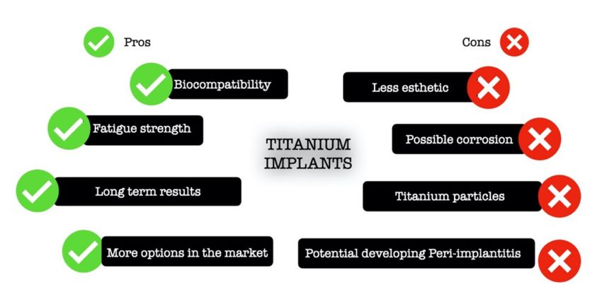
Appl. Sci. 2021, 11, 6776                                                                                            2 of 15

                            chloride ion, resulting in the metal surface breakdown. Even in a passive environment,
                            the basal implant corrosion rate is 0.02 mm/year to 0.13 mm/year [5]. Moreover, another
                            drawback of titanium implants is their dark color. This can be an issue in a patient with a
                            thin gingival phenotype, mainly in the esthetic area [6–8]. The grey shadow of titanium
                            may be visible through the peri-implant mucosa, thus impairing the esthetic outcome [9].
                                  In order to overcome the drawbacks of titanium implants, zirconia implants were
                            implemented. These implants have been used in Europe since the early 2000s and, subse-
                            quent to FDA approval in 2011, it has been more often used in the USA [10]. Furthermore,
                            implants are a popular solution for edentulous areas, and with the plethora of information
                            available, patients search for different implant alternatives and come to dental offices
                            requesting other options to the conventional titanium. In this matter, clinicians are raising
                            questions such as whether the zirconia implants are reliable? Are they a better option
                            compared with titanium implants? This narrative review aims to clarify these questions
                            and review the long-term results of zirconia implants compared with titanium implants.
                            Furthermore, this article offers a concise review of the literature regarding osteointegration,
                            soft-tissue, microbiota, and peri-implantitis of the two materials, mainly focusing on clini-
                            cal outcomes. Lastly, this review’s ultimate objective is to provide qualified information to
                            support the decision-making process of clinicians regarding implant material.

                            2. What Is a Zirconia Implant?
                                  Ceramics are a common material used in dentistry owing to their biology compatibility
                            and long-term durability. The ceramic composites currently in use in medical and dental
                            devices originated from structural materials used in the aerospace and military industry [5].
                            The first ceramic implant attempt was made of aluminum oxide. However, because of its
                            unsatisfactory toughness and low long-term survival rates, it was withdrawn from the
                            market in the early 1990s [11]. Nowadays, among all dental ceramics, zirconium dioxide
                            (ZrO2 ), also known as zirconia, has arisen as a multipurpose material thanks to its biological,
                            mechanical, and optical properties. Zirconia does not exist as a pure oxide. Therefore, it
                            needs to be extracted from minerals such as zirconate (ZrO2-SiO2 , ZrSiO4 ) and baddeleyite
                            (ZrO2 ) [12]. In order to meet structural demands, zirconia is doped with stabilizers to
                            achieve high strength and fracture toughness. Zirconia structures are characterized in
                            three crystal forms: monoclinic, cubic, and tetragonal. At room temperature, zirconia is
                            in its monoclinic structure and changes into the tetragonal structure at 117 ◦ C and into a
                            cubic phase at 237 ◦ C. The cubic phase is unstable and can break into pieces. However,
                            to stabilize the zirconia, CaO, MgO, and Y2 O3 (Yttrium) are added to its structure [13].
                            The properties that make tetragonal zirconia polycrystal a suitable biomedical material
                            are low porosity, high bending, high density, compression strength including low thermal
                            conductivity, high flexural strength, favorable fracture resistance, as well as wear and
                            corrosion resistance [14,15]. Moreover, the opaque white color of zirconia, as well as its
                            biocompatibility, osseointegration, and low affinity to bacterial plaque, make this composite
                            a good candidate for dental implants [14].
                                  The enhanced aesthetics of zirconia is attributed to its ability to mask dark substrates
                            with good opacity in the visible and infrared spectrum and controlled translucency. The
                            masking ability is due to its grain size being greater than the length of light, high refractive
                            index, low absorption coefficient, and high density with low residual porosity, as well as
                            the presence of various additives, stabilizers, and pigments [16].
                                  On the other hand, zirconia also has some disadvantages as aging or degradation at
                            low temperatures. In the occurrence of water or water vapor, there is a slow transformation
                            from the tetragonal phase into the monoclinic phase. It may lead to slow development
                            of roughness, thus producing progressive deterioration of the material. Aging occurs
                            as a result of compressive stresses and microcracking. It may be predisposed by its
                            manufacturing processes, such as the macroscopic form and the surface features of an
                            implant, but this has not yet been fully explained [16,17].
Appl. Sci. 2021, 11, 6776 3 of 15
The mechanical and physical properties of zirconia implants depend upon its com-
position, nature of crystals, metastable polymorphic structure, the ratio of the monoclinic
to tetragonal phase, percentage of stabilizing metal oxide, the aging process, the macro
and micro design of the implant, the nature of the finish line on the implant abutment, the
characteristics of implant abutment, and the amount of occlusal load [16].
Likewise, the implants made of titanium, the zirconia implants, also undergo a surface
modification to improve their biological responses. The most common are sandblasting, acid
etching, selective infiltration technique (coating the surface with glass infiltration and then
heating it in temperature higher than glass transition temperature followed by the infiltration
of molten glass between the material grains), polishing, laser treatment, and ultraviolet
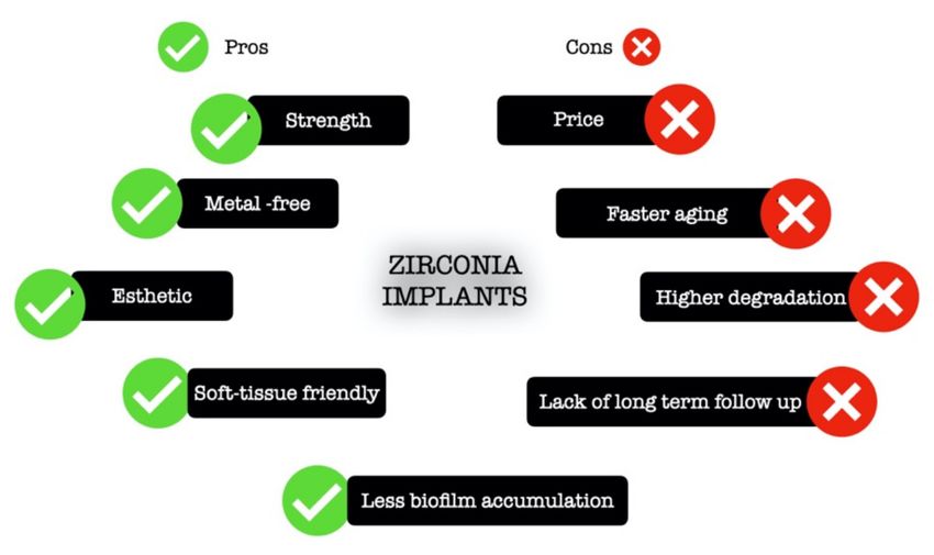
light [13]. Figure 1 presents the advantages and disadvantages of zirconia implants.
Figure 1. Schematic illustration of the advantages and drawbacks of zirconia implants.
Nowadays, the main clinical indication of a zirconia implant is the anterior esthetic
zone cases, especially in those with a scalloped, thin phenotype gingival architecture.
Another indication is patients with metal allergies and chronic diseases resulting from
this allergy. Further, owing to the lower bacteria accumulation propriety of zirconia
implants, this ceramic might be a preferred material for patients with a history of peri-
implantitis [7,18–23].
3. What Is a Titanium Implant?
Titanium is the ninth most common material found in nature [24]. The most com-
mon oxide is titanium dioxide (TiO2 ), which can be seen as a natural mineral source in
three different crystalline forms called anatase, brookite, and rutile [24–26]. This material
undergoes an allotropic transformation and changes from one crystallographic form to
another. At room temperature, it has a hexagonal close-packed structure called the α phase.
However, at high temperatures, or precisely at 883 ◦ C, it changes to the β phase where
it has a body-centered cubic structure [24,27]. Moreover, titanium can be combined with
various other elements to increase its strength and provide resistance against corrosion [25].
Thanks to titanium biocompatibility, this material is broadly used in medicine and dentistry.
Overall, titanium is considered inert and safe for human use with minimal side effects in
the human body. Figure 2 presents the advantages and disadvantages of titanium implants.
However, the true inertness of titanium has been questioned [28]. Lately, some reports
of titanium allergy are described in the literature, and the prevalence of this allergic
reaction is estimated to be 0.6% [4]. Likewise, one of the significant drawbacks of this
metal is corrosion, leading to the release of titanium particles. Therefore, it is hypothesized
that the oral cavity would be more susceptive to titanium corrosion than other parts of
the body because of the oral environment condition such as the presence of some level
of infection (gingivitis or periodontitis), the use of fluoride-containing toothpaste andAppl. Sci. 2021, 11, 6776 4 of 15
mouthwashes, and higher glucose levels, as well as the presence of saliva containing
corrosive compounds such as hydrogen, chloride ions, sulfide compounds, dissolved
oxygen, and free radicals [29–34]. Besides, it has been shown that peri-implant tissues have
100–300 parts per million (ppm) of titanium concentration [26,35], and this number can
be considerably higher if implantoplasty procedures are performed [36]. Nowadays, the
effect of free titanium particles in the body is still not clear in the literature; however, some
reports indicate that these particles could be harmful [37,38]. In the orthopedic field, for
example, the most common reason for titanium prosthesis replacement is aseptic loosening,
a local periprosthetic osteolysis around joint replacements triggered by wear debris in the
absence of bacteria, an inherently mechanical problem [38,39]. More precisely, in the oral
cavity, free titanium particles can trigger DNA damage response in oral epithelial cells and
the release of titanium parts by ultrasonic scaling can activate the inflammatory response,
by secretion of IL-1β, IL-6, and TNF-α [40–42]. Furthermore, titanium corrosion may lead
to reduced implant strength, compromising its stability [36,43].
Figure 2. Schematic illustration of advantages and drawbacks of titanium implants.
Titanium alloys are categorized into five micro-structure categories: α, near α, α + β,
near β, and β. The alloying elements are classified as α-stabilizers, for instance, aluminum
and oxygen, or β-stabilizers, such as vanadium, iron, nickel, and cobalt [44]. The most
common titanium alloys are so-called commercially pure titanium and Ti-6Al-4V, also called
TC4, Ti64, or ASTM Grade 5. These alloys are standard in dentistry owing to their high
specific strength and excellent corrosion resistance [45]. The commercially pure titanium
is classified as α micro-structure. It is available in four different grades numbered 1 to 4,
according to the purity and the processing oxygen content, with 1 being the least in terms
of oxygen content and 4 the highest. Moreover, the grades differ in corrosion resistance,
ductility, and strength. On the other hand, Ti-6Al-4V is categorized as α + β alloy and is
composite of 90% titanium, 6% aluminum, and 4% vanadium [46]. It has higher strength
and elongation at failure compared with commercially pure titanium. However, aluminum
and vanadium can be released by this alloy, causing problems in bone mineralization and
allergy reactions [47,48].
4. Osteointegration
Osseointegration is a term from the Latin language where osseous means “bony”
and integrare “to make whole”, and it is defined as a “function ankylosis” [49]. Similar
to in titanium implants, the osseointegration of zirconia implants is a dynamic process
composed of primary stability and secondary stability. Immediately after implant place-
ment, mechanical fixation of the implant provides primary stability, which is direct contact
between the bony walls and the implant. It is considered biomechanical stability as there is
no biological activity. On the other hand, the secondary biological stability results from
the cellular activity with new bone deposition on the implant surface, giving rise to the
functional ankylosis [50,51].Appl. Sci. 2021, 11, 6776 5 of 15
At the histological level, osteointegration is an active process that evolves basically
two phases: the establishment of the implant in the recipient site and its maintenance.
A classical study performed by Berglundh and colleagues [52] elucidates the implant
wound healing progression. Briefly, after the titanium is placed in the drilled site, the
implant bed is filled with blood from the marginal tissue, and threads of the implant
assure the primary stability in direct contact with the surrounding bone. A water molecule
forms a pellicle in the implant surface, which facilitates the adhesion of intracellular matrix
proteins provided by the blood cells and interstitial fluid [53]. These proteins assist cell
adhesion, migration, and differentiation. Next, when hemostasis is achieved and the blood
clot is formed, the inflammatory cells are attracted to the wound site, and give rise to the
granulation tissues, and fibroblast-like cells can be encountered in the implant zone area.
One week after the implant placement, the provisional matrix replaces the granulation
tissues, and the first signs of woven bone are present. Between one and two weeks, the
bone tissues between the threads, responsible for primary mechanical stability, began to
be reabsorbed by osteoclasts, and new viable bone became deposited. Between eight to
twelve weeks, the mature bone can be found to be surrounded by bone marrow [52,54,55].
Overall, titanium and zirconia implants present the same bone tissue integration [56].
However, microstructure and implant surface modification methods play a key role in
osseointegration. These implant modifications are made to improve mechanical properties
of the implants like strength, ductility, biological function, bacterial adhesion, wear, and
fracture toughness. This has been well demonstrated in the literature for titanium im-
plants [57–61]. Basically, the surface modifications can be prepared by physical or chemical
methods. Among physical modifications can be cited maching, grit-blasting, acid-etching,
laser ablation, and nanocomposite. Regarding the chemical modification, examples that
can be mentioned are crystalline deposition, oxidation, fluoride treatment, UV treatment,
and hydroxyapatite addition [62,63]. Surfaces with proper roughness and hydrophilicity
are prone to promote more osseointegration than other surfaces. The most common surface
modifications in clinical practice are SLA, SLActive, Osseotite, and TiUnite. SLA has a
rough surface produced by sandblasting with large grit followed by mixed acid etching
with hydrochloric and sulfuric materials. The SLActive surface is prepared by rinsing
SLA-treated implants under a nitrogen atmosphere and storing them in NaCl solution
rather than placing them in dry storage. The Osseotite surface is a rough surface with a
uniform micro-texture produced by the dual-acid (HCl-H2 SO4 ) etching method. Lastly,
TiUnite results from the oxidation process in which implants are treated in a galvanic cell
containing phosphoric acid electrolyte. The TiUnite surface is characterized by a thick TiO2
layer enriched with highly crystalline calcium phosphate [19,64–66]. A recent network
meta-analysis [67] concluded that, regarding osseointegration in the early healing stages,
SLActive shows better results. However, over time, TiUnite is the best option among the
four implants evaluated. Concerning stability, all implants are eligible with ISQ values
greater than 60. Additionally, a previous systematic review evaluation of 27 randomized
clinical trials including 38 different implant types concluded that relatively smooth (turned)
surfaces were less prone to lose bone as a result of chronic infection (peri-implantitis) than
implants with much rougher surfaces (titanium-plasma-sprayed) [68]. Considering the
effect of nanostructure at the cellular level, a systematic review concluded that this type of
structure has a remarkable advantage in osteoblast proliferation [69].
Specifically regarding zirconia implants, according to Martins and colleagues [70],
limited bone remodeling with machined-zirconia implants containing yttria over time
can decrease the secondary stability of this type of implant compared with machined and
resorbable blast media titanium ones.
In the same way as happens in the titanium implants, the surface modification of
the zirconia implants may dictate differences in their osseointegration. Preclinical studies
have revealed bone apposition on zirconia implants with various surface modifications,
including sandblasting, etching [71], sintering, and coating [72], and even subtle alterations
in the surface may have an enormous benefit in the osseointegration.Appl. Sci. 2021, 11, 6776 6 of 15
Owing to the increase in demand for zirconia implants, mainly in an esthetic-driven
patient, clinicians and researchers question themselves about the efficiency of this type
of implant and its non-inferiority in relation to conventional titanium implants. Mostly
preclinical, but also some randomized clinical trials and systematic reviews have been pub-
lished to answer these questions. Table 1 summarizes the results from existing systematic
reviews analyzing the osseointegration of zirconia implants.
Table 1. Summary of systematic reviews (SRs) analyzing the osseointegration of zirconia implants.
Reference Aim Main Results Conclusions
Zirconia implants may
The mean bone–implant contact percentage
SR with seven animals’ become an alternative to
was above 60%. In studies comparing zirconia
studies to report the clinical titanium implants, but cannot
Wenz et al., vs. titanium, the first was comparable to or
success of zirconia implants currently be recommended for
2008 [73] even better than the second. Surface
and osseointegration routine clinical use, as no
modifications may further improve initial bone
compared with titanium. long-term clinical data
healing and resistance to removal torque.
are available.
SR with 14 clinical studies The overall survival rate of zirconia was 92% Zirconia ceramics could
Hashim et al., and at least 1 year follow up after one year of function. Early failure of potentially be the alternative
2016 [10] to analyze osseointegration zirconia implants oscillated between 1.8 and to titanium for a non-metallic
of zirconia implants. 100 %, with 77% of overall early failure rate. implant solution.
The survival rate and
SR of two RCTs and seven
The survival rate after 12 months ranged marginal bone loss of zirconia
prospective clinical trials
Pieralli et al., between 85% and 100 with a mean of 95.6%. dental implants after one year
with zirconia implants
2017 [74] The mean marginal bone loss after 12 months are comparable to available
restored with single crowns
follow up was 0.79 mm. data of two-piece
and fixed dental prostheses.
titanium implants.
Titanium implants showed a bone to implant
(BIC) contact of 60.70% after the mean
follow-up time. In comparison, zirconia
implants showed a reduced BIC of −3.47%.
No significant difference of
Titanium implants showed a removal torque
zirconia or titanium on the
SR with 54 studies analyzing analysis of 102.71 Ncm after the mean
Pieralli et al., results analyzed could be
osseointegration of zirconia follow-up time. No significant difference was
2018 [75] found. Moreover, the results
implants in animal studies. found for zirconia implants (−7.31 Ncm, p =
might depend on the animal
0.44). In general, smooth implant surfaces
model studied.
(−43.85 Ncm, p = 0.03) and implant loading
(+40.78 Ncm, p < 0.01) significantly influenced
the removal torque. A roughness of Ra >2 m
and Sa > 2 m resulted in a positive effect.
SR with 19 articles regarding The marginal bone remodeling of 0.8 mm and
Zirconia presents results
survival rate and marginal 1.01 mm at 1 year and 2 years post-loading,
Borges et al., similar to titanium. However,
bone loss (MBL), with a respectively. Overall failure rate of 6.8% in 2.75
2020 [76] the studies demonstrate a
minimum follow-up of years. Early failure was 3.4%, late failure was
short follow-up period.
12 months. 1.7%, and implant fracture was 1.7.
A study with human biopsies from 22 failed zirconia implants (failed from trauma
or extensive bone loss compatible with peri-implantitis) showed dense bone with stable
laminar structure in tight contact with the implant surface and no gaps. The bone–implant
area ranged from 58.1 to 93.7% [77].
5. Soft Tissue Reaction
In natural dentition, the consensus is that there are not necessarily specific keratinized
gingiva dimensions to maintain periodontal health under good oral hygiene [78–80]. On
the other hand, the peri-implant soft tissue is different from that around a natural tooth.
In implants, as its surface is directly attached to the bone, there are not periodontal fibers
anchoring in the bone, and collagen fibers run parallel to the implant surface without directAppl. Sci. 2021, 11, 6776 7 of 15
anchorage [81,82]. This soft tissue architecture is considered unsteady and provides an
inferior biologic seal [83]. As a consequence, the soft tissues surrounding the implants
are more prone to develop inflammatory disease processes. Furthermore, implants with a
thin phenotype and inadequate keratinized mucosa width (less than 2 mm) have a higher
prevalence of peri-implantitis and peri-implant mucositis [83–85], demonstrating that soft
tissue plays a crucial role in implant health.
Currently, several in vitro studies show that zirconia implants have better biological
reactions in periodontal tissues when compared with titanium [13]. For instance, fibrob-
lasts proliferate faster and are more equally distributed in zirconia disks compared with
titanium [86]. Moreover, this material enhances collagen and extracellular matrix proteins’
release [87,88]. Furthermore, analyzing the results of zirconia interaction with epithelial
cells, it looks like zirconia increases the differentiation and proliferation of these cells,
facilitating the healing process and protective scarring around the implants [13]. More-
over, the zirconia implants present longer junctional epithelium and a higher density of
collagen fibers, which might increase the soft-tissue closure and decrease the inflammatory
infiltration around zirconia implants [56].
Regarding the orientation of collagen fibers around implants, the same way as in
titanium pieces, the adherence of collagen fibers into zirconia implants occurs in a parallel
and parallel-oblique way [89]. Furthermore, the supracrestal tissue adhesion (also formerly
known as biology width or supracrestal tissue attachment) is more prolonged in titanium
implants (mean of 5 mm) compared with zirconia implants (4.5 mm), meaning that zirconia
implants have a significantly lower sulcus depth. Nevertheless, the epithelium length
mean was 2.9 mm in both groups. On the other hand, a significant difference was found
in the connective tissue of both materials (2.4 mm around zirconia implants and 1.5 mm
around titanium implants), demonstrating a higher organization of collagen fibers. These
results suggest that a more mature and pronounced soft tissue integration happens with
zirconia implants compared with titanium [53].
6. Microbiota and Peri-Implantitis
Peri-implantitis is an inflammatory process around an osseointegrated implant, in-
cluding soft tissue inflammation and progressive loss of supporting bone beyond biological
bone remodeling [1]. This inflammatory process is the leading cause of loss of implants
in the long term. Peri-implantitis results from biofilm dysbiosis deposits surrounding
the peri-implants’ mucosa, meaning that bacteria deposition is essential to develop the
inflammation reaction. In addition, research considering bacterial adhesion on titanium
has revealed that the corrosion of titanium increases plaque accumulation. On this matter,
zirconia implants have been suggested as a peri-implant mucosa-friendly alternative [90].
Recent studies show intriguing results promoting the advantage of zirconia implants
in this matter regarding biofilm formation and bacterial adhesion in zirconia implants.
An in vitro study aimed to compare the biofilm formation in the zirconia or titanium
disks using three-species biofilm or human plaque samples in an anaerobic flow chamber
mode. The researchers found that zirconia showed a reduction in biofilm thickness and
mass compared with titanium, but similar biofilm metabolism was found. These results
suggest that zirconia implants can form less plaque, leading to less peri-implant inflam-
mation than titanium implants [20]. Considering the bacterial adhesion in zirconia, the
Grossner–Schreiber group [22] found that bacterial counts were higher with titanium discs
than zirconia, while Scarano et al. [23] showed that bacterial adhesion was significantly
higher with pure titanium surfaces as compared with zirconium oxide surfaces, as data
also supported by Sadid-Zadeh and collaborators find [87]. Moreover, these results are sup-
ported by an animal model. That is, a study comparing ligature-induced peri-implantitis
around zirconia or titanium implants in dogs. They found that the first presented less
crestal peri-implant bone loss and no implant failure. In contrast, the second shows one
implant loss due to peri-implantitis [21]. Additionally, a recent systematic review withAppl. Sci. 2021, 11, 6776 8 of 15
meta-analysis indicated that zirconia accumulated less initial oral biofilm parameters and
less roughness in the zirconia surface [91].
On the other hand, taking into consideration the role of host response in peri-implantitis,
a recent study [92] compared the expression of immunoinflammatory markers in the tissues
around ceramic and titanium implants. The authors found a similar number of inflam-
matory cells (T- and B-cells) in mucosa surrounding ceramic implants when compared
with titanium ones, concluding that the cellular response to the inflammatory challenge
provoked by the biofilm around implants is the same, independent of the material with
which the implant is made.
Besides, the hypothesis has been raised in the literature that not only biofilm is re-
sponsible for peri-implantitis in titanium implants, but also foreign body reaction, allergies,
cement excess, or even metallosis [93]. Considering all the factors discussed above, it could
be assumed that zirconia implants present lower prevalence rates when compared with
titanium implants. Nevertheless, according to a systematic review and meta-analysis [94],
owing to the heterogeny of assessment and short-term evaluation of the zirconia implants’
clinical studies, non-conclusive results are found regarding inflammatory parameters in
peri-implant mucosa around zirconia implants. On the other hand, one prospective study
that followed up zirconia implants for 7.8 years found no difference between teeth (control
group) and implant regarding bleeding index, attachment level, or bacterial colonization.
Interestingly, zirconia implants presented less plaque accumulation and recession, but
more probing depths when compared with natural dentition [95]. Moreover, another
systematic review [96] concluded that zirconia implants had less plaque accumulation and
less inflammation around the peri-implant mucosa.
7. Long-Term Results
The objective of a dental implant is to replace a missing tooth in a functional and
esthetic way. Thus, for an implant to be considered successful, it needs to be permanently
osseointegrated and healthy. Thus, long-term results are essential to determine which
material is superior. In this way, to compare titanium versus zirconia implants is an unfair
evaluation because, on one side, it encounters a long-term result of the stabilized treatment
option—titanium implants—against a novel material with short-term results and specific
indications—zirconia.
Regarding the overall success of the implants, both materials present a satisfactory
survival rate. According to Buser et al. [97], titanium implants show an overall 10-year
implant survival rate of 98.8% and a success rate of 97.0%. On the other hand, the recent
systematic review and meta-analysis, which evaluate randomized clinical trials of at least 1
year follow-up showed that zirconia implants present a survival rate of 91.5% to 98.3%, a
success rate of 91.6%, and marginal bone loss ranging from 0.7 mm to 0.98 mm [94].
Table 2 presents the main findings of systematic reviews that analyzed titanium versus
zirconia implants.Appl. Sci. 2021, 11, 6776 9 of 15
Table 2. Summary of systematic reviews comparing zirconia and titanium implants.
Reference Aim Main Results Conclusions
No difference was found in the rate of
osseointegration between the different
Animal studies show similar results between titanium and zirconia implants. Clinical implant materials in animal experiments.
Andreioitelli et al., SR with 25 studies describing
outcomes presented a survival rate from 84% after 21 months to 98% after 1 year for Regarding the clinical studies, the authors
2009 [98] clinical animal studies.
zirconia implants. concluded that only cohort investigations
with questionable scientific value were found
at that time.
Titanium: in rabbits, BIC = 32%, after 3 weeks and 80% in 4 weeks; in minipigs, 24% after
4 weeks and 83% after 12 weeks.
Removal torque—in rabbits, 42 Ncm in the tibia after 6 weeks 36 to 74 Ncm in the femur
SR with 16 animal studies BIC and removal torque did not show a
Manzano et al., after 6 weeks; in minipigs, 245 Ncm after 4 weeks and 105 Ncm after 12 weeks.
comparing the removal torque significant statistical difference between
2014 [99] Zirconia—in rabbits, 72% after 4 weeks;
and bone–implant contact. titanium and zirconia implants.
in minipigs, 71% after 12 weeks.
Removal torque—in rabbits, 12 Ncm in the tibia after 6 weeks to 98 Ncm in the femur
after 6 weeks; in minipigs, 112 Ncm after 4 weeks and 26 Ncm after 12 weeks.
The range of BIC was 31.80% to 87.85% for titanium, 33.74% to 84.17% for machined
zirconia, 41.35% to 67.4% for blasted zirconia, and 51.1% to 71.4% for surface etched
SR and meta-analysis of
Hafezeqoran and zirconia in the studies. No significant statistical difference in the BIC values between
15 preclinical studies analyzing Acid-etched zirconia implants may present a
Koodaryan 2017 titanium machined zirconia or blasted zirconia implants was found. However,
bone to implant contact and successful osseointegration.
[100] acid-etched zirconia significantly increased BIC compared with titanium implants
removal torque.
(p = 0.032). More favorable removal torque values for untreated zirconia implants than
those of machined zirconia implants, which were statistically significant (p < 0.001).
Zirconia has shown a similar %BIC to that of Ti implants in most of the studies. Cracks
Siddiqi et al., 2017 The use of zirconia implants to restore
SR of 29 animal studies. and material fracture have been attributed as the major technical complications related to
[101] edentulous area should be carefully evaluated.
Zr implants possibly leading to early implant failure.
The early fracture of a one-piece zirconia implant, especially in the posterior region, is a
sensitive and critical factor to be considered regarding its use and acceptance in all Zirconia implants are a favorable alternative
Sivaraman et al., clinical situations. As most clinical studies on zirconia implants are short-term, to titanium with greater soft tissue response,
Critical review
2018 [16] substantial evidence supported by long-term clinical trials is warranted before biocompatibility, and aesthetics with
zirconia-based implant systems can completely replace titanium for comparable osseointegration.
prosthetic rehabilitation.Appl. Sci. 2021, 11, 6776 10 of 15
Table 2. Cont.
Reference Aim Main Results Conclusions
Outcome measurements:
Titanium—BIC = 59.1%
Removal torque = 102.6 Ncm
Zirconia and titanium implants demonstrate a
Push in = 25.1 N
similar soft and hard tissue integration
SR and meta-analysis of 37 Zirconia—BIC = 55.9%
capacity. However, titanium tended to show a
Roehling et al., preclinical studies comparing Removal torque = 71.5 Ncm
faster initial osseointegration process
2019 [21] titanium and zirconia implants Push in = 22.0 N
compared with zirconia. Nevertheless, animal
regarding soft and hard tissue. Similar qualitative soft tissue integration was reported for zirconia and titanium
species and study protocols can significantly
implants. However, faster maturation processes of epithelial and connective tissues
influence the outcomes.
around zirconia implants were assumed. Quantitatively, similar bone width dimensions
were evaluated for titanium (3.5 mm) and zirconia (3.2 mm), whereas the loading
protocol significantly influenced the outcomes.
SR of six studies comparing
ArRejaie et al., Zirconia implants showed higher crestal bone loss compared with titanium. Variable
one-piece titanium vs. High heterogeneity of results.
2019 [102] results regarding failure rate were found.
one-piece zirconia implants.
Zirconia implants showed better esthetic outcomes, less plaque accumulation, and less
It seems like zirconia implants present better
SR with 15 articles evaluating inflammation around the peri-implant mucosa than titanium. Moreover, zirconia
Comisso et al., clinical performance than titanium ones.
aesthetic, clinical benefits, and presented a lower probing depth and an ideal papilla crown proportion with an
2021 [96] However, more studies are needed to confirm
survival rates. increased papillary height. For survival and success rates, no difference was found
these data.
between the materials.
* BIC = bone implant contact.Appl. Sci. 2021, 11, 6776 11 of 15
8. Conclusions
To conclude, answering the question posed in the title of this review, it is unlikely
that zirconia implants will replace the titanium implants’ use entirely owing to the high
success rates, long-term stability, and satisfactory osseointegration of the titanium implants
found on the market nowadays. However, based on the literature, it seems like zirconia
implants are reliable substitutes for cases in which titanium is not an option or in cases
where aesthetics is in high demand. Moreover, it is essential for the clinician to be aware
of the advantages and drawbacks of this implant option to make the best decision for the
treatment of the edentulous area. Furthermore, possibly, in the future, zirconia implants
can be the first choice for a patient with a history of periodontitis/peri-implantitis, but more
studies with longer follow-up are necessary to prove that zirconia has better performance
against bacterial challenges.
Based on the findings of this review, it is possible to conclude that
• Titanium implants are still the standard material for the replacement of a missing tooth;
• Titanium particles are potentially harmful to the peri-implant tissue; however, more
studies are necessary for definitive conclusions;
• Osteointegration of zirconia and titanium implants are similar and are influenced by
microstructure and the treatment of the surface;
• Some clinical studies show better soft tissue response with zirconia implants, suggest-
ing that this material can provide a protective effect against inflammation;
• Some studies suggest that zirconia implant seems to be less susceptive to peri-
implantitis than titanium implants;
• There is a lack of studies with a long-term follow up of zirconia implants.
Author Contributions: L.P.W., H.-L.C. and H.-L.W. contributed to the conception, acquisition, and
interpretation of the review data. L.P.W. was responsible for initial draft preparation. H.-L.C. and
H.-L.W. were responsible for the final review and supervision. All authors have read and agreed to
the published version of the manuscript.
Funding: This research received no external funding.
Institutional Review Board Statement: Not applicable.
Informed Consent Statement: Not applicable.
Data Availability Statement: Not applicable.
Conflicts of Interest: The authors declare no conflict of interest.
References
1. Berglundh, T.; Armitage, G.; Araujo, M.G.; Avila-Ortiz, G.; Blanco, J.; Camargo, P.M.; Chen, S.; Cochran, D.; Derks, J.;
Figuero, E.; et al. Peri-implant diseases and conditions: Consensus report of workgroup 4 of the 2017 World Workshop on
the Classification of Periodontal and Peri-Implant Diseases and Conditions. J. Clin. Periodontol. 2018, 45, S286–S291. [CrossRef]
2. Kordbacheh Changi, K.; Finkelstein, J.; Papapanou, P.N. Peri-implantitis prevalence, incidence rate, and risk factors: A study of
electronic health records at a U.S. dental school. Clin. Oral Implants Res. 2019, 30, 306–314. [CrossRef]
3. Lee, J.H.; Kim, Y.T.; Jeong, S.N.; Kim, N.H.; Lee, D.W. Incidence and pattern of implant fractures: A long-term follow-up
multicenter study. Clin. Implant Dent. Relat. Res. 2018, 20, 463–469. [CrossRef] [PubMed]
4. Vijayaraghavan, V.; Sabane, A.V.; Tejas, K. Hypersensitivity to titanium: A less explored area of research. J. Indian Prosthodont. Soc.
2012, 12, 201–207. [CrossRef]
5. Kelly, J.R.; Denry, I. Stabilized zirconia as a structural ceramic: An overview. Dent. Mater. 2008, 24, 289–298. [CrossRef]
6. Wang, T.; Wang, L.; Lu, Q.; Fan, Z. Changes in the esthetic, physical, and biological properties of a titanium alloy abutment
treated by anodic oxidation. J. Prosthet. Dent. 2019, 121, 156–165. [CrossRef]
7. Kim, A.; Campbell, S.D.; Viana, M.A.; Knoernschild, K.L. Abutment Material Effect on Peri-implant Soft Tissue Color and
Perceived Esthetics. J. Prosthodont. 2016, 25, 634–640. [CrossRef]
8. Thoma, D.S.; Ioannidis, A.; Cathomen, E.; Hämmerle, C.H.; Hüsler, J.; Jung, R.E. Discoloration of the Peri-implant Mucosa Caused
by Zirconia and Titanium Implants. Int. J. Periodontol. Restor. Dent. 2016, 36, 39–45. [CrossRef] [PubMed]
9. Cionca, N.; Hashim, D.; Mombelli, A. Zirconia dental implants: Where are we now, and where are we heading? Periodontol. 2000
2017, 73, 241–258. [CrossRef]Appl. Sci. 2021, 11, 6776 12 of 15
10. Hashim, D.; Cionca, N.; Courvoisier, D.S.; Mombelli, A. A systematic review of the clinical survival of zirconia implants.
Clin. Oral Investig. 2016, 20, 1403–1417. [CrossRef] [PubMed]
11. Koth, D.L.; McKinney, R.V.; Steflik, D.E.; Davis, Q.B. Clinical and statistical analyses of human clinical trials with the single crystal
aluminum oxide endosteal dental implant: Five-year results. J. Prosthet. Dent. 1988, 60, 226–234. [CrossRef]
12. Vagkopoulou, T.; Koutayas, S.O.; Koidis, P.; Strub, J.R. Zirconia in dentistry: Part 1. Discovering the nature of an upcoming
bioceramic. Eur. J. Esthet. Dent. 2009, 4, 130–151. [PubMed]
13. Kunrath, M.F.; Gupta, S.; Lorusso, F.; Scarano, A.; Noumbissi, S. Oral Tissue Interactions and Cellular Response to Zirconia
Implant-Prosthetic Components: A Critical Review. Materials 2021, 14, 2825. [CrossRef]
14. Covacci, V.; Bruzzese, N.; Maccauro, G.; Andreassi, C.; Ricci, G.A.; Piconi, C.; Marmo, E.; Burger, W.; Cittadini, A. In vitro
evaluation of the mutagenic and carcinogenic power of high purity zirconia ceramic. Biomaterials 1999, 20, 371–376. [CrossRef]
15. Piconi, C.; Maccauro, G. Zirconia as a ceramic biomaterial. Biomaterials 1999, 20, 1–25. [CrossRef]
16. Sivaraman, K.; Chopra, A.; Narayan, A.I.; Balakrishnan, D. Is zirconia a viable alternative to titanium for oral implant? A critical
review. J. Prosthodont. Res. 2018, 62, 121–133. [CrossRef] [PubMed]
17. Lughi, V.; Sergo, V. Low temperature degradation -aging- of zirconia: A critical review of the relevant aspects in dentistry.
Dent. Mater. 2010, 26, 807–820. [CrossRef] [PubMed]
18. Apratim, A.; Eachempati, P.; Krishnappa Salian, K.K.; Singh, V.; Chhabra, S.; Shah, S. Zirconia in dental implantology: A review.
J. Int. Soc. Prev. Commun. Dent. 2015, 5, 147–156. [CrossRef] [PubMed]
19. Bornstein, M.M.; Valderrama, P.; Jones, A.A.; Wilson, T.G.; Seibl, R.; Cochran, D.L. Bone apposition around two different
sandblasted and acid-etched titanium implant surfaces: A histomorphometric study in canine mandibles. Clin. Oral. Implants Res.
2008, 19, 233–241. [CrossRef] [PubMed]
20. Roehling, S.; Astasov-Frauenhoffer, M.; Hauser-Gerspach, I.; Braissant, O.; Woelfler, H.; Waltimo, T.; Kniha, H.; Gahlert, M.
In Vitro Biofilm Formation on Titanium and Zirconia Implant Surfaces. J. Periodontol. 2017, 88, 298–307. [CrossRef]
21. Roehling, S.; Schlegel, K.A.; Woelfler, H.; Gahlert, M. Zirconia compared to titanium dental implants in preclinical studies—A
systematic review and meta-analysis. Clin. Oral Implants Res. 2019, 30, 365–395. [CrossRef]
22. Grössner-Schreiber, B.; Teichmann, J.; Hannig, M.; Dörfer, C.; Wenderoth, D.F.; Ott, S.J. Modified implant surfaces show different
biofilm compositions under in vivo conditions. Clin. Oral Implants Res. 2009, 20, 817–826. [CrossRef] [PubMed]
23. Scarano, A.; Piattelli, M.; Caputi, S.; Favero, G.A.; Piattelli, A. Bacterial adhesion on commercially pure titanium and zirconium
oxide disks: An in vivo human study. J. Periodontol. 2004, 75, 292–296. [CrossRef] [PubMed]
24. Barksdale, J. Titanium. In The Encyclopedia of the Chemical Elements; Hampel, C.A., Ed.; Reinhold Book Corporation: New York,
NY, USA, 1968; pp. 732–738.
25. Fage, S.W.; Muris, J.; Jakobsen, S.S.; Thyssen, J.P. Titanium: A review on exposure, release, penetration, allergy, epidemiology, and
clinical reactivity. Contact Dermat. 2016, 74, 323–345. [CrossRef] [PubMed]
26. Kaur, M.; Singh, K. Review on titanium and titanium based alloys as biomaterials for orthopaedic applications. Mater. Sci. Eng. C
Mater. Biol. Appl. 2019, 102, 844–862. [CrossRef]
27. Donachie, M.J. Titanium a Technical Guide; ASM International: Columbus, OH, USA, 1988.
28. Kotsakis, G.A.; Olmedo, D.G. Peri-implantitis is not periodontitis: Scientific discoveries shed light on microbiome-biomaterial
interactions that may determine disease phenotype. Periodontol. 2000 2021, 86, 231–240. [CrossRef] [PubMed]
29. Koike, M.; Fujii, H. The corrosion resistance of pure titanium in organic acids. Biomaterials 2001, 22, 2931–2936. [CrossRef]
30. Barão, V.A.; Mathew, M.T.; Assunção, W.G.; Yuan, J.C.; Wimmer, M.A.; Sukotjo, C. The role of lipopolysaccharide on the
electrochemical behavior of titanium. J. Dent. Res. 2011, 90, 613–618. [CrossRef]
31. Barão, V.A.; Mathew, M.T.; Yuan, J.C.; Knoernschild, K.L.; Assunção, W.G.; Wimmer, M.A.; Sukotjo, C. Influence of corrosion on
lipopolysaccharide affinity for two different titanium materials. J. Prosthet. Dent. 2013, 110, 462–470. [CrossRef]
32. Nakagawa, M.; Matsuya, S.; Udoh, K. Effects of fluoride and dissolved oxygen concentrations on the corrosion behavior of pure
titanium and titanium alloys. Dent. Mater. J. 2002, 21, 83–92. [CrossRef]
33. Noguti, J.; de Oliveira, F.; Peres, R.C.; Renno, A.C.; Ribeiro, D.A. The role of fluoride on the process of titanium corrosion in oral
cavity. Biometals 2012, 25, 859–862. [CrossRef] [PubMed]
34. Lindholm-Sethson, B.; Ardlin, B.I. Effects of pH and fluoride concentration on the corrosion of titanium. J. Biomed. Mater. Res. A
2008, 86, 149–159. [CrossRef] [PubMed]
35. Suárez-López Del Amo, F.; Garaicoa-Pazmiño, C.; Fretwurst, T.; Castilho, R.M.; Squarize, C.H. Dental implants-associated release
of titanium particles: A systematic review. Clin. Oral Implants Res. 2018, 29, 1085–1100. [CrossRef] [PubMed]
36. Stavropoulos, A.; Bertl, K.; Eren, S.; Gotfredsen, K. Mechanical and biological complications after implantoplasty—A systematic
review. Clin. Oral Implants Res. 2019, 30, 833–848. [CrossRef] [PubMed]
37. Wang, J.X.; Fan, Y.B.; Gao, Y.; Hu, Q.H.; Wang, T.C. TiO2 nanoparticles translocation and potential toxicological effect in rats after
intraarticular injection. Biomaterials 2009, 30, 4590–4600. [CrossRef] [PubMed]
38. Fretwurst, T.; Nelson, K.; Tarnow, D.P.; Wang, H.L.; Giannobile, W.V. Is Metal Particle Release Associated with Peri-implant Bone
Destruction? An Emerging Concept. J. Dent. Res. 2018, 97, 259–265. [CrossRef] [PubMed]
39. Marshall, A.; Ries, M.D.; Paprosky, W. How prevalent are implant wear and osteolysis, and how has the scope of osteolysis
changed since 2000? J. Am. Acad. Orthop. Surg. 2008, 16, S1–S6. [CrossRef] [PubMed]Appl. Sci. 2021, 11, 6776 13 of 15
40. Suárez-López Del Amo, F.; Rudek, I.; Wagner, V.P.; Martins, M.D.; O’Valle, F.; Galindo-Moreno, P.; Giannobile, W.V.; Wang, H.L.;
Castilho, R.M. Titanium Activates the DNA Damage Response Pathway in Oral Epithelial Cells: A Pilot Study. Int. J. Oral
Maxillofac. Implants 2017, 32, 1413–1420. [CrossRef]
41. Pettersson, M.; Kelk, P.; Belibasakis, G.N.; Bylund, D.; Molin Thorén, M.; Johansson, A. Titanium ions form particles that activate
and execute interleukin-1β release from lipopolysaccharide-primed macrophages. J. Periodont. Res. 2017, 52, 21–32. [CrossRef]
[PubMed]
42. Eger, M.; Sterer, N.; Liron, T.; Kohavi, D.; Gabet, Y. Scaling of titanium implants entrains inflammation-induced osteolysis.
Sci. Rep. 2017, 7, 39612. [CrossRef]
43. Okazaki, Y.; Rao, S.; Ito, Y.; Tateishi, T. Corrosion resistance, mechanical properties, corrosion fatigue strength and cytocompatibil-
ity of new Ti alloys without Al and V. Biomaterials 1998, 19, 1197–1215. [CrossRef]
44. Nicholson, J.W. Titanium Alloys for Dental Implants: A Review. Prosthesis 2020, 2, 11. [CrossRef]
45. Liu, X.; Chen, S.; Tsoi, J.K.H.; Matinlinna, J.P. Binary titanium alloys as dental implant materials—A review. Regen. Biomater. 2017,
4, 315–323. [CrossRef]
46. Niinomi, M. Mechanical biocompatibilities of titanium alloys for biomedical applications. J. Mech. Behav. Biomed. Mater. 2008, 1,
30–42. [CrossRef] [PubMed]
47. Klein, G.L. Aluminum toxicity to bone: A multisystem effect? Osteoporos. Sarcopenia 2019, 5, 2–5. [CrossRef]
48. Thyssen, J.P.; Jakobsen, S.S.; Engkilde, K.; Johansen, J.D.; Søballe, K.; Menné, T. The association between metal allergy, total hip
arthroplasty, and revision. Acta Orthop. 2009, 80, 646–652. [CrossRef] [PubMed]
49. Listgarten, M.A.; Lang, N.P.; Schroeder, H.E.; Schroeder, A. Periodontal tissues and their counterparts around endosseous
implants. Clin. Oral Implants Res. 1991, 2, 1–19. [CrossRef]
50. Bosshardt, D.D.; Chappuis, V.; Buser, D. Osseointegration of titanium, titanium alloy and zirconia dental implants: Current
knowledge and open questions. Periodontol. 2000 2017, 73, 22–40. [CrossRef] [PubMed]
51. Monje, A.; Ravidà, A.; Wang, H.L.; Helms, J.A.; Brunski, J.B. Relationship Between Primary/Mechanical and Secondary/Biological
Implant Stability. Int. J. Oral Maxillofac. Implants 2019, 34, s7–s23. [CrossRef] [PubMed]
52. Berglundh, T.; Abrahamsson, I.; Lang, N.P.; Lindhe, J. De novo alveolar bone formation adjacent to endosseous implants.
Clin. Oral Implants Res. 2003, 14, 251–262. [CrossRef]
53. Kazemian, M.; Hoseini, S.H.; Ghorbanzade, S. A brief overview of cellular and molecular mechanisms of osseointegration. Int. J.
Contemp. Dent. Med. Rev. 2015. [CrossRef]
54. Lang, N.P.; Salvi, G.E.; Huynh-Ba, G.; Ivanovski, S.; Donos, N.; Bosshardt, D.D. Early osseointegration to hydrophilic and
hydrophobic implant surfaces in humans. Clin. Oral Implants Res. 2011, 22, 349–356. [CrossRef] [PubMed]
55. Davies, J.E. Understanding peri-implant endosseous healing. J. Dent. Educ. 2003, 67, 932–949. [CrossRef] [PubMed]
56. Liñares, A.; Grize, L.; Muñoz, F.; Pippenger, B.E.; Dard, M.; Domken, O.; Blanco-Carrión, J. Histological assessment of hard and
soft tissues surrounding a novel ceramic implant: A pilot study in the minipig. J. Clin. Periodontol. 2016, 43, 538–546. [CrossRef]
57. Buser, D.; Nydegger, T.; Oxland, T.; Cochran, D.L.; Schenk, R.K.; Hirt, H.P.; Snétivy, D.; Nolte, L.P. Interface shear strength of
titanium implants with a sandblasted and acid-etched surface: A biomechanical study in the maxilla of miniature pigs. J. Biomed.
Mater. Res. 1999, 45, 75–83. [CrossRef]
58. Buser, D.; Schenk, R.K.; Steinemann, S.; Fiorellini, J.P.; Fox, C.H.; Stich, H. Influence of surface characteristics on bone integration
of titanium implants. A histomorphometric study in miniature pigs. J. Biomed. Mater. Res. 1991, 25, 889–902. [CrossRef]
59. Carmo Filho, L.C.D.; Marcello-Machado, R.M.; Castilhos, E.D.; Del Bel Cury, A.A.; Faot, F. Can implant surfaces affect implant
stability during osseointegration? A randomized clinical trial. Braz. Oral Res. 2018, 32, e110. [CrossRef] [PubMed]
60. López-Valverde, N.; Flores-Fraile, J.; Ramírez, J.M.; Sousa, B.M.; Herrero-Hernández, S.; López-Valverde, A. Bioactive Surfaces vs.
Conventional Surfaces in Titanium Dental Implants: A Comparative Systematic Review. J. Clin. Med. 2020, 9, 47. [CrossRef]
61. Wennerberg, A.; Albrektsson, T.; Chrcanovic, B. Long-term clinical outcome of implants with different surface modifications.
Eur. J. Oral Implantol. 2018, 11, S123–S136.
62. Xue, T.; Attarilar, S.; Liu, S.; Liu, J.; Song, X.; Li, L.; Zhao, B.; Tang, Y. Surface Modification Techniques of Titanium and its Alloys
to Functionally Optimize Their Biomedical Properties: Thematic Review. Front. Bioeng. Biotechnol. 2020, 8, 603072. [CrossRef]
63. Kligman, S.; Ren, Z.; Chung, C.H.; Perillo, M.A.; Chang, Y.C.; Koo, H.; Zheng, Z.; Li, C. The Impact of Dental Implant Surface
Modifications on Osseointegration and Biofilm Formation. J. Clin. Med. 2021, 10, 1641. [CrossRef] [PubMed]
64. Xiropaidis, A.V.; Qahash, M.; Lim, W.H.; Shanaman, R.H.; Rohrer, M.D.; Wikesjö, U.M.; Hall, J. Bone-implant contact at calcium
phosphate-coated and porous titanium oxide (TiUnite)-modified oral implants. Clin. Oral Implants Res. 2005, 16, 532–539.
[CrossRef]
65. Roccuzzo, M.; Aglietta, M.; Bunino, M.; Bonino, L. Early loading of sandblasted and acid-etched implants: A randomized-
controlled double-blind split-mouth study. Five-year results. Clin. Oral Implants Res. 2008, 19, 148–152. [CrossRef] [PubMed]
66. Browaeys, H.; Defrancq, J.; Dierens, M.C.; Miremadi, R.; Vandeweghe, S.; Van de Velde, T.; De Bruyn, H. A retrospective analysis
of early and immediately loaded osseotite implants in cross-arch rehabilitations in edentulous maxillas and mandibles up to
7 years. Clin. Implant Dent. Relat. Res. 2013, 15, 380–389. [CrossRef] [PubMed]
67. Hao, C.P.; Cao, N.J.; Zhu, Y.H.; Wang, W. The osseointegration and stability of dental implants with different surface treatments
in animal models: A network meta-analysis. Sci. Rep. 2021, 11, 13849. [CrossRef] [PubMed]Appl. Sci. 2021, 11, 6776 14 of 15
68. Esposito, M.; Ardebili, Y.; Worthington, H.V. Interventions for replacing missing teeth: Different types of dental implants.
Cochrane Database Syst. Rev. 2014, Cd003815. [CrossRef]
69. Goldman, M.; Juodzbalys, G.; Vilkinis, V. Titanium surfaces with nanostructures influence on osteoblasts proliferation: A system-
atic review. J. Oral Maxillofac. Res. 2014, 5, e1. [CrossRef]
70. Martins, R.; Cestari, T.M.; Arantes, R.V.N.; Santos, P.S.; Taga, R.; Carbonari, M.J.; Oliveira, R.C. Osseointegration of zirconia
and titanium implants in a rabbit tibiae model evaluated by microtomography, histomorphometry and fluorochrome labeling
analyses. J. Periodont. Res. 2018, 53, 210–221. [CrossRef]
71. Hoffmann, O.; Angelov, N.; Zafiropoulos, G.G.; Andreana, S. Osseointegration of zirconia implants with different surface
characteristics: An evaluation in rabbits. Int. J. Oral Maxillofac. Implants 2012, 27, 352–358. [PubMed]
72. Lee, J.; Sieweke, J.H.; Rodriguez, N.A.; Schüpbach, P.; Lindström, H.; Susin, C.; Wikesjö, U.M. Evaluation of nano-technology-
modified zirconia oral implants: A study in rabbits. J. Clin. Periodontol. 2009, 36, 610–617. [CrossRef]
73. Wenz, H.J.; Bartsch, J.; Wolfart, S.; Kern, M. Osseointegration and clinical success of zirconia dental implants: A systematic review.
Int. J. Prosthodont. 2008, 21, 27–36.
74. Pieralli, S.; Kohal, R.J.; Jung, R.E.; Vach, K.; Spies, B.C. Clinical Outcomes of Zirconia Dental Implants: A Systematic Review.
J. Dent. Res. 2017, 96, 38–46. [CrossRef] [PubMed]
75. Pieralli, S.; Kohal, R.J.; Lopez Hernandez, E.; Doerken, S.; Spies, B.C. Osseointegration of zirconia dental implants in animal
investigations: A systematic review and meta-analysis. Dent. Mater. 2018, 34, 171–182. [CrossRef] [PubMed]
76. Borges, H.; Correia, A.R.M.; Castilho, R.M.; de Oliveira Fernandes, G.V. Zirconia Implants and Marginal Bone Loss: A Systematic
Review and Meta-Analysis of Clinical Studies. Int. J. Oral Maxillofac. Implants 2020, 35, 707–720. [CrossRef] [PubMed]
77. Kohal, R.J.; Schwindling, F.S.; Bächle, M.; Spies, B.C. Peri-implant bone response to retrieved human zirconia oral implants after a
4-year loading period: A histologic and histomorphometric evaluation of 22 cases. J. Biomed. Mater. Res. B Appl. Biomater. 2016,
104, 1622–1631. [CrossRef]
78. Jepsen, S.; Caton, J.G.; Albandar, J.M.; Bissada, N.F.; Bouchard, P.; Cortellini, P.; Demirel, K.; de Sanctis, M.; Ercoli, C.; Fan, J.; et al.
Periodontal manifestations of systemic diseases and developmental and acquired conditions: Consensus report of workgroup 3
of the 2017 World Workshop on the Classification of Periodontal and Peri-Implant Diseases and Conditions. J. Periodontol. 2018,
89, S237–S248. [CrossRef]
79. Kennedy, J.E.; Bird, W.C.; Palcanis, K.G.; Dorfman, H.S. A longitudinal evaluation of varying widths of attached gingiva.
J. Clin. Periodontol. 1985, 12, 667–675. [CrossRef]
80. Miyasato, M.; Crigger, M.; Egelberg, J. Gingival condition in areas of minimal and appreciable width of keratinized gingiva.
J. Clin. Periodontol. 1977, 4, 200–209. [CrossRef]
81. Avila-Ortiz, G.; Gonzalez-Martin, O.; Couso-Queiruga, E.; Wang, H.L. The peri-implant phenotype. J. Periodontol. 2020, 91,
283–288. [CrossRef]
82. Lindhe, J.; Berglundh, T. The interface between the mucosa and the implant. Periodontol. 2000 1998, 17, 47–54. [CrossRef]
[PubMed]
83. Carcuac, O.; Abrahamsson, I.; Albouy, J.P.; Linder, E.; Larsson, L.; Berglundh, T. Experimental periodontitis and peri-implantitis
in dogs. Clin. Oral Implants Res. 2013, 24, 363–371. [CrossRef]
84. Carcuac, O.; Berglundh, T. Composition of human peri-implantitis and periodontitis lesions. J. Dent. Res. 2014, 93, 1083–1088.
[CrossRef] [PubMed]
85. Gharpure, A.S.; Latimer, J.M.; Aljofi, F.E.; Kahng, J.H.; Daubert, D.M. Role of thin gingival phenotype and inadequate keratinized
mucosa width (You can also read