www.myjo.org Volume 3 Issue 1 2021 - College of Ophthalmologists
←
→
Page content transcription
If your browser does not render page correctly, please read the page content below
www.myjo.org

                    Volume 3 • Issue 1 • 2021

KUGLER PUBLICATIONS
www.kuglerpublications.com
ABOUT THE COVER IMAGE
All hands on deck belongs to the personal
collection of Dr. Chan Li Yen, consultant
at the Department of Ophthalmology,
Hospital Kuala Lumpur, Malaysia. A fitting
image to commemorate the appointment of
our new editorial board members.
                                             www.myjo.org
Chief editor
                                                                Liza Sharmini Ahmad Tajudin
                                                                Deputy editor
                                                                Norlina Ramli
                                                                Editorial board
                                                                Khairidzan Mohd Kamal
                                                                Lee Mun Wai
                                                                Sabong Srivannaboon
The Malaysian Journal of Ophthalmology (MyJO) is the            Mae-Lynn Catherine Bastion
official journal for the Malaysian Society of Ophthalmol-       Kenneth Choong-Sian Fong
ogy (MSO), College of Ophthalmologists Malaysia, and
                                                                Norfariza Ngah
Malaysian Universities Conjoint Committee in Ophthal-
mology (MUCCO). MUCCO is the national board responsible         Teh Wee Min
for training ophthalmologists in Malaysia, comprising the       Jay Kumar Chhablani
Universiti Kebangsaan Malaysia, Universiti Malaya, and          Mimiwati Zahari
Universiti Sains Malaysia, as well as the Ministry of Health.   Azhany Yaakub
                                                                Puspha Raman
MyJO aims to provide a platform for ophthalmologists,           Mohtar Ibrahim
clinicians, researchers, trainees, students, optome-            Chandramalar T.
trists, and eye care providers to publish their work and to     Santhirathelagan
promote knowledge enhancement among ophthalmolo-
                                                                Shatriah Ismail
gists and eye care providers in Malaysia.
                                                                Safinaz Mohd Khialdin
I: https://myjo.org                                             Wan Hazabbah Wan Hitam
E: hello@myjo.org                                               Satoshi Kashii
                                                                Gangadhara Sundar
                                                                Othmaliza Othman
Copyright                                                       Mohamad Aziz Salowi
                                                                Amir Samsudin
Authors who publish in MyJO agree to the following
terms:                                                          Chan Jan Bond
a. Authors retain copyright and grant the journal MyJO          Tengku Ain Kamalden
    right of first publication, with the work twelve (12)       Bariah Mohd Ali
    months after publication simultaneously licensed            Su Xinyi
    under a Creative Commons Attribution License that           Ch’ng Tun Wang
    allows others to share the work with an acknowl-
    edgement of the work’s authorship and initial               Advisory board
    publication in MyJO.                                        Aung Tin
b. After 12 months from the date of publication,
                                                                Fang Seng Kheong
    authors are able to enter into separate, additional
    contractual arrangements for the non-exclusive dis-         Peng Khaw
    tribution of MyJO’s published version of the work,          Stephanie Watson
    with an acknowledgement of its initial publication          Timothy YY Lai
    in MyJO.
                                                                Bahasa Malaysia Translator
                                                                Azhany Yaakub
                                                                Tuan Jamil Tuan Muhammad
ISSN
                                Open access policy
Online: 2665-9565               MyJO provides immediate open access to its content after
Print: 2665-9557                (free) registration, on the principle that making research
                                freely available to the public supports a greater global
                                exchange of knowledge. There are no fees required to
Malaysia
                                publish in the journal.
Online: 2716-5329
Print: 2716-5248
                                Article referencing
Publisher
Malaysian Society of            As a member of Crossref, MyJO references articles by
Ophthalmology                   DOIs included in the first page header of each article, i.e.,
                                https://doi.org/10.35119/myjo.v3i1.217. Please use DOIs
Unit #UG33, PJ Midtown,
                                when referencing MyJO articles.
Jalan Kemajuan, Seksyen
13, 46200 Petaling Jaya,
Selangor
                                Disclaimers
admin@mso.org.my
                                All published articles, including editorials and letters,
Kugler Publications             represent the opinions of the authors and do not reflect
                                the off icial policy of MyJO, its sponsors, the publisher or
P.O. Box 20538
                                the institution with which the author is aff iliated, unless
1001 NM Amsterdam               this is clearly specified. Although every effort has been
The Netherlands                 made to ensure the technical accuracy of the contents
info@kuglerpublications.com     of MyJO, no responsibility for errors or omissions is
www.kuglerpublications.com      accepted. MyJO and the publisher do not endorse or
                                guarantee, directly or indirectly, the quality or eff icacy
Manuscript submissions          of any product or service described the advertisements
Author guidelines and           or other material that is commercial in nature in any
templates are available via     issue. All advertising is expected to conform to ethical
the website, through which      and medical standards. No responsibility is assumed
all manuscripts should be       by MyJO or the publisher for any injury and/or damage
submitted. For inquiries        to persons or property as a matter of products liability,
please contact us via e-mail.   negligence or otherwise, or from any use or operation of
                                any methods, products, instructions, or ideas contained
Publication frequency           in the material herein. Because of rapid advances in the
MyJO is published four          medical sciences, independent verification of diagnoses
issues per year (quarterly)     and drug dosages should be made.
electronically.
Advertising inquiries
MyJO offers online and
in print sponsorship and
advertising opportunities.
Please contact Kugler
Publications for inquiries.
Sponsors
Table of contents

Editorial
Eye acupuncture in Malaysia: the need for guidelines, regulation
and enforcement                                                  6
Mohamad Aziz Salowi

Original articles
Cornea and external eye diseases
Recurrent pterygium in Bintulu, Sarawak (Malaysian Borneo):
determining its risk factors                                                    9
Loshni Murugia, Chong Ka Lung, Lim Lik Thai, Rohanah Alias

Wies procedure for correcting involutional entropion of the lower
lid in geriatrics                                               21
Erum Shahid, Uzma Fasih, Arshad Shaikh

Brief report
Public health and health economics in ophthalmology
A series of acupuncture-related ocular injuries in Malaysia                    30
Ainal Adlin Naffi, Ling Kiet Phang, Mohanarasan Ratanam, Peter Kong Vui Yin,
Kenneth Fong Choong Sian, Mae-Lynn Catherine Bastion

Case reports/Case series
Cataract and refractive surgery
Modified sewing machine technique in combination with
lens aspiration and anterior vitrectomy for large iridodialysis
repair                                                                         39
Iu Kwang Kwok, Nadia Yaacob, Sylves Patrick, Azhany Yakub

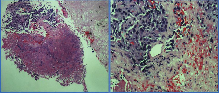
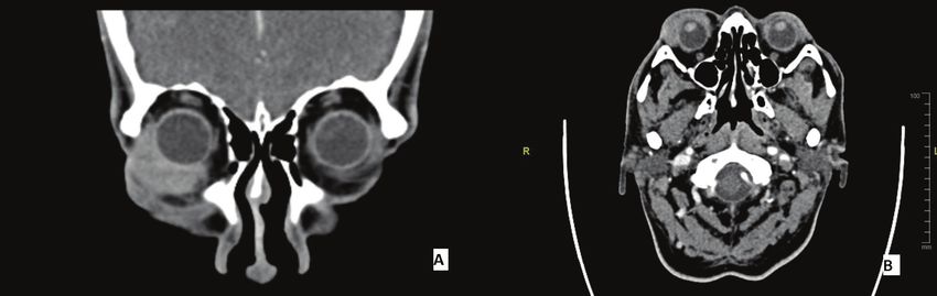
Neuro-ophthalmology and ocular problems in systemic diseases
Optic neuritis as the ocular manifestation of dengue infection:
a case report                                                   46
Indra Tri Mahayana, Andreas Surya Anugrah, Ika Kartika, Natalia Christina Angsana,
Tatang Talka Gani

A tale of ptosis, pharmacological tests, and Pancoast tumour                     52
Muhammad Fadhli Ab Hamid, Mimiwati Zahari, Norlina Mohd Ramli

Orbit and oculoplasty
Ocular marginal zone B-cell lymphoma of mucosa-associated
lymphoid tissue masquerading as chalazion: a case report                         57
Nik Ahmad Syafiq Mat Zaidan, Azida Juana Wan Ab Kadir, Fazliana Ismail, Sagili
Chandrasekhara Reddy

Pediatric ophthalmology and strabismus
Evans syndrome in child with sudden blindness                                    64
Muhammad Fadhli Ab Hamid, Choo May May, Nurliza Khaliddin, Norlina Mohd Ramli
© Malaysian Journal of Ophthalmology 2020; 1:6-8
                                                Editorial
                                                https://doi.org/10.35119/myjo.v3i1.217

Eye acupuncture in Malaysia: the
need for guidelines, regulation and
enforcement
Mohamad Aziz Salowi1,2

1
 Department of Ophthalmology, Selayang Hospital, Selangor, Ministry of Health,
Malaysia; 2Department of Ophthalmology, Faculty of Medicine, Universiti Sultan Zainal
Abidin, Kuala Terengganu, Malaysia

Acupuncture originates from the practice of the ancient Chinese more than 2000
years ago.1 It was initially performed using sharp stones, bone, or bamboo, before
the discovery of metals. In modern days, the needles are made of stainless steel
and can vary from 5 to 23 cm in length.1 Eye acupuncture involves the application
of these needles to multiple acupoints around the orbital area. 2 In eye care,
acupuncture has been reported to be used in treating myopia, glaucoma, retinitis
pigmentosa, and cranial nerve paralysis. 3-6 It is increasingly being used for the
treatment of dry eye.7 Eye acupuncture has also been reported to be used as the
primary treatment or adjuvant therapy to treat other ailments not related to the
eye.8,9 The usage of needles in eye-acupuncture exposes individuals to ocular
injury due to the close proximity of the eyeball or critical orbital structures to the
acupoints. Poor sterilization practices and the usage of recycled instruments may
lead to infection or transmission of sexually transmitted diseases.10 Therefore,
anatomical knowledge of the orbit and eyeball as well as adherence to standard
ethical practices are mandatory for all acupuncturists. Reported adverse effects
are not uncommon. They range from superficial conjunctival haemorrhage
to penetrating ocular injury, resulting in traumatic cataract, subretinal track,
vitreous haemorrhage, proliferative vitreoretinopathy, or endophthalmitis.6,11-15
In this current issue, Ainal et al. highlighted six cases of ocular injuries related to
acupuncture in their brief report.15
   In the local setting, Traditional and Complementary Medicine (T/CM) has
been integrated into the Malaysian Healthcare System. Although other types of
acupuncture services are available and listed for general information in the Annual
Report and the Consumer Guideline, eye acupuncture services are not available
yet in public hospitals.16,17 The National Guidelines for these non-eye acupuncture
services are available for both healthcare providers and consumers.17,18 Essential
information, such as the location of the acupuncturist, contact number, type of
acupuncture services offered, and complaint or feedback line, is also available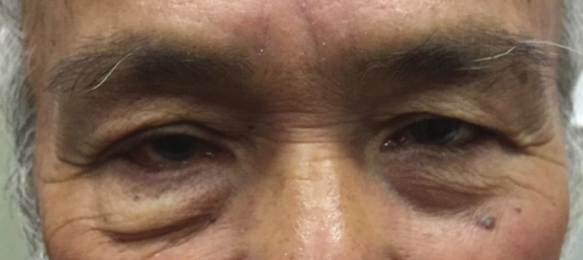
Eye acupuncture in Malaysia                                                         7

to the public.17 Like other T/CM services in the country, they are governed by the
National Policies and Law on Traditional and Complementary Medicine.
   The National Policy was developed by the Traditional and Complementary
Medicine Division of the Malaysian Ministry of Health (MOH) in 2002.19 It states that
the T/CM system should be an essential component of the Healthcare System,
coexisting with modern medicine and contributing towards enhancing the health
and quality of life of all Malaysians. The Act on Traditional and Complementary
Medicine (Act 775: 2016) came to full enforcement on August 1, 2016. It allows the
Traditional and Complementary Medicine Council to regulate the T/CM services
in Malaysia. It also requires T/CM practitioners to register in the T/CM Practi-
tioner Bodies, the National Bodies registered with the Registrar of Societies and
appointed by the MOH. This appointment allows this body to self-regulate its
practitioners through codes of ethics and practice regulated by the T/CM-MOH
standing committee and endorsed by the T/CM council.20
   The case series included in this issue of Malaysian Journal of Opthalmology
highlights the possible hidden magnitude of the problem in the country. Within
seven months (from April 2019 to October 2019), six cases of ocular injury following
eye acupuncture treatment presented to different ophthalmologists in the
country. The number could probably be higher if the writer extended the duration
of the case series reporting or if the related enforcement body performed an active
case tracing at the same time. The multiple incidences of acupuncture-related eye
injury occurring within a short period suggests a possible error in the notification
system (either within the healthcare system or within the community), if the noti-
fication system existed at all. It also shows a lack of coordination and possible lack
of awareness among acupuncturists, ophthalmologists, and the T/CM Practitioner
Body regarding the risk of unmonitored eye acupuncture practice to the public.
   When properly regulated and monitored, eye acupuncture may contribute
favourably to the community’s eye healthcare. T/CM Practitioner Bodies and
Councils, as organizations mentioned in the Act, need to monitor and evaluate the
process of registering acupuncturists, develop standards and ethics frameworks,
and regulate the practice of eye acupuncture. The regulation shall include
stringent certification and penalties for breach of professional conduct and rules.
The T/CM MOH Division shall develop a policy and guidelines on acupuncture
involving the eye as well as provide a platform to host notification and encourage
communication among the practitioners. The Prevention of Blindness Committee
in the MOH shall work together with the T/CM MOH Division in planning to prevent
further occurrence of acupuncture-related ocular injuries in the community. Most
importantly, the public must be informed through the media and social media
platforms regarding the standard conducts in eye acupuncture and the potential
risks of the procedure.
8                                                                                            M.A. Salowi

References

1.    Koh EK. Acupuncture. Br J Gen Pract. 1973;23:265-72.
2.    Shao Y, Wang P, Wang Q, et al. Eye-acupuncture with rehabilitation therapy for stroke. Medicine (Bal-
      timore). 2020;99(18):e20096.
3.    Dabov S, Goutoranov G, Ivanova R, Petkova N. Clinical application of acupuncture in ophthalmology.
      Acupunct Electrother Res. 1985;10(1-2):79-93..
4.    Law SK, Lowe S, Law SM, et al. Prospective Evaluation of Acupuncture as Treatment for Glaucoma.
      Am J Ophthalmol. 2015;160(2):256-65.
5.    Ji XJ, Zhou LY, Si CQ, et al. [Efficacy observation on electroacupuncture in the treatment of oculo-
      motor impairment caused by ophthalmic nerve injury]. Zhongguo Zhen Jiu. 2013;33(11):975-979.
6.    You TT, Youn DW, Maggiano J, et al. Unusual ocular injury by an acupuncture needle. Retin Cases
      Brief Rep. 2014;8(2):116-119.
7.    Dhaliwal DK, Zhou S, Samudre SS, et al. Acupuncture and dry eye: current perspectives. A double-blind-
      ed randomized controlled trial and review of the literature. Clin Ophthalmol. 2019;13:731–740.
8.    Lin Q, Hu YL, Han CW, Li Y. [Eye acupuncture for treatment of renal and ureteral colic]. Zhongguo
      Zhen Jiu. 2007;27(9):663-664.
9.    Shaoa Y, Wang M, Liu B, et al. Eye-acupuncture as adjuvant therapy for stroke: A bibliometric analysis
      of clinical studies. Journal of Traditional Chinese Medical Sciences 2019;6(3):263-69.
10.   Woo PCY, Lin AWC, Lau SKP, et al. Acupuncture transmitted infections. BMJ. 2010;340:c1268.
11.   Denstedt J, Schulz DC, Diaconita V, et al. Acupuncture resulting in eye penetration and proliferative
      vitreoretinopathy - Surgical and medical management with intraocular methotrexate. Am J Ophthal-
      mol Case Rep. 2020;18:100605.
12.   Fielden M, Hall R, Kherani F, et al. Ocular perforation by an acupuncture needle. Can J Ophthalmol.
      2011;46(1):94-95.
13.   Kao TE, Kuo YW, Wu KY. Acupuncture-related penetrating eye injury. Kaohsiung J Med Sci.
      2017;33(9):473-474.
14.   Shuang H, Yichun K. A case of perforating injury of eyeball and traumatic cataract caused by acu-
      puncture. Indian J Ophthalmol. 2016;64(4):326-327.
15.   Naffi AA, Ling KP, Ratanam M, Kong VYP, Fong CSK, Bastion MLC. A series of acupuncture-related
      ocular injuries in Malaysia. Malaysian Journal of Ophthalmology. 2021;3(1):30-38.
16.   Ministry of Health Malaysia. Annual Report. Traditional and Complementary Division. Kuala Lumpur;
      Ministry of Health: 2019.
17.   Ministry of Health Malaysia. Consumer Guideline: FOR PROPER USE OF TRADITIONAL AND COM-
      PLEMENTARY MEDICINE SERVICES IN MALAYSIA: Traditional and Complementary Medicine Division
      Ministry of Health Malaysia, 2019.
18.   Ministry of Health Malaysia. Practice Guideline on Acupuncture. Kuala Lumpur: Traditional and Com-
      plementary Medicine Division; 2017.
19.   Ministry of Health Malaysia. National Policy on Traditional and Complementary Medicine. Kuala
      Lumpur: Traditional and Complementary Medicine Division; 2002.
20.   Malaysia Government. Laws of Malaysia - Act 775: Traditional and Complementary Medicine Act
      2016: Percetakan Nasional Malaysia Berhad 2016.
© Malaysian Journal of Ophthalmology 2021; 1:9-20
Original article
http://doi.org/10.35119/myjo.v3i1.172
Recurrent pterygium in Bintulu,
Sarawak (Malaysian Borneo):
determining its risk factors
Loshni Murugia1, Chong Ka Lung1, Lim Lik Thai2,3, Rohanah Alias4
1
Ophthalmology Department, Hospital Bintulu, Sarawak, Malaysia; 2Universiti
Malaysia Sarawak (UNIMAS), Sarawak, Malaysia; 3Sarawak General Hospital,
Sarawak, Malaysia; 4Ophthalmology Department, Hospital Kuala Lumpur, Kuala
Lumpur, Malaysia
Abstract
Introduction: Pterygium may give rise to astigmatism in addition to causing
blindness in advanced stages, reflecting the importance of timely surgical interven-
tion. Despite various operative approaches, the recurrence rate continues to range
from 2% to 89%. Therefore, it is essential to investigate the risk factors influencing
recurrence to improve therapeutic strategies.
Purpose: To determine the risk factors of pterygium recurrence among the
multiethnic cohort of patients of Bintulu, Sarawak (Malaysian Borneo).
Study design: Retrospective cohort.
Materials and methods: This study was conducted in Bintulu Hospital, Sarawak
(Malaysian Borneo) and involved patients who underwent pterygium excision
with conjunctival autografting between April 1, 2016 and May 31, 2019. Patients
completed at least a year of follow-up for recurrence detection. Collected data
included presence of recurrence, sociodemographics, outdoor activities, habits,
first-degree family history, pterygium type and location, as well as laterality.
Chi-squared test, Fisher’s exact test, and logistic regression analysis were used.
Results: A total of 161 eyes that underwent pterygium excision in 137 patients were
identified. Percentage of recurrence was found to be 42%. The mean age during
excision was 59.3 ± 11.5 years; age group showed no significance in pterygium
recurrence (p = 0.447). Male gender showed statistical significance (p = 0.045,
OR 1.90, CI 1.01, 3.58) in chi-squared test but not in logistic regression. Ethnicity,
Correspondence: Dr.Loshni Murugia, MD, Ophthalmology Department, Hospital Bintulu,
Jalan Bukit Nyabau, 97000, Bintulu, Sarawak, Malaysia.
E-mail:loshni66@gmail.com10 L Murugia et al. marital status, income, and education level showed no association with recurrence (p > 0.05). Anatomic factors like location (p = 0.353) and laterality (p = 0.955) also showed no association. Smoking (p = 0.867) and alcohol intake (p = 0.397) were insignificant, similar to first-degree family history (p = 0.137). Activities involving sun exposure (p < 0.001, OR 18.34, 95% CI 5.59, 60.17) and recurrent pterygium type (p = 0.001, OR 7.40, 95% CI 1.81, 30.21) supported a positive association with recurrence. Medication adherence (p < 0.001, OR 3.61, 95% CI 1.07, 12.21) and the use of sun protection (p < 0.001, OR 7.90, 95% CI 3.25, 19.19) showed a statistically significant decrease in recurrence. Conclusion: Activities involving sun exposure, use of sun protection, medication adherence, and recurrent pterygium type have shown to be statistically significant in influencing recurrence after excision and conjunctival autograft. Keywords: conjunctival grafting, pterygium, pterygium excision, recurrence, risk factors Pterigium berulang di Bintulu, Sarawak (Borneo Malaysia): penentuan faktor risiko Abstrak Pendahuluan: Pterigium dapat menimbulkan astigmatisme selain menyebabkan kebutaan pada tahap lanjut, yang menggambarkan pentingnya campur tangan pembedahan tepat pada masanya. Walaupun terdapat pelbagai pendekatan pembedahan, kadar pengulangan terus berkitar antara 2% hingga 89%. Oleh itu, adalah mustahak untuk menyelidik faktor risiko yang mempengaruhi kes berulang dan memperbaiki strategi terapi. Tujuan: Untuk menentukan faktor risiko berulang pterigium di antara kohort pesakit pelbagai etnik Bintulu, Sarawak (Borneo Malaysia). Reka bentuk kajian: Kumpulan retrospektif. Bahan dan kaedah: Kajian ini dilakukan di Hospital Bintulu, Sarawak (Borneo Malaysia) dan melibatkan pesakit yang menjalani eksisi pterigium dengan cantuman autograf konjunktiva antara 1 April 2016 dan 31 Mei 2019. Pesakit menyelesaikan sekurang-kurangnya satu tahun susulan lanjut untuk pengesanan kes berulang. Data yang dikumpulkan termasuk kehadiran berulang, sosiodemografi, aktiviti luar, kebiasaan, sejarah keluarga darjah pertama, lokasi dan jenis serta kelateralan pterigium. Uji Chi-kuadrat, uji tepat Fisher, dan analisis regresi logistik digunakan. Dapatan: Sebanyak 161 mata yang menjalani eksisi pterigium pada 137 pesakit dikenal pasti. Peratusan berulang didapati 42%. Umur min semasa eksisi adalah 59.3
Recurrent pterygium in Bintulu, Sarawak: determining risk factors                 11

 ± 11.5 tahun; kumpulan umur tidak menunjukkan kepentingan dalam kambuhan
 pterigium (p = 0.447). Jantina lelaki menunjukkan kepentingan statistik (p = 0.045,
 OR 1.90, CI 1.01, 3.58) dalam ujian chi-square tetapi tidak dalam regresi logistik.
 Etnik, status perkahwinan, pendapatan, dan tahap pendidikan tidak menunjukkan
 hubungan dengan kambuhan (p > 0.05). Faktor anatomi seperti lokasi (p = 0.353)
 dan lateral (p = 0.955) juga tidak menunjukkan perkaitan. Merokok (p = 0.867) dan
 pengambilan alkohol (p = 0.397) tidak signifikan, serupa dengan sejarah keluarga
 darjah pertama (p = 0.137). Aktiviti yang melibatkan pendedahan cahaya matahari
 (p < 0.001, OR 18.34, 95% CI 5.59, 60.17) dan jenis pterigium berulang (p = 0.001,
 atau 7.40, 95% CI 1.81, 30.21) menyokong hubungan positif dengan berulang.
 Kepatuhan ubat (p
12                                                                    L Murugia et al.

and excimer laser treatment.14 Despite surgery being the single most practical
way to treat pterygium, recurrence rates continue to range from 2% to 89% with
various operative approaches.15,16 Therefore, it is essential to investigate the risk
factors involved in pterygium recurrence in order to improve therapeutic strategies,
especially in light of the fact that it has tendency to proliferate more aggressive-
ly during recurrence.1 In addition, various authors have addressed the need for
research on the effect of ethnicity and other risk factors as prognosticators of post-
operative pterygium recurrence in a controlled fashion.14,17 Ethnicity in particular
has consistently been overlooked as a potential predictor due to the assumed
homogeneity of the research subjects.17,18
   To the best of our knowledge, a study regarding the risk factors of pterygium
recurrence has yet to be done in an equatorial population. In our case, Bintulu is
located 3° north of the equator and its multiethnic population, living in the same
geographical area, provides an exceptional opportunity to study it from a single
health facility. This study was intended to investigate the postoperative recurrence
of pterygium in association with demographic, environmental, habitual, anatomic,
and familial predisposition factors.

Materials and methods
This study was conducted in compliance with the ethical principles outlined in the
Declaration of Helsinki as well as the Malaysian Good Clinical Practice Guideline
and obtained ethical clearance from the country’s Medical Research and Ethics
Committee. This retrospective cohort study involved patients who underwent
pterygium excision with conjunctival autografting and completed at least 1 year of
follow-up in Bintulu Hospital. Recruitment of subjects was done from patients who
underwent the procedure between April 1, 2016 and May 31, 2019. These patients
were identified and approached during their regular follow-up at the clinic.
   Written informed consent was obtained and patients were interviewed to
obtain demographic data (age during excision, gender, ethnicity, education
level, income, marital status), information on lifestyle as well as habits (activities
involving sun exposure, smoking, use of sun protection, postoperative medication
adherence, alcohol consumption), and familial predisposition. Household income
was calculated from the total income of each household member. Households
were grouped into quintiles according to income.19 Sun exposure was considered
significant when outdoor activities involved sun exposure for more than 5 hours per
day.4,20 Non-smokers were categorised as those who had never smoked or smoked
less than five packs in their lifetime.4,20 Sun protection included the use of hats,
sunglasses, umbrellas, or shades during outdoor activities and was categorised
using Likert-scale answers.21,22Medication adherence was defined as taking 80%
or more of the prescribed medication doses.23Prescribed medications referred to
Recurrent pterygium in Bintulu, Sarawak: determining risk factors                   13

Table 1. Univariate and multivariate analysis of variables in patients with and without
recurrence
                        Recurrence
 Characteristics                                    AOR (95% CI)          P-value
                        Yes (%)        No
 Age during excision
 60                    33 (41.3%)     47
 Gender
 Male                   41 (50.6%)     40           1.90 (1.01,3.58) (COR) 0.045ᵃ
 Female                 28 (35%)       52
 Ethnicity
 Malay                  7 (43.8%)      9                                  0.607ᵃ
 Chinese                10 (38.5%)     16
 Iban                   34 (39.5%)     52
 Melanau                13 (52.0%)     12
 Others                 5 (62.5%)      3
 Marital status
 Single                 3 (37.5%)      5                                  0.485ᵃ
 Married                52 (41.9%)     72
 Widower/Widow/
 Divorcee               14 (48.3%)     15
 Education level

 No formal education
                        21 (41.2%)     30                                 0.953ᵃ
 Primary education      20 (44.4%)     25

 Secondary education
                        23 (41.8%)     32
 Tertiary education     5 (50%)        5
 Income
 1st quintile           16 (38.1%)     26                                 0.642ᵃ
 2 quintile
  nd
                        15 (38.5%)     24
 3 quintile
  rd
                        23 (50%)       23
 4th quintile           15 (44.1%)     19
14 L Murugia et al.
Recurrence
Characteristics AOR (95% CI) P-value
Yes (%) No
Location
Nasal 47 (40.2%) 70 0.353ᵇ
Temporal 3 (75%) 1
Both 19 (47.5%) 21
Laterality
Unilateral 14 (42.4%) 19 0.955ᵃ
Bilateral 55 (43%) 73
Smoking
Smoker 15 (44.1%) 19 0.867ᵃ
Non-smoker 54 (42.5%) 73
Alcohol Intake
Abstinent 66 (43.7%) 85 0.397ᵇ
Light drinking 1 (25%) 3
Moderate drinking 0 (0%) 3
Unsafe drinking 2 (66.7%) 1
First-degree family history of pterygium
Yes 28 (50.9%) 27 0.137ᵃ
No 41 (38.7%) 65
Activities involving sun exposure
Yes 30 (85.7%) 5 18.34 (5.59,60.17) < 0.001
No 39 (31%) 87
Medication adherence
Yes 46 (34.6%) 87
No 23 (82.1%) 5 3.61 (1.07,12.21) < 0.001
Use of sun protection
Never/Rarely/
Sometimes 39 (68.4%) 18 7.90 (3.25,19.19) < 0.001
Often/Always 30 (28.8%) 74Recurrent pterygium in Bintulu, Sarawak: determining risk factors 15
Recurrence
Characteristics AOR (95% CI) P-value
Yes (%) No
Pterygium type
Primary 54 (38%) 88
Recurrent 15 (78.9%) 4 7.40 (1.81,30.21) 0.001
Total 69 (42%) 92
ᵃPearson chi-square test
ᵇFisher’s test
AOR: adjusted odds ratio; COR: crude odds ratio
lubricant and steroid eye drops given postoperatively for 1 month. Patients were
deemed “abstinent” if they consumed less than 1 unit of alcohol a week. “Light”
drinking was defined as between 1 and < 7 units a week and “moderate” drinking as
7 to < 14 units a week. “Unsafe” drinking was defined as > 14 units a week.24,25
Information regarding date of operation, duration of follow-up, age during
surgery, laterality and location of pterygium, presence of recurrence, type of
pterygium, and date of recurrence detection was obtained from the hospital
information system. Recurrence of pterygium was defined as any fibrovascular
growth extending across the limbus onto the cornea at the site of previous surgical
excision.26Patients who did not consent for this research and those with incomplete
data were excluded.
All the information obtained was analysed for associations with pterygium
recurrence using SPSS version 26. The analytical statistics used were chi-square test
and Fisher’s exact test. Subsequently, variables that showed significant association
in these tests were further analysed via multivariate logistic regression. Variables
with 95% confidence interval (CI) and p-value < 0.05 were considered statistically
significant factors for pterygium recurrence.
Results
A total of 161 eyes from 137 patients who underwent pterygium excision from April
2016 to May 2019 were included in this study. Male-to-female ratio was noted to be
almost 1:1(81 to 80). The mean age during excision was 59.3 ± 11.5 years. Percentage
of recurrence was noted to be 42%.
Table 1 summarises the risk factors investigated and the presence of recurrence.
Most pterygium excisions (49.7%) were performed in patients above 60 years of age.
This demographic factor showed no significant difference when incorporated into
Fisher’s test (p = 0.447). Male gender was statistically significant (p = 0.045), with a crude
odds ratio (OR) of 1.90 and CI of 1.01 to 3.58 when analysed using chi-squared test;16 L Murugia et al.
however, once integrated in binary logistics, the value became non-significant.
Other sociodemographic data, which included ethnicity, marital status, and
education level, showed a p > 0.05. Income, which was divided equally in quintiles,
also appeared to be non-significant (p = 0.642). Location and laterality as anatomic
factors also showed no significant difference, with p-values of 0.353 and 0.955,
respectively. The initial data was analysed with Fisher’s test, while the latter with
chi-squared test. Being known risk factors for many diseases, smoking (p = 0.867)
and alcohol intake (p = 0.397) showed no association with recurrence in this study.
Positive family history in first-degree relatives was analysed with chi-squared test
and resulted in non-association with recurrence (p = 0.137).
Moving forward, all factors which showed significant difference when analysed
with univariate analysis were assimilated into logistic regression to control
confounding factors. Data on activities involving sun exposure yielded p <
0.001 with OR 18.34 (95% CI 5.59, 60.17), supporting a positive association with
recurrence. Medication adherence (p < 0.001, OR 3.61, 95% CI 1.07, 12.21) and
use of sun protection (p < 0.001, OR 7.90, 95% CI 3.25, 19.19) showed statistically
significant decrease in recurrence. Recurrent pterygium type were 7.40 times more
likely to cause recurrence compared to primary pterygium type (p = 0.001, 95% CI
1.81, 30.21).
Discussion
The aim of this study was to determine the risk factors that contribute to pterygium
recurrence after excision and conjunctival grafting in a multiethnic cohort of
patients in Bintulu, Sarawak (Malaysian Borneo). Given its considerably high
recurrence rate, it is essential for us to identify its risk factors in order to establish
different therapeutic strategies as well as modify surveillance follow-up frequency
and duration for those who are deemed to have higher tendency for recurrence.
Intensive counselling on lifestyle and aggressive treatment approach may be
administered in anticipation of recurrence. We performed our study retrospective-
ly to analyse anatomy, sociodemographics, environment, lifestyle, and pterygium
type as potential factors for recurrence.
Pterygium recurrence often develops in a short span of time after excision;
therefore, sun exposure was assumed to be a rather negligible risk factor.1 This
assumption was refuted in our study, which proved that sunlight exposure is a
significant risk factor, further supporting similar studies conducted in Croatia,
Spain, and Korea.27-29 In addition, lack of sun protection also appeared to be signifi-
cantly associated with recurrence in this study. In fact, the burden may be higher in
regions located in the “pterygium belt”, in this case, our study site, Bintulu. Multiple
studies have addressed the effect of UVB on limbal cells leading to productionRecurrent pterygium in Bintulu, Sarawak: determining risk factors 17 of interleukin (IL)-6, IL-8, and growth factors which is linked to inflammation, blood vessel formation, cellular proliferation, and antiapoptosis.30-32 Although the pathogenesis of recurrence due to sunlight has never been well documented, the previously mentioned molecular pathogenesis may possibly apply to any type of pterygium. Recurrent pterygium itself was more likely associated with further recurrence as compared to primary pterygium in this study. A possible explanation for this could be the degree of expression of cyclooxygenase-2 (COX-2), which is said to be increased in stromal fibroblasts of recurrent pterygium in contrast to primary pterygium and is believed to be part of the pathogenesis of pterygium itself through its antiapoptotic nature.33-35 The use of corticosteroids and artificial tears has been important in reducing the postoperative complications of pterygium, including recurrence.39-40 In our study site, both corticosteroids and artificial tears were given postoperatively; our results showed poor medical adherence be a significant risk factor in recurrence. Without proper consistent usage of these medications, the protective factor is no longer in place. This theory is supported by a recent study in Spain, which showed that tapering doses of corticosteroids within 5 weeks as compared to 4 months improved medication compliance and therefore resulted in less complications such as recurrence.40 Male gender showed significance in univariate analysis, but lost significance once incorporated into binary regression. This may be due to the disproportion- ately greater number of males exposed to the sun, possibly because of the nature of their occupation in Bintulu. Male gender as a predictive factor, however, seemed inconclusive as there have been mixed results in multiple studies.1,17,28,38,39 Younger age has been documented to be more prone towards recurrence in a few studies.1,38,40 Surprisingly, our study contradicts this, possibly due to an uneven distribution number of patients whereby most of the patients in our study were more than 60 years old. This study has several limitations. The first is the lack of standardized surgical technique for pterygium excision due to the surgeries being performed by multiple surgeons using different surgical techniques, which may have influenced postop- erative outcomes. The second is the patients who were lost to follow-up, who may have experienced recurrences but could not be included in our study. Finally, due to its retrospective nature, we were unable to obtain information on pterygium morphology prior to excision, which has been reported to influence recurrence in several studies. In conclusion, activities involving sun exposure, the use of sun protection, medication adherence, and recurrent pterygium type have shown to be statistical- ly significant in influencing recurrence after excision and conjunctival autograft in our multiethnic cohort of patients in Bintulu, Sarawak (Malaysian Borneo).
18                                                                                        L Murugia et al.

Acknowledgements
We wish to express our gratitude to Madam Nurakmal Baharum, Clinical Research
Centre (CRC, Kuala Lumpur, Malaysia), for her valuable assistance in the statistical
analysis of this study. The authors have no conflict of interest or financial disclosures
to declare.

References
1.    Ha SW, Park JH, Shin IH, Kim HK. Clinical analysis of risk factors contributing to recurrence of pteryg-
      ium after excision and graft surgery. Int J Ophthalmol. 2015;8(3):522–527.
2.    Feng QY, Hu ZX, Song XL, Pan HW. Aberrant expression of genes and proteins in pterygium and their
      implications in the pathogenesis. Int J Ophthalmol. 2017;10(6):973–981.
3.    Lü P, Chen XM. Prevalence and risk factors of pterygium. Int J Ophthalmol. 2008;8(5):871–874.
4.    Alemayehu TK, Addis Y, Bizuneh ZY, Tegegne MM, Alemayehu AM. Prevalence and associated
      factors of pterygium among adults living in KollaDiba Town, Northwest Ethiopia. Clin Ophthalmol.
      2020;14:245–255.
5.    Detels R, Dhir SP. Pterygium: A Geographical Study. Arch Ophthalmol. 1967;78(4):485–491.
6.    Marmamula S, Khanna RC, Rao GN. Population-based assessment of prevalence and risk factors for
      pterygium in the South Indian state of Andhra Pradesh: The Andhra Pradesh eye disease study. Inves-
      tigOphthalmol Vis Sci. 2013;54(8):5359–5366.
7.    Ang M, Li X, Wong W, Zheng Y, Chua D, et al. Prevalence of and racial differences in pterygium: A mul-
      tiethnic population study in Asians. Ophthalmology. 2012;119(8):1509–1515.
8.    Gazzard G, Saw SM, Farook M, et al. Pterygium in Indonesia: Prevalence, severity and risk factors. Br
      J Ophthalmol. 2002;86(12):1341–1346.
9.    Tano T, Ono K, Hiratsuka Y, Otani K, et al. Prevalence of pterygium in a population in Northern
      Japan: The Locomotive Syndrome and Health Outcome in Aizu Cohort Study. Acta Ophthalmol.
      2013;91(3):232–237.
10.   Hashemi H, Khabazkhoob M, Yekta A, Jafarzadehpour E, Ostadimoghaddam H, Kangari H. The preva-
      lence and determinants of pterygium in rural areas. J Curr Ophthalmol. 2017;29(3):194–198.
11.   Maharjan I, Shreshth E, Gurung B, Karmacharya S. Prevalence of and associated risk factors
      for pterygium in the high-altitude communities of Upper Mustang, Nepal. Nepal J Ophthalmol.
      2014;6(1):65–70.
12.   Taylor SL, Coates ML, Vallejos Q, et al. Pterygium among Latino migrant farmworkers in North
      Carolina. Arch Environ Occup Health. 2006;61(1):27-32.
13.   Ramalingam M, Joshi N, Nair J, Ali NAM. Outcome of surgical management of pterygium in Brunei
      Darussalam. Brunei Int Med J. 2011;7(1):8–14.
14.   Hirst LW. The treatment of pterygium. SurvOphthalmol. 2003;48(2):145-180
15.   He SY, Sun H, Huang Y, et al. Identification and Interaction Analysis of Significant Genes and MicroR-
      NAs in Pterygium. BioMed Res Int. 2019;19:1–12.
Recurrent pterygium in Bintulu, Sarawak: determining risk factors 19
16. Fernandes M, Sangwan VS, Bansal AK, et al Outcome of pterygium surgery: analysis over 14 years.
Eye (Lond). 2005;19(11):1182-1190.
17. Kandavel R, Kang JJ, Memarzadeh F, Chuck RS. Comparison of pterygium recurrence rates in hispanic
and white patients after primary excision and conjunctival autograft. Cornea. 2010;29(2):141–145.
18. Campagna G, Adams M, Wang L, Khandelwal S, Al-Mohtaseb Z. Comparison of pterygium recurrence
rates among different races and ethnicities after primary pterygium excision by surgeons in training.
Cornea. 2018;37(2):199–204.
19. Institute for Public Health (IPH) 2015. National Health and Morbidity Survey 2015 (NHMS 2015). Vol.
II: Non-Communicable Diseases, Risk Factors & Other Health Problems; 2015.
20. Pyo EY, Mun GH, Yoon KC. The prevalence and risk factors for pterygium in South Korea: the Korea
National Health and Nutrition Examination Survey (KNHANES) 2009-2010. Epidemiol Health.
2016;38:e2016015.
21. Peters CE, Koehoorn MW, Demers PA, Nicol AM, Kalia S. Outdoor Workers’ Use of Sun Protection at
Work and Leisure. Saf Health Work. 2016;7(3):208–12.
22. Brokalaki H, Patelarou E, Vardavas C, Elefsiniotis IS, Giakoumidakis KA, Brokalaki E. Current patterns
of the sun protection measures adopted by nurses and the risk factors influencing their compliance.
Open J Nurs. 2011;1(3):43–50.
23. Morrison A, Stauffer ME, Kaufman AS. Defining medication adherence in individual patients. Patient
Prefer Adherence. 2015;9:893–897.
24. Topiwala A, Allan CL, Valanova V, et al. Moderate alcohol consumption as risk factor for adverse brain
outcomes and cognitive decline: longitudinal cohort study. BMJ. 2017;357:j2353.
25. Department of Health. UK Chief Medical Officers’ Low Risk Drinking Guidelines. Dep Heal Engl
[Internet]. 2016;(August):1–11. Available from: https://www.gov.uk/.
26. Han SB, Jeon HS, Kim M, et al. Risk factors for recurrence after pterygium surgery: An image analysis
study. Cornea. 2016;35(8):1097–103.
27. Sekelj S, Dekaris I, Kondža-Krstonijević E, Gabrić N, Predović J, Mitrović S. Ultraviolet light and pte-
rygium. Coll Antropol. 2007;31(SUPPL. 1):45–47.
28. Torres-Gimeno A, Martínez-Costa L, Ayala G. Preoperative factors influencing success in pterygium
surgery. BMC Ophthalmol. 2012;12(1):1-7.
29. Chun YH, Paik J-S, Oh JH, Kim H-S, Na K-S. Association between pterygium, sun exposure, and
serum 25-hydroxyvitamin in a nationally representative sample of Korean adults. Lipids Health Dis.
2018;17(1):1–8.
30. Crǎiţoiu S, Ciprian L, Rodica M, Mihai A, Anca EI. Etiopathogenic aspects in development and evolu-
tion of pterigyum. Oftalmologia. 2008;52(2):29–34.
31. Di Girolamo N, Kumar RK, Coroneo MT, Wakefield D. UVB-mediated induction of interleukin-6
and -8 in pterygia and cultured human pterygium epithelial cells. InvestigOphthalmol Vis Sci.
2002;43(11):3430–3437.
32. Ginger-Eke HA, Ogbonnaya CE, Ezisi CN. Pterygium: Recent trends and perspectives – A review of
pathogenesis and current management options. Niger J Ophthalmol. 2018;26(2):89-98.
33. Karahan N, Baspinar S, Ciris M, Baydar C, Kapucuoglu N. Cyclooxygenase-2 expression in primary
and recurrent pterygium. Indian J Ophthalmol. 2008;56(4):279–283.20 L Murugia et al.
34. Chiang CC, Cheng YW, Lin CL, Lee H, Tsai FJ, Tseng SH, et al. Cyclooxygenase 2 expression in pterygi-
um. Mol Vis. 2007;13:635–638.
35. Maxia C, Perra MT, Demurtas P, et al. Relationship between the expression of cyclooxygenase-2 and
survivin in primary pterygium. Mol Vis. 2009;15:458–463.
36. Kampitak K, Leelawongtawun W, Leeamornsiri S, Suphachearaphan W. Role of artificial tears in
reducing the recurrence of pterygium after surgery: a prospective randomized controlled trial. Acta
Ophthalmol. 2017;95(3):e227–9.
37. Sabater-Cruz N, Dotti-Boada M, Rios J, et al. Postoperative treatment compliance rate and com-
plications with two different protocols after pterygium excision and conjunctival autografting. Eur
J Ophthalmol [Internet]. 2020 [cited 2020 Aug 6]. Available from: https://pubmed.ncbi.nlm.nih.
gov/32338523/. doi: 10.1177/1120672120917335.
38. Anguria P, Ntuli S, Carmichael T. Young patient’s age determines pterygium recurrence after surgery.
Afr Health Sci. 2014;14(1):72–76.
39. Varssano D, Fischer N. Pterygium Excision With Conjunctival Autograft: True Survival Rate Statistics.
2013;32(9):1243–50.
40. Huerva V, March A, Martinez-Alonso M, Muniesa MJ, Sanchez C. Pterygium surgery by means of con-
junctival autograft: Long term follow-up. Arq Bras Oftalmol. 2012;75(4):251–255.© Malaysian Journal of Ophthalmology 2021; 1:21-29
Original article
https://doi.org/10.35119/myjo.v3i1.170
Wies procedure for correcting
involutional entropion of the lower
lid in geriatrics
Erum Shahid, Uzma Fasih, Arshad Shaikh
Ophthalmology Department, Karachi Medical and Dental College, Abbasi Shaheed
Hospital, Karachi, Pakistan
Abstract
Objective: To evaluate the anatomic outcome and recurrence rate of the Wies
procedure for treating involutional entropion of the lower lid in geriatrics.
Materials and methods: This retrospective case series was conducted in the Ophthal-
mology department of a tertiary care hospital from January 1, 2016 to December 31,
2017. Geriatric patients (≥ 65 years) who had undergone the Wies procedure, i.e.,
transverse lid split and everting sutures for correction of involutional entropion of
the lower lid were included. All the surgeries were done under local anaesthesia by a
single ophthalmologist. The follow-up period was 12 months. A successful outcome
was defined as restoration of lid margin to its position with no lash touching the
cornea and no recurrence within 12 months.
Results: Eighteen eyes of 13 patients with a mean age of 67.6 ± 2.2 SD years were
included. There were 11 males (61%) and 7 females (39%). Bilateral entropion
correction was done in five patients. Nine right eyes and nine left eyes were included.
Anatomical success was 94.4% at 12 months. Recurrence was seen in one (5.6%)
patient at 12 months.
Conclusion: The Wies procedure for correction of involutional entropion with
horizontal lid laxity in the geriatric population provided good anatomic results in
our study. The recurrence rate was minimal within 1 year. The recurrence rate can
be reduced by an accurate initial entropion assessment.
Keywords: entropion, geriatric, involutional entropion, lower lid, Wies procedure
Correspondence: Dr Erum Shahid, MCPS, FCPS, Assistant Professor, Ophthalmology
Department, Karachi Medical and Dental College, Abbasi Shaheed Hospital, Karachi,
Pakistan.
E-mail: drerum007@yahoo.com22 E Shahid, U Fasih and A Shaikh Prosedur Wies untuk membaiki entropion involusi kelopak mata bawah dalam geriatrik Abstrak Objektif: Untuk menilai hasil anatomi dan kadar pengulangan prosedur Wies untuk merawat entropion kelopak mata dalam geriatrik. Bahan dan kaedah: Siri kes retrospektif ini dilakukan di jabatan Oftalmologi hospital rawatan tertier dari 1 Januari 2016 hingga 31 Disember 2017. Pesakit geriatrik (≥ 65 tahun) yang telah menjalani prosedur Wies, iaitu pemisahan kelopak mata melintang dan jahitan untuk pembetulan entropion inklusi penutup bawah dimasukkan. Semua pembedahan dilakukan di bawah anestesia setempat oleh pakar oftalmologi tunggal. Tempoh susulan adalah 12 bulan. Kejayaan prosedur ditakrifkan sebagai pemulihan margin ke kedudukannya tanpa bulu mata menyentuh kornea dan tidak berulang dalam 12 bulan. Hasil: Lapan belas mata dari 13 pesakit dengan usia sekitar 67.6 ± 2.2 SD tahun dimasukkan. Terdapat 11 lelaki (61%) dan 7 perempuan (39%). Pembetulan entropion kedua belah mata dilakukan pada lima pesakit. Sembilan mata kanan dan sembilan mata kiri. Kejayaan anatomi adalah 94.4% pada 12 bulan. Pengulangan entropion berlaku dan dilihat pada satu (5.6%) pesakit pada 12 bulan. Kesimpulan: Prosedur Wies untuk membetulkan entropion involusi akibat kelonggaran kelopak mata mendatar pada populasi geriatrik memberikan hasil anatomi yang baik dalam kajian kami. Kadar entropion berulang adalah minimum dalam tempoh 1 tahun. Kadar berulang dapat dikurangkan dengan penilaian awal awal yang tepat. Kata kunci: entropion, entropion inklusi, geriatrik, penutup bawah, prosedur Wies Introduction For the first time in history, life expectancy has exceeded 60 years.1 The geriatric population in low-income countries is estimated to reach approximately 840 million by 2025.2 This global rise in the aging population has also made its impact on Pakistan: life expectancy has risen by three decades in the last 50 years and will reach nearly 70 years by 2023.3 Pakistan stands as the fifth most populous country in the world and has a geriatric population currently to be more than 8 million.4 With increasing life expectancy, an increase in ocular problems in the geriatric population is also to be expected. Various eyelid diseases such as ectropion, entropion, dermatochalasis, etc., are frequently encountered in the elderly.5 As the elderly resort to medical and surgical methods for treatment of these associated
Wies procedure for lower-lid entropion in geriatrics                                23

eyelid diseases, surgical treatments are constantly being improved.6
   Entropion refers to an inward rotation of an eyelid.7 It is further classified into
congenital, cicatricial, involutional, and spastic. Involutional or senile entropion is
the most commonly encountered type of entropion.6 Entropion causes chronic con-
junctivitis, punctate epithelium erosions, ocular irritation, and blepharospasm in the
elderly. If left untreated, it may cause vision loss and ocular damage leading to dry
eye, corneal ulceration, and microbial keratitis.7 Senile entropion has a prevalence
of 2.1% in the geriatric population and is more common in women.7 Several aetio-
logical factors are thought to play an important role in the development of senile
entropion. Horizontal lid laxity, vertical lid laxity, overriding of the preseptal
orbicularis oculi muscle onto the pretarsal muscle, and appositional pressure of the
lids during eyelid closure are the anatomical changes that occur with aging, leading
to entropion formation.8
   Everting sutures is the most commonly used technique for lower-lid entropion
correction, dating back to Hippocrates.9,10 However, everting sutures provide only
temporary relief for involutional entropion.11 The Wies procedure, which combines
transverse full-thickness blepharotomy and everting sutures, is the other method
used for entropion without horizontal lid laxity.12
   In developing countries, a sizeable proportion of the geriatric population treated
in public hospitals is chronically ill and belongs to lower socioeconomic groups,
facing significant financial constraints. These elderly patients are frequently using
multiple topical eye drops with preservatives, which make them prone to recurrent
corneal ulcers and worsen their entropion. Various surgical techniques for entropion
correction have been reported in the literature with different success rates.7-12 These
studies have been conducted worldwide, but local data is missing. To target this
dearth of data in Pakistan, in this study we evaluated the anatomic outcome and
recurrence rate of the Wies procedure for treating involutional entropion in geriatric
patients coming to a tertiary care hospital.

Materials and methods
This is a retrospective case series conducted in the Ophthalmology department of a
tertiary care hospital. The study adheres to the tenets of the Declaration of Helsinki.
As it is a retrospective study, ethical approval by the institutional review board
has been waived. We reviewed the records of 13 patients aged 65 years or above
with 18 lower-lid involutional entropion who underwent the Wies procedure with
or without horizontal lid laxity, from January 1, 2016 to December 31, 2017. Their
records were examined for their history, examination, management, and surgical
complications. They were followed in an eye outpatient department for recurrence
rate and success rate up to 12 months. Written informed consent was taken from all
the patients before the surgical procedure.
24                                                  E Shahid, U Fasih and A Shaikh

   Preoperative assessment was taken on a separate form. Patient history
regarding symptoms, visual acuity, and slit-lamp examination was collected. The
lid, lashes, conjunctiva (for scarring), cornea (for punctate epithelial erosions),
epithelial defects, and ulceration were assessed. Entropion was evaluated by the
following tests:13
   1. Squeeze test: The patient is asked to squeeze their eye on looking down.
        This reveals a rotated lid margin with eyelashes touching the globe in
        primary position.
   2. Reverse ptosis: In downgaze, the lower lid will not be as low as the lid on
        the unaffected side.
   3. Medial canthal tendon laxity: The lacrimal punctum migrates laterally on
        lateral traction over the lid or more than 5 mm displacement lateral to the
        nasal limbus.
   4. Lateral canthal tendon laxity: There is a displacement of the lateral
        canthus medially on medial traction. The sharpness of the lateral canthus
        replaced by rounding off suggests marked laxity.
   5. Distraction test: When the lower lid is pulled away from the globe, a
        distance between the posterior lid margin and globe of > 6mm is abnormal,
        normal being 2 to 4 mm.
   6. Snap back test: This test specifically checks for horizontal lid laxity. When
        the lower lid is pinched, pulled, and released, it returns to its normal
        position without a blink. Slow return indicates mild laxity, incomplete
        return unless the patient blinks indicates moderate laxity, and incomplete
        return even after blinking indicates severe laxity.

Surgical technique
All the surgeries were done under local anaesthesia by a single ophthalmic
surgeon (ES). Strict aseptic measures were taken. The skin was cleaned with
povidone 10%. One to 2 ml of 2% lignocaine containing 1 in 200,000 units of
adrenaline was injected in the subcutaneous tissues along the whole length of
the respective lid. The conjunctiva was anaesthetized with topical anaesthesia
(0.5% proparacaine). A straight, full-thickness horizontal incision was made with
help of the surgical blade number 15. An incision was made 4 mm below the
lid margin to avoid cutting of the tarsal plate. An entropion clamp was used to
protect the globe from accidental perforation. Everting sutures with single-arm
vicryl 5/0 were passed 2 mm below the lash line into the anterior lamella of the
upper cut end then into the posterior lamella of the lower cut end. The needle
engaged the lower lid retractors, which are seen as a subconjunctival, infratarsal
white band at the lowest point of the inferior conjunctival fornix. It was then
passed back through the anterior lamella of the upper cut end 2 mm away from
the entry point. The point of entry and point of exit of the sutures were 2 mm
below the lash line and 2 mm apart. These two ends of the suture were left to
Wies procedure for lower-lid entropion in geriatrics 25 be tied at the end. Two more similar transverse everting sutures were passed in every patient. The cut ends of the everting sutures were tied after applying all sutures starting from the lateral side of the lid to the medial side. The medial suture was tied in the last to avoid medial ectropion. The skin wound was closed with interrupted black silk 4/0. Slight overcorrection was aimed at the end of the procedure. Antibiotic drops were instilled in the conjunctival sac after completion of surgery and ointment was applied in the sac and at the suture line. The wound was closed with eye padding, which was removed on the first postoperative day. Postoperatively, systemic antibiotic ciprofloxacin 500 mg, analgesic (mefenamic acid tablet), and anti-inflammatory (serratiopeptidase) were given twice a day for 5 days. Antibiotic steroid combination eye drops and ointment (tobramycin and dexamethasone) were prescribed for 2 weeks. Lubricants were also prescribed three times a day. The silk sutures were removed after 7–10 days depending on wound healing. Vicryl was removed after a month if required, otherwise it was left in place for disintegration. Patients were examined postoperatively on day 1, 1 week, 2 weeks, 1 month, and 3 months for recurrence of symptoms. All the patients were followed up for a period of 12 months. They were assessed for overcorrection, undercorrection, and wound infection. A successful outcome was defined as restoration of the lid margin to its position with no lash touching the cornea and no recurrence within a 12-month period. Results This study had a total number of 18 eyelids of 13 patients. The mean age of the patients was 67.6 ± 2.2 standard deviation (SD) years. The minimum age was 65 years; the maximum age was 75 years. There were 11 males (61.1%). Bilateral entropion correction was done in five patients. The mean duration of symptoms was 5.3 ± 2.8 SD months, with a minimum of 1 month and a maximum of 12 months. The right eye and left eye were seen in 9 (50%) cases each. One patient had a history of grafting at the medial canthus 5 years prior due to basal cell carcinoma. The patients’ demographic features are summarized in Table 1. Postoperative complications including overcorrection (secondary ectropion), undercorrection (residual entropion), and wound infection were not seen in any patient. Ecchymosis was seen in three (16.7%) patients. Postoperative anatomic success was seen in 94.4% of patients, defined as no lash touching the cornea. Recurrence was seen in one patient (5.6%).
26                                                     E Shahid, U Fasih and A Shaikh

Discussion

Several surgical techniques have been described in the literature for correction
of involutional entropion, ranging from skin patches,14 botulinum toxins,15 tissue
glue,16 to everting sutures.9,10 Everting sutures are most commonly used worldwide
but they provide only temporary entropion correction.11 Wright et al. recommended
everting sutures on the patient’s first appointment as they can be employed quickly,
safely, and cheaply.17
   The present study evaluated the Wies procedure for anatomic success and
recurrence rate. We found 100% anatomic success postoperatively at 6 months and
94.4% at 12 months with minimal complications. Rosbach et al. reported success
rates of 91.2% for primary entropion surgery and 88.9% for recurrent entropion
in a case series with a mean follow-up of 34 months.18 El‑Sobky et al. reported a
success rate of 85.7% after the Wies procedure in 15 patients at the 6-month follow-
up.19 The results of our study are similar to these results. Detailed examination of
every patient is very important to decide whether the procedure is suitable for the
individual patient.
   In our study, only one patient (5.6%) developed recurrence at 12 months. Serin
et al. reported 29.0% recurrence after the Wies procedure with a mean follow-up
period of 18.4 months.20 The average time of recurrence reported in this study was
4.8 months;20 these early recurrences could be due to missing horizontal lid laxity at
the initial assessment. Borboradis et al. reported a recurrence rate of 17%,21 while
Karki and Sharma reported a recurrence rate of 29%22 by 12 months of follow-up.
In our study, the single recurrence at 12 months could be due to the development
of significant horizontal lid laxity a few months after the surgery. This patient was
later treated with horizontal lid shortening comprising vertical full-thickness lid
resection.
   The Wies procedure is a combination of transverse full-thickness blepharotomy
and everting sutures. Everting sutures correct vertical lid laxity by passing through
the retractor layer and tightening lower lid retractors. Blepharotomy creates a scar
between the skin, conjunctiva, and preseptal and pretarsal orbicularis. Combined,
these procedures prevent overriding of the preseptal onto the pretarsal orbicularis
oculi muscle.20 The Wies procedure does not address horizontal lid laxity, which is
due to canthal tendon laxity and tarsal plate laxity.23 Lid shortening procedures like
lateral tarsal strip or full-thickness wedge resection can overcome horizontal lid
laxity.24 The general consensus states that recurrence will be higher when horizontal
laxity is not addressed by horizontal tightening.25,26 Simple advancement of dehisced
retractors may be satisfactory for effective repair in absence of horizontal lid laxity.
The recurrence rate can be decreased by correcting horizontal laxity in case of a
positive snap back test.27 Therefore, we suggest that initial assessment is critical to
exclude horizontal lid laxity before performing the Wies procedure for good surgical
outcome and prevention of early recurrence.
You can also read