A BESPOKE SCREENING PLATFORM TO STUDY MONO(ADP-RIBOSYLATION) - TIM J. WIGLE, PHD WWW.RIBONTX.COM
←
→
Page content transcription
If your browser does not render page correctly, please read the page content below

A Bespoke Screening Platform to Study
Mono(ADP-Ribosylation)
Tim J. Wigle, PhD

 twigle@ribontx.com
 www.ribontx.com
 0
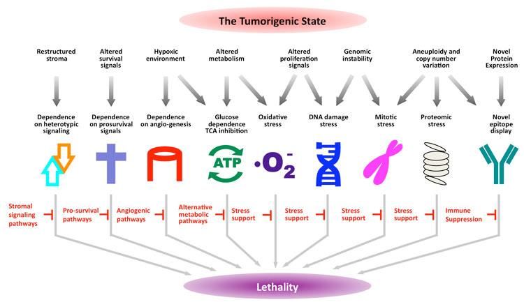
Blocking Cancer’s Fundamental Stress Response to Develop New Therapies

• The “tumorigenic state” creates
 multiple types of stress and cancers
 depend on overcoming stress to
 survive
• Blocking stress support pathways
 represents a new approach to treat
 cancer
• NAD+-using enzymes, e.g., PARPs,
 have evolved to regulate stress
 pathways

 Luo et al, Cell (2009)

 1
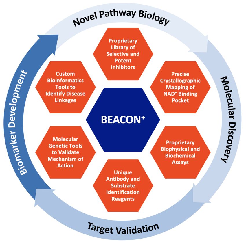
Proprietary BEACON+ Platform:
Unlocking the Biochemical Roles of NAD+-Utilizing Enzymes for the Treatment of Cancer

 • Proprietary collection of biochemical tools
 and technologies to elucidate NAD+-utilizing
 enzyme biology
 • Proprietary small molecule library from
 which to design and develop selective and
 potent NAD+-utilizing enzyme inhibitors
 • Broad library of crystal structures to
 precisely map the NAD+ binding pocket
 • Custom bioinformatics analyses to identify
 novel therapeutic targets and their linkage
 to cancer
 • Sophisticated molecular genetic tools to
 validate drug mechanism of action

 Blocking the Enzyme Activity Component Of NAD+
Common “PARP” Misconception

 “PARP” Inhibitor =

 3
Setting the Record Straight on “PARP” Inhibitors

“PARP1/2” Inhibitor =

 4
The PARP Enzyme Family is Sub-Divided Based on the Type of ADP-
Ribosylation Performed
 Substrate

 NAD+

 nicotinamide

 Substrate modified with Substrate modified with
 mono(ADP-ribose) (MAR) poly(ADP-ribose) (PAR)
 5
MonoPARPs Are an Underexplored Enzyme Class

 Common themes of monoPARP publications:
 • Cellular stress response
 • Anti-viral response
 • Innate and extrinsic immunity
 • Inflammation

 Potent and selective inhibitors would facilitate
 studies on role of monoPARPs in human diseases
 → Screening assays needed
 Wigle et al., SLAS Discovery (2019) 6

Challenges Associated with Developing Biochemical & Cell-Based Assays to
 Screen MonoPARP Enzymes
Lack of validated substrates No selective anti-MAR
for in vitro enzyme assays antibodies for assay
• No X-ray or NMR structures of development
 monoPARPs bound to substrates NAD+
 • Unclear what are best antigens
 exist in PDB
 and how to make them
• Reported recombinant protein
 • Unclear if antigens are stable in
 substrates mixed with enzyme are
 not modified to detectable levels
 ? ? animals
 substrate monoPARP ADP-ribosylated • MAR-binding protein domains
• Reported self-modification is
 substrate (“MAR readers”) have modest
 virtually undetectable
 affinity for MAR and context-
 dependent binding
 ?
 ̶ +
 Unclear how monoPARPs are activated
 • Cellular stress linked to activation but mechanisms unclear

 7
Strategy for First Generation Biochemical MonoPARP Screening Platform:
Forced Self-Modification of Immobilized Enzymes
 Immobilization overcomes weak KM for self-modification

 Dissociation Enhanced Lanthanide Fluorescence Immunoassay (DELFIA) of Immobilized MonoPARPs

 Wigle et al., SLAS Discovery (2019) 8
Example of DELFIA Assay Development for PARP16 Self-Modification
 Produced pure protein Product formation Velocity vs. [enzyme]
 Dye front & linearity vs. time
 MW marker
 PARP16 (95% pure)
 A

 MW marker

 Biotin-NAD+ KMapp IC50 of unlabeled NAD+ Automation & uniformity

 Wigle et al., SLAS Discovery (2019) 9
SPR Shows PARP16 NAD+-Competitive Ligand Binding Affinity Correlates to
Enzyme Inhibition

 Wigle et al., SLAS Discovery (2019) 10
Self-Modification DELFIA Assays Are a Scalable Approach to Family-Wide
PARP Assay Development & Screening
 * *

 = DELFIA assay developed

 = not enzymatically active

 * = assay uses a substrate (not self-modification)

 *

 11
SPR Shows Ligand Binding Correlates Well to Enzyme Inhibition Across the
Entire PARP Family

 Wigle et al., SLAS Discovery (2019)

 12
In Vitro Self-Modification Assays Reveal PARP1 and PARP2 Inhibitors Are
Not Very Potent Against MonoPARPs

 ib
 rib
 par
 rib

 rib
 ib

 apa
 apa
 par

 azo

 ipa
 ruc
 ola

 vel
 nir

 tal
 PARP1 0 0 0.01 0 0

 PARP2 0 0.02 0.01 0 0.01

 PARP3 0.02 2.5 0.02 0.02 0.06

 PARP4 0.09 0.09 0.04 0.41

 PARP5a

 PARP6 0.12 7.4 10.4 0.09 21.5 IC50 (µM) < 0.01
 PARP7 0.2 20.6 4.4 3.5 31 0.01 < IC50 (µM) < 0.1
 PARP8 0.18 40 49 0.1 > 100 0.1 < IC50 (µM) < 1
 PARP9 > 100 > 100 > 100 > 100 > 100 1 < IC50 (µM) < 10
 PARP10 4.4 2.4 0.66 14.4 33.3 10 < IC50 (µM) < 100
 PARP11 18.8 96.8 9.9 5.7 89.7 IC50 (µM) > 100
 PARP12 1.2 16.2 0.86 5.9 5.8

 PARP14 100 14.2 7.5 31.8 100

 PARP15 7.2 48.4 28.3 100 100

 PARP16 1.4 8.2 3.3 0.16 58.5

 Wigle et al., SLAS Discovery (2019) 13
Cross Screening Panel Used to Determine Selectivity of Novel MonoPARP
 Starting Points
 Literature polyPARP inhibitors
 Every compound is screened
 HTS of 500,000 Cross Screening Panel against the panel of PARPs to
 compounds vs. PARP14 understand the determinants
 of selectivity and potency
 PARP1

 PARP2

 PARP3 Selective
 Inhibitor
 PARP4
 monoPARPs polyPARPs
 PARP5a 3 4 6 7 8…. 16 1… 5b

 SPR fragment screen
identifies 19 fragments Medicinal Chemistry
 bound to PARP16 PARP16
 (confirmed by X-ray)

 14
A Limitation of Self-Modification Format:
High Amounts of Enzyme Needed Restrict Resolution of Potency

 Enzyme concentration Length of Assay
 PARP Assay
 (µM) (min)
 PARP1 0.002 60
 PARP2 0.002 120
 PARP3 0.0025 120 [ ]
 PARP4 0.075 180
 50 =
 2
 PARP5a 0.01 120
 PARP6 0.003 180 • Some self-modification assays use high amounts of
 PARP7 0.075 240 enzyme, limiting the ability to resolve potent inhibitors
 PARP8 0.05 180
 PARP9 0.008 180
 • More sensitive in vitro assays are needed
 PARP10 0.015 180 • Cellular assays needed to characterize inhibitors of
 PARP11 0.008 180 increasing potency
 PARP12 0.015 180
 PARP14 0.05 180
 PARP15 0.001 1440
 PARP16 0.15 180

 15
Investigating Active Site Probe Displacement to Generate More Sensitive
Assays

 16
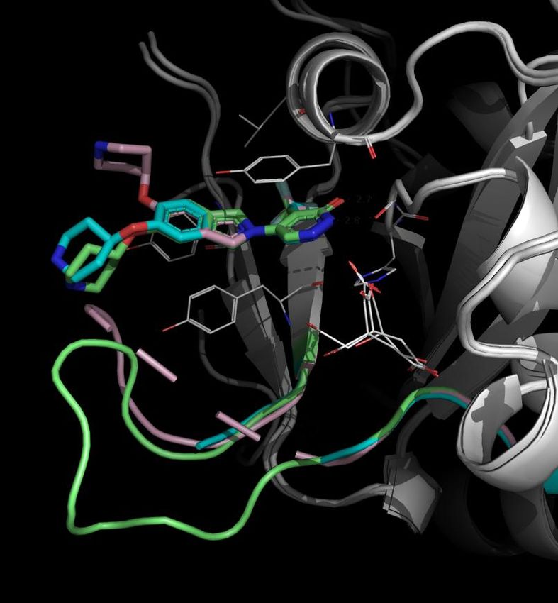
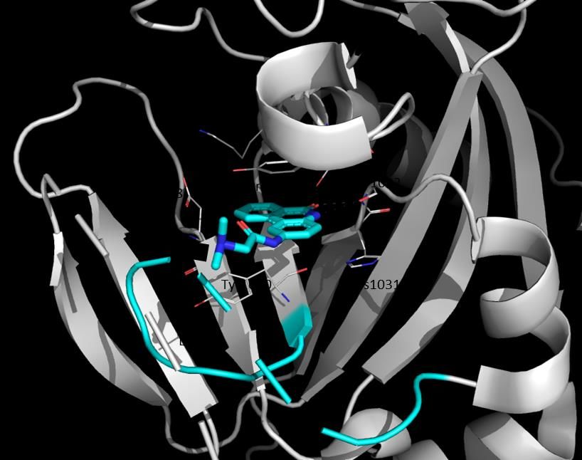
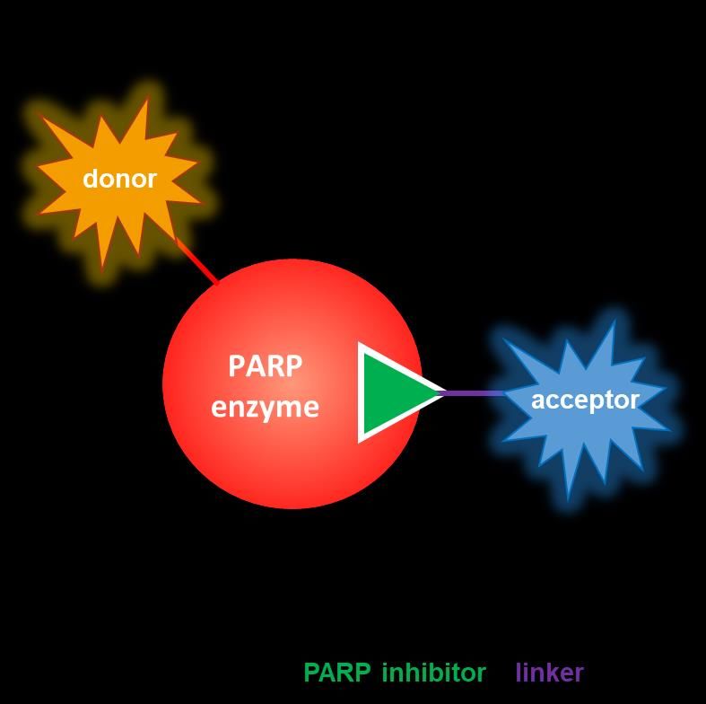
Active Site Probes Designed to Enable Orthogonal Assay Development
 Design of potent monoPARP probe ligands SPR assays show that probes
 retain high affinity to most PARPs

 60

 47
 98
 108

 111
 111
 RBN010860/PARP16 (2.1 Å)

 N0

 N0
 N0
 RB

 RB
 RB
Flexible “tail” of PARP1 0.7 0.7 0.1

 RBN010860 PARP2 0.52 0.26 0.26

points to solvent PARP3 > 10 > 10 > 10

 PARP4 0.56 0.24 0.31

 PARP5a
 Tyr193 9.8 > 10 3.9

 Gly153 PARP6 0.05 0.04 0.08

 PARP7 0.05 0.04 0.08 Kd (µM) < 0.1
 PARP8 0.2 0.65 0.32 0.1 < Kd (µM) < 1
 PARP9 > 10 > 10 > 10 1 < Kd (µM) < 10
 PARP10 0.03 0.05 0.21 Kd (µM) > 10
 PARP11 0.03 0.01 0.04

 PARP12 0.11 0.16 0.19

 PARP13 > 10 > 10 > 10

 PARP14 0.06 0.03 0.23
 D-loop PARP15
 linker + SE590 0.14 0.23 0.66

 PARP16 0.01 0.02 0.02

 linker + biotin
 Wigle et al., Cell Chemical Biology (2020) 17
Development of a Sensitive PARP7 In Vitro TR-FRET Probe Displacement
 Assay
 Simultaneous Titration of All Components

 Emission
 665 nm
 Excitation Emission
 320 nm 615 nm
 TR-FRET
 Ulight-labeled Probe (nM)
 anti-His antibody
 His
 Eu

Europium-labeled
 streptavidin PARP

 Biotin-labeled
 TR-FRET probe

 Sensitive to tool compounds Automated assay is robust

 Wigle et al., Cell Chemical Biology (2020) 18
In Vitro TR-FRET Probe Displacement Assays Correlate to In Vitro Enzyme
Inhibition Assays and Improve Potency Limit for Several PARPs

 For some monoPARPs, probe
 displacement assay extends the
 range of measurable potency
 by 1 – 2 orders of magnitude
 over self-modification assay

 = theoretical potency limit of assay

 Wigle et al., Cell Chemical Biology (2020) 19
Development of NanoBRET Assays to Measure Cellular Target Engagement
for PARP7

 substrate Product + light

 Excitation
 NanoLuc 450 nm

 Emission
 PARP 610 nm

 SE590-labeled
 NanoBRET probe

 Wigle et al., Cell Chemical Biology (2020) 20
Correlation of NanoBRET to Biochemical Assays Across Multiple
MonoPARPs

 Wigle et al., Cell Chemical Biology (2020) 21
Less Potent Outliers in PARP14 NanoBRET Have Low Permeability Compounds highlighted with red have low permeability measured by MDCK-MDR1 assay PARP14 NanoBRET IC50 (M) 10 1 0.1 0.01 0.001 0.001 0.01 0.1 1 10 PARP14 Biochemical IC50 (M) Wigle et al., Cell Chemical Biology (2020) 22
NanoBRET Can Be Used to Measure Inhibitor Residence Time in Cells

 Overexpress Add excess PARP inhibitor @ 10X IC50 and Wash out unbound inhibitor then add NanoBRET
NanoLuc-tagged PARP equilibrate to saturate all binding sites probe and measure signal increase in real-time

 Wigle et al., Cell Chemical Biology (2020) 23
Cellular Residence Time Analysis by NanoBRET Gives Similar Results to SPR
 for Moderately Slow-Off PARP14 Inhibitors

SPR Kd = 47 nM
 t1/2 = 32 s
 Kd = 2 nM Kd = 2 nM
 t1/2 = 29 min t1/2 = 19 min

 Cellular half-life
 1 min
NanoBRET 14 min
 12 min

 Wigle et al., Cell Chemical Biology (2020) 24
Cellular Residence Time Analysis by NanoBRET Gives Similar Results to SPR
 for Very Slow-Off PARP7 Inhibitors

SPR
 Kd = 44 nM Kd = 5 nM Kd = 0.2 nM
 t1/2 = 18 s t1/2 = 82 min t1/2 = 216 min

NanoBRET

 Wigle et al., Cell Chemical Biology (2020) 25
NanoBRET Probe for polyPARP Enzymes
 Designed a potent PARP1/3 probe SPR confirms probe binds Comparison of NanoBRET &
 to PARP1 and PARP3 enzyme inhibition assay
PARP5b/PJ-34
 [PDB: 4BJB]

 be
 pro
 RP
 yPA
 ent
 par

 pol
 PARP1 0.02 0.1

 PARP2 1.6 4.8
 Known PARP1-DNA
 PARP3 0.07 0.53 trapping compounds
 PARP4 0.72 1.4

 PARP5a 3.9 > 10

 PARP6 3.9 5

 PARP7 > 10 > 10 Kd (µM) < 0.1
 PARP8 > 10 > 10 0.1 < Kd (µM) < 1
 PARP9 > 10 > 10 1 < Kd (µM) < 10
 PARP10 > 10 > 10 Kd (µM) > 10
 PARP11 > 10 > 10

 PARP12 > 10 > 10

 PARP13 > 10 > 10

 PARP14 > 10 > 10

 PARP15 > 10 > 10

 PARP16 > 10 > 10

 Wigle et al., Cell Chemical Biology (2020) 26
Off-The-Shelf NanoBRET Probes for PARP1 Also Available Profiling Example PARP1-NanoLuc 40 Tracer Only 100 A-966492 Occupancy (%) Tracer + excess BRET Ratio (mBu) 30 AG-14361 unlabeled Olaparib 20 50 ME0328 UPF 1069 10 0 0 10 -4 10 -3 10 -2 10 -1 10 0 10 -4 10 -2 10 0 10 2 [Tracer], M [Test Compound], M 27
Measuring monoPARP Enzyme Inhibition in Cells: The Next Frontier

 Potent inhibitors discovered
 using self-modification enzyme New anti-MAR/PAR antibody
Developed self-modification
 assays used to generate active obtained and cellular MARylation
 enzyme assays
 site probes for TR-FRET and assay development initiated
 NanoBRET assays

 28
Characterization of a Novel Antibody that Binds to MAR & PAR
 MAR/PAR antibody gives more robust signals than protein-based reagents in spot blot

 Pan-ADPr
 • Macrodomain of Af1521 from the archaebacteria Archaeoglobus fulgidus fused to rabbit IgG
 • Binds MAR and PAR
 Mono-ADPr
 • Macrodomain of human PARP14 fused to rabbit IgG
 • Binds MAR only
 MAR/PAR
 • Rabbit IgG antibody
 • Binds MAR and PAR
 • Not available until 2018
 Lu et al., Biochemical Pharmacology (2019) 29
MAR/PAR Antibody Binds MARylated Substrates with Higher Affinity than
MAR “Reader” Protein Domains
 MAR “Reader” Protein Reagent MAR/PAR antibody

 Kd = 5 µM Kd = 0.2 µM

MARylated peptide • MAR/PAR antibody
 binds with higher
 affinity in SPR assay

 • Evidence of context-
 dependent binding
 High affinity binding detected for MAR “reader”
 No binding detected (data not fitted)
 protein reagent

 MARylated BSA

 30
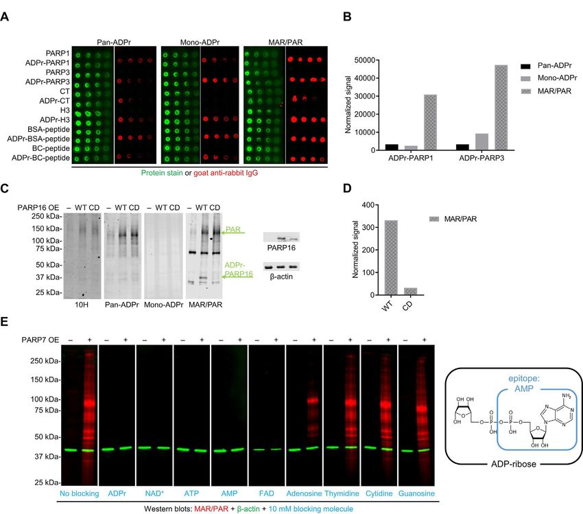
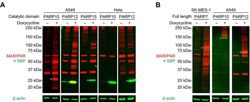
Overexpression of MonoPARPs Leads to Differential MARylation Banding
Patterns on MAR/PAR Western Blot
 Overexpression of catalytic domains Overexpression of full-length PARPs

 • Not all PARPs lead to MAR changes under these conditions
 • Cell-line and construct dependencies observed
 Lu et al., Biochemical Pharmacology (2019) 31
MAR/PAR Antibody Enables Multiple High-Throughput Methods of Detecting Inhibition of PARP7 Enzymatic Activity in Cells Increase in MAR observed after Conversion to In-Cell Western: Compound Dose Response by ICW Format PARP7 stable overexpression by Western PARP7i - + Optimization of Signal Window Compound Compound 6 5 MAR/PAR M A R A b /Ab 2 / A b2° Ab * S ig n a l W in d o w 4 1 :1 0 0 0 /1 :1 0 0 0 1 :4 0 0 0 /1 :1 0 0 0 3 1 :1 0 0 0 /1 :2 0 0 0 2 1 :4 0 0 0 /1 :2 0 0 0 1 0 MAR Ab + anti-β-actin MAR/DNA -- DNA MAR/PAR Conversion to High Content Microscope Conversion from ICW to Immunofluorescence (IF) on High Content Microscope Robust Measurement of MAR Inhibition by IF 120 1 Im m u n o f lu o r e s c e n c e 100 0 .1 80 % In h ib it io n 0 .0 1 60 40 0 .0 0 1 20 0 .0 0 0 1 0 0 .0 0 0 1 0 .0 0 1 0 .0 1 0 .1 1 0 .0 0 0 0 1 0 .0 0 0 1 0 .0 0 1 0 .0 1 0 .1 1 -2 0 In - c e ll W e s t e r n Com pound, M 32
PARP7 NanoBRET Assay Correlates with Cellular Enzyme Inhibition and
Phenotypic Screening Funnel Assays

 NanoBRET vs. Biochemical Cellular Enzyme Inhibition vs. NanoBRET Phenotypic vs. Cellular Enzyme Inhibition

 33
BEACON+ Platform Generates Suite of Screening Assays for PARP Family

 *
 *

 *
 Biochemical - DELFIA
 Biophysical - TR-FRET
 Biophysical - SPR
 X-Ray crystal structure
 Cell biophysical - NanoBRET
 Cell MARylation/biochemical assay
 Potent and selective tool inhibitor
 * Literature tool inhibitor

 34
BEACON+ Platform Generates Selective Inhibitors Across the Entire
PARP Family

• PARP1 inhibitors do not inhibit monoPARPs
• No potent and selective monoPARP inhibitors existed in the literature prior to Ribon
• Ribon has developed multiple selective monoPARP inhibitors

 35
Targeting Stress Support Pathways: Activating Both Tumor Intrinsic Killing
and Activation of the Immune System

 Cancer cells ( ) must balance Inhibiting ( ) stress support … leading to extrinsic activation of the
 cellular stresses ( ) with pathways initiates a tumor- immune system ( ), adding stress
 stress support pathways ( ) intrinsic effect on proliferation… and enhancing tumor killing ( ).
 in order to proliferate.
RBN-2397, a Small Molecule Inhibitor of PARP7: Eliminates Stress Support in
Tumors and Activates Anti-Tumor Immune Response
• PARP7 is a stress-induced protein and is amplified in multiple tumor types including squamous
 carcinoma of the lung and head and neck cancer
• RBN-2397 is a potent and selective inhibitor of PARP7 and causes complete regressions and anti-
 tumor immunity in preclinical tumor models by restoring nucleic acid sensing and induction of
 interferon signaling
• RBN-2397 is in a phase 1 clinical trial in cancer patients and is well-tolerated with preliminary
 evidence of clinical activity
 PARP7 is genetically amplified in RBN-2397 inhibits PARP7 and restores RBN-2397 causes dose-dependent
 several cancer indication (TCGA) nucleic acid sensing and Type I complete regressions in preclinical
 interferon expression tumor models
Summary
• BEACON+ Platform contains suite of de novo biochemical and biophysical assays for monoPARP
 enzymes that do not rely on knowledge of the substrates for each enzyme; assays correlate well with
 each other
 • Self-modification enzyme assays of immobilized protein detected by DELFIA
 • SPR assays
 • NAD+-competitive active site probes
 • Detected in vitro by TR-FRET
 • Detected in cells by NanoBRET

• Newly available MAR/PAR antibody enabled observation of changes in global MARylation detected by
 in-cell Western and immunofluorescence when monoPARP enzymes were overexpressed
 • Assay does not require knowledge of substrates
 • Correlates with phenotypic effect as shown with PARP7 inhibitors in NCI-H1373 cells

• Screening platform used to develop tool compounds for multiple monoPARP enzymes, including
 PARP7 inhibitor (RBN-2397) in phase 1 clinical trial in oncology

 38
Acknowledgements
• Danielle Blackwell
• Dave Church
• Hetvi Desai
• Heike Keilhack
• Kevin Kuntz
• Alvin Lu
• Mario Niepel
• Ahmed Mady
• Christina Majer
• Nick Perl
• Yue Ren
• Victoria Richon
• Andy Santospago
• Laurie Schenkel
• Kerren Swinger
• Melissa Vasbinder
• Rest of the Ribon Team
• Matt Robers and Promega Team
• Multiple CRO Partners

 39
You can also read