A RARE CASE OF A FUNCTIONING GONADOTROPH TUMOR ACCOMPANIED BY ERYTHROCYTOSIS IN AN ELDERLY MAN
←
→
Page content transcription
If your browser does not render page correctly, please read the page content below
CASE REPORT                                                            Проблемы эндокринологии / Problems of Endocrinology | 37

A RARE CASE OF A FUNCTIONING GONADOTROPH TUMOR ACCOMPANIED
BY ERYTHROCYTOSIS IN AN ELDERLY MAN

© Elizaveta O. Mamedova*, Liliya S. Selivanova, Kristina A. Potapova, Svetlana A. Buryakina, Vilen N. Azizyan,
Andrey Yu. Grigoriev, Zhanna E. Belaya

Endocrinology Research Center, Moscow, Russia

Functioning gonadotroph adenomas are rare pituitary tumors secreting one or two gonadotropins (follicle-stimulating
hormone (FSH) and/or luteinizing hormone (LH)), which are hormonally active. In the majority of cases, gonadotroph
tumors are endocrinologically “silent” and make up more than a half of non-functioning pituitary adenomas. In this article
we describe a rare clinical case of LH/FSH-secreting pituitary macroadenoma with bitemporal hemianopsia in a 62-year-
old man. The patient underwent transnasal transsphenoidal adenomectomy, leading to remission. The distinctive feature
of this case is the presence of secondary erythrocytosis due to endogenous hyperandrogenism, which required several
blood exfusions to normaliza the level of hematocrit before surgery. It is noteworthy that clinical signs of erythrocytosis
were present long before visual impairment. This clinical case demonstrates difficulties in the early diagnosis of functioning
gonadotroph adenomas.

KEYWORDS: pituitary adenoma; gonadotroph tumor; LH; FSH; hyperndrogenism; erythrocytosis.

РЕДКИЙ СЛУЧАЙ ГОРМОНАЛЬНО-АКТИВНОЙ ГОНАДОТРОПИНОМЫ,
АССОЦИИРОВАННОЙ СО ВТОРИЧНЫМ ЭРИТРОЦИТОЗОМ, У МУЖЧИНЫ В ПОЖИЛОМ
ВОЗРАСТЕ
© Е.О. Мамедова*, Л.С. Селиванова, К.А. Потапова, С.А. Бурякина, В.Н. Азизян, А.Ю. Григорьев, Ж.Е. Белая

Национальный медицинский исследовательский центр эндокринологии, Москва, Россия

Гормонально-активные гонадотропиномы — это редкие опухоли гипофиза, секретирующие один или два гонадо-
тропных гормона (фолликулостимулирующий гормон (ФСГ) и/или лютеинизирующий гормон (ЛГ)), которые обладают
биологической активностью. В большинстве случаев гонадотропиномы являются «молчащими» и составляют более
половины гормонально-неактивных аденом гипофиза. В статье представлено описание редкого клинического на-
блюдения: ЛГ/ФСГ-секретирующей макроаденомы гипофиза с развитием битемпоральной гемианопсии у 62-летне-
го мужчины. Пациенту была проведена трансназальная транссфеноидальная аденомэктомия, позволившая достичь
ремиссии заболевания. Отличительной особенностью данного случая являлось наличие вторичного эритроцитоза,
развившегося вследствие эндогенной гиперандрогении, что потребовало проведения процедур эксфузий крови
с целью нормализации уровня гематокрита перед проведением оперативного вмешательства. Примечательно, что
клинические признаки эритроцитоза у пациента были выявлены задолго до развития зрительных нарушений. Пред-
ставленный клинический случай демонстрирует сложность ранней диагностики гормонально-активных гонадотро-
пином.
КЛЮЧЕВЫЕ СЛОВА: аденома гипофиза; гонадотропинома; ЛГ; ФСГ; гиперандрогения; эритроцитоз.

    BACKGROUND                                                          adenomas [5], while functioning gonadotroph tumors are
                                                                        described in individual cases or small case series.
    Functioning gonadotroph adenomas are rare pituitary tu-                 Clinical signs of functioning gonadotroph tumors de-
mors secreting one or two gonadotropins (follicle-stimulat-             pend on sex and age. In children these tumors cause pre-
ing hormone (FSH) and/or luteinizing hormone (LH)), which               cocious puberty [6-8]. In women of reproductive age
are hormonally active [1]. It is noteworthy that in the ma-             the prolonged exposure of ovaries to FSH leads to menstrual
jority of cases gonadotroph tumors are endocrinologically               irregularity, causes infertility, cystic lesions in ovaries, ovari-
“silent”, i.e. they have FSH and/or LH positively stained dur-          an hyperstimulation syndrome, chronic pain in pelvis or ab-
ing immunohistochemical (IHC) staining while they do not                domen [9-15]. In the postmenopausal period signs caused
secrete these hormones or secrete them in a biologically                by the tumor mass-effect (chiasmal syndrome, headaches,
inactive form (alpha-subunit, beta-subunit of FSH or LH) and            hypopituitarism) become more presented. The high level
therefore are not accompanied by hormonal hypersecretion                of gonadotropins in postmenopausal women does not im-
signs [1-4]. Overall «silent» gonadotroph tumors occur quite            pact the ovarian function and estrogen production that is
often and make up to 64% of all non-functioning pituitary               why the secretion of functioning hormones by the pituitary

© Endocrinology Research Centre, 2021            Received: 15.05.2021. Accepted: 13.06.2021.
Проблемы эндокринологии 2021;67(3):37-44          doi: https://doi.org/10.14341/probl12758             Problems of Endocrinology. 2021;67(3):37-44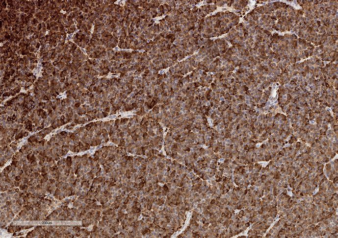
3 8 | Проблемы эндокринологии / Problems of Endocrinology КЛИНИЧЕСКИЙ СЛУЧАЙ
tumor does not cause the ovarian hyperstimulation syn- ventricle; it was adjacent to anterior communicating arteries,
drome [1]. In men testicular enlargement, decrease in libi- extended laterally into cavernous sinuses with complete en-
do and erectile dysfunction can be observed; isolated visual casement of intracavernous internal carotid arteries (Knosp
function impairment without signs of hypogonadism is less IV); it was adjacent to medial basal parts of temporal lobes
common [2, 3, 9, 16]. When the tumor hypersecretes LH, and straight gyri. The anterior part of the mass bulged into
there can be testosterone blood level increase [3, 17]. the basilar sinus, the posterior part destroyed the clinoid plate
The main treatment modality for gonadotroph tumors and extended into the prepontine cistern with its moderate
is transsphenoidal adenomectomy which allows achieving narrowing (fig. 1). The normal pituitary and its stalk were not
remission at early stages [1]. If total tumor resection is im- differentiated. Findings: pituitary macrodenoma with supra-,
possible (invasive growth, cavernous sinuses invasion) or infra-, retro- parasellar (D, S) extension (Knosp IV).
the tumor is recurrent, radiotherapy may be an option [1]. Ophthalmological examination findings: visual acuity
The phamacological therapy (somatostatin analogues, do- OD=0.2 incorrigible, OS=up to 0.8 left of field; bilateral par-
pamine agonists, temozolomide) is not a method of choice tial optic nerve atrophy. Perimetry discovered the narrowing
to treat gonadotroph tumors due to the limited data of its of the visual field (fig. 2A). Hormonal tests excluded endog-
efficiency [1]. enous hypercortisolism and acromegaly (late-night salivary
In this article we describe a rare clinical case of a LH/FSH- cortisol was 5.11 nmol/L (0.5-9.65), IGF-1 was 104.6 ng/ml
secreting pituitary macroadenoma with bitemporal hemia- (16-245)) and confirmed secondary hypothyroidism and
nopsia and secondary erythrocytosis in a 62-year-old man, secondary hyperprolactinemia (table 1, before surgery).
the diagnostic process and treatment results. The study of gonadotroph function determined the increase
of FSH level while LH and testosterone were at the up-
CASE DESCRIPTION per limit of the reference range (table 1, before surgery).
The blood tests also showed high red blood cell count, in-
Patient G., 62 y.o., was admitted to the Neuro crease of hemoglobin, hematocrit (table 1 before surgery),
endocrinology and Bone Diseases Department of the Federal i.e. significant erythrocytosis in the setting of relatively nor-
State Budgetary Institution «Endocrinology National Medical mal values of other blood count results. Biochemical blood
Research Center» under the Russian Ministry of Health tests showed the creatinine level of 117.2 µmol/L (eGFR-EPI
in September 2020 with complaints of significant visual im- 58 ml/min/1.73 m2), signs of hyperuricemia, dyslipidemia;
pairment, blurry vision, narrowing of the visual field, and in- other tests demonstrated no significant clinical findings. It
termittent headache. is noteworthy that during the previous 5-6 years the patient
The patient started experiencing these symptoms noticed plethora of his face, neck, trunk, but despite annual
in December 2019. In February 2020 the patient went to a lo- checkups the patient was not aware of erythrocytosis.
cal ophthalmologist who detected partial optic atrophy and Ultrasound (US) of scrotal organs was performed
early cataract of both eyes. The ophthalmologist suspected on September 16, 2020: right testis volume was 22 ml, left
a pituitary disorder and recommended magnetic resonance testis volume was 20.7 ml. The ultrasound of abdomen
imaging (MRI) of the brain. Earlier, according to the patient, on September 21, 2020 detected signs of hepatomegaly
during annual checkups (the last one in 2019) no significant (the thickness of the right lobe was 13.4 cm, the left lobe
visual function abnormalities were found. Brain MRI showed was 6.2 cm), fatty liver disease, chronic calculous cholecysti-
a mass lesion in the sellar region, 44x34x43 mm in size. For tis, normal spleen size of 10.1x5.2 cm.
further examination and surgical treatment the patient Thus, based on the laboratory tests (high levels of LH,
was admitted to the Federal State Budgetary Institution FSH, testosterone, secondary erythrocytosis), pituitary mass
«Endocrinology National Medical Research Center» under lesion verified by MRI, LH/FSH-secreting pituitary macroade-
the Russian Ministry of Health with the referral diagnosis noma was suspected in the patient.
«Non-functioning pituitary adenoma». Taking into account the gonadotropin-secreting pitui-
During the initial medical examination ocular injections, tary macroadenoma with chiasm compression, neurosur-
plethora of the face, neck, trunk and palms were noticed; gery was chosen as the optimal treatment option. To prepare
no clinical signs of acromegaly and hypercortisolism were the patient for surgery in order to reduce the risk of throm-
found. The patient was hypersthenic, with height 174.4 cm, bosis, whole blood exfusions were performed with saline
weight 102 kg (BMI = 33.5 kg/m2). Subcutaneous adipose replacements (3 procedures were made) to achieve periph-
tissue was evenly distributed with no swelling. Male pat- eral blood target values (Hb up to 150-160 g/L, Ht u nder
tern of hair distribution, secondary sex characteristics were 51%). In September 2020 the patient underwent transna-
properly developed. The liver on palpation was +1 cm from sal transsphenoidal adenomectomy in the Federal State
the costal margin, the liver edge was round, firm, non-ten- Budgetary Institution «Endocrinology National Medical
der. The rest of the organs and systems were unremarkable. Research Center» under the Russian Ministry of Health.
There were no significant past medical and family history; no Intraoperatively neurosurgeons detected a yellow-brown
smoking or alcohol consumption according to the patient. tumor of moderately dense consistency which was fully
According to the brain MRI of September 17, 2020 the se- removed. Histological findings showed a solid alveolar pi-
ries of sagittal, frontal and axial T1- and T2-weighted images tuitary basophilic adenoma (fig. 3). The IHC study revealed
(WI) clearly showed a mass with clear and smooth margins the expression of LH in 80% of tumor cells (fig. 4) and of FSH
in the sella turcica of the following size: width 40 mm, height (fig. 5) in 30% of tumor cells; weak reaction to somatostatin
41 mm, A-P dimension 33 mm. The structure was heteroge- receptors type 2A (fig. 6) and type 5 (fig. 7) (IRS score 3) was
neous due to fluid inclusions. The mass had suprasellar exten- found. The Ki-67 index was 3% (fig. 8). Thus, the diagnosis
sion with the compression of the optic chiasm and the third of LH/FSH-secreting pituitary adenoma was verified.
Проблемы эндокринологии 2021;67(3):37-44 doi: https://doi.org/10.14341/probl12758 Problems of Endocrinology. 2021;67(3):37-44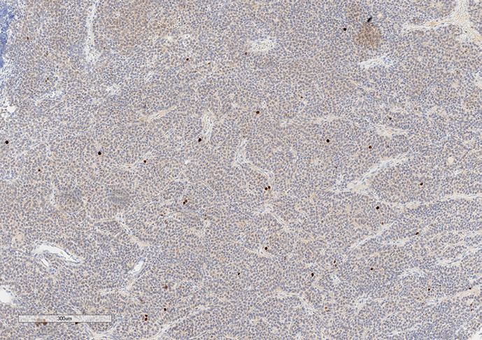
CASE REPORT                                                                      Проблемы эндокринологии / Problems of Endocrinology | 39

    The development of adrenal insufficiency in the early post-                   day after surgery in satisfactory condition. Six months af-
operative period was noted (blood cortisol in the morning                         ter surgery the patient was in remission: a laboratory study
was 175.6 nmol/L; table 1, after surgery), and thus hydrocor-                     showed that hypogonadotropic hypogonadism persisted as
tisone replacement therapy was initiated. To correct the sec-                     well as secondary adrenal insufficiency and secondary hy-
ondary hypothyroidism (table 1, after surgery) the consecu-                       pothyroidism (table 1, 6 months after surgery). The patient
tive treatment with levothyroxine was started with the initial                    was prescribed hydrocortisone, levothyroxine and testoster-
dose of 50 µg. It was recommended to start pharmacological                        one. Ophthalmological examination findings: visual acuity
therapy for secondary hypogonadism (table 1, after surgery)                       OD=0.4, OS=0.8; bilateral partial optic atrophy (OS>OD), pos-
with testosterone gel after performing prostate US. During                        itive changes in visual field on perimetry (fig. 2B). The brain
the first week after surgery a tendency to hyponatremia was                       MRI on March 22, 2021 showed in the sella turcica, cavernous
noted (up to 134 mmol/L), but water restriction (up to 1 L/day                    sinuses and in basilar bone sinuses a cystic solid mass lesion
with gradual extension to 1.5-2 L/day) allowed the sodium                         of heterogeneous structure (contained a cyst of 11x16x13 mm
level to normalize. Blood pressure and heart rate stayed with-                    size) which incoherently accumulated the contrast media;
in the normal range. The patient was discharged on the 13th                       the size of the lesion: vertical — 20 mm, transversal — 27 mm,

 а                                                                                 b

 c                                                                                 d
               Figure 1. Brain MRI of patient G. Pituitary macroadenoma with supra-, infra- and retro- parasellar (D, S,) extension (Knosp IV):
a) T2WI (weighted image), axial projection. Pituitary adenoma (arrow); b) T1WI, coronal projection; c) T2WI, coronal projection. Optic chiasm compression
(arrow); d) T1WI, sagittal projection. The adenoma infrasellar extension into the basilar sinus (thick arrow) and retrosellar extension into the prepontine
                                                                    cistern (thin arrow).

Проблемы эндокринологии 2021;67(3):37-44                    doi: https://doi.org/10.14341/probl12758                 Problems of Endocrinology. 2021;67(3):37-44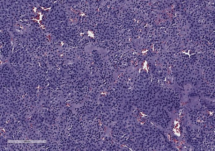
4 0 | Проблемы эндокринологии / Problems of Endocrinology                                                                 КЛИНИЧЕСКИЙ СЛУЧАЙ

                                  OS                                                                           OD

   А

   B

         Figure 2. Perimetry findings before surgery (A, upper row) and 6 months after surgery (B, lower row). OS — left eye, OD — right eye.

Table 1. Laboratory parameters before and after surgery

                                                                                                        6 months after
 Studied parameter, measurement units                      Before surgery             After surgery                             Reference range
                                                                                                           surgery
                                                              Complete blood count
Red blood cells, c/L                                         6.66x10¹²         5.15x10¹²                    4.71x10¹²               4.3-5.8x10¹²
Mean corpuscular volume, MCV, fL                                 85.1              87.2                        83.9                     82-98
Hemoglobin, g/L                                                  196               152                         137                    132-172
Hematocrit, %                                                   56.7               44.9                        39.5                     40-51
Leukocytes, c/L                                               7.38x10⁹          8.52x10⁹                    4.54x10⁹                 3.9-10x109
Platelets, c/L                                                 207109            249109                      207109                 148-339x109
ESR, mm/h                                                          9                19                          18                       2-20
                                                          Hormonal blood and urine tests
LH, IU/L                                                         10.3            0.513                        0.216                    2.5-11
FSH, IU/L                                                       22.5               4.19                        0.87                    1.6-9.7
Testosterone, nmol/L                                              28              0.62                        0.271                   11-28.2
TSH, mIU/L                                                      0.795            0.088                        1.071                   0.25-3.5
Free T4, pmol/L                                                 6.95              5.78                         7.09                     9-19
IGF-1, ng/ml                                                    104.6                -                        52.36                    16-245
Prolactin, mU/L                                                562.1                 -                        178.1                   78-380
Cortisol blood in the morning, nmol/L                           293.7             175.6                       237.3                   171-536
Daily urinary free cortisol, nmol/day                              -                 -                         52.7                   100-379

Проблемы эндокринологии 2021;67(3):37-44                   doi: https://doi.org/10.14341/probl12758            Problems of Endocrinology. 2021;67(3):37-44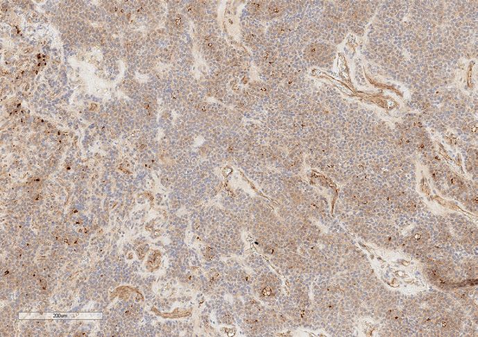
CASE REPORT Проблемы эндокринологии / Problems of Endocrinology | 41
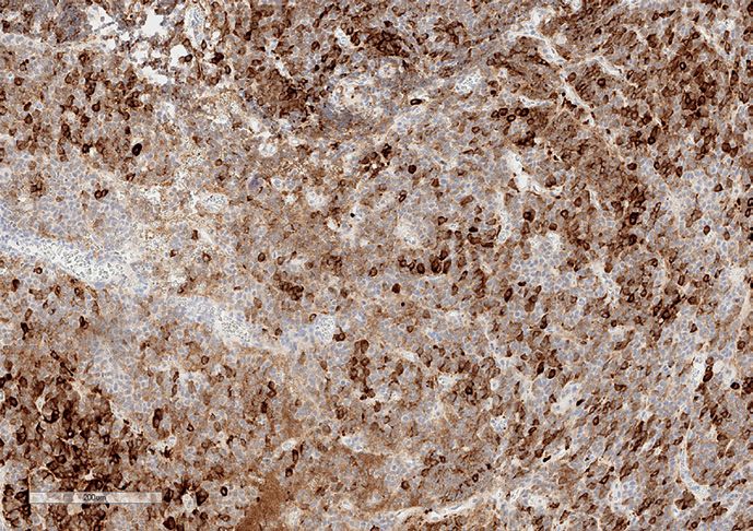
Figure 3. Tumor microstructure. Figure 4. Luteinizing hormone expression by tumor cells.
Stained with hematoxylin and eosin. Magnification x100. Magnification x100.
Figure 5. Follicle stimulating hormone expression by tumor cells. Figure 6. SSTR2A expression by tumor cells. Magnification x100.
Magnification x100.
Figure 7. SSTR5 expression by tumor cells. Magnification x100. Figure 8. Tumor proliferative index (via Ki-67 expression).
A-P dimension — 25 mm; pituitary tissue was not clearly DISCUSSION
differentiated at the right edge of the lesion, stalk deviated
to the right. Findings: the condition after the removal of a gi- The clinical case demonstrates difficulties in the early
ant pituitary adenoma, mass lesion in the sellar region with diagnosis of functioning gonadotroph adenomas. The pe-
infra- and parasellar (D, S, Knosp III) extension. Given that culiarity of this case is combined hypersecretion of FSH and
the laboratory study demonstrated remission, MRI data were LH with predominance of LH secretion by the tumor which
considered as postoperative changes, and MRI dynamic fol- led to the development of hyperandrogenism. Cases
low-up was recommended. of functioning gonadotroph tumors in men described
Проблемы эндокринологии 2021;67(3):37-44 doi: https://doi.org/10.14341/probl12758 Problems of Endocrinology. 2021;67(3):37-44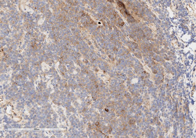
4 2 | Проблемы эндокринологии / Problems of Endocrinology                                                              КЛИНИЧЕСКИЙ СЛУЧАЙ

in the literature (individual cases or small case series) mostly             ADDITIONAL INFORMATION
had FSH-hypersecretion by the tumor which led to testicu-
lar enlargement (while LH and testosterone levels were low                    Funding sources. The study was performed on the authors’ initiative
or normal); a vast majority of tumors were macroadenomas                without attracting funding.
causing visual impairment [2, 4, 8, 9, 18-20]. Some cases de-                 Conflict of interest. The authors declare no obvious and potential con-
scribed had combined FSH and LH hypersecretion [3, 16, 17,              flicts of interest related to the content of this article.
21-24]. In some singular presented cases a tumor secreted                     Contribution of the authors. Elizaveta O. Mamedova — patient’s
only LH [25, 26]. It is noteworthy that in most cases of com-           attending doctor, literature data analysis, writing; Liliya S. Selivanova —
bined FSH and LH hypersecretion causing testosterone level              histology and IHC studies; Kristina A. Potapova — writing; Svetlana A.
increase there were no clinical signs of hyperandrogenism               Buryakina — MRI examination and interpretation; Vilen N. Azizyan — sur-
[3, 16, 21, 23]. In other cases, clinical signs of hyperandro-          gical treatment, editing; Andrey Yu. Grigoriev — surgical treatment, edit-
genism included: spermatogenesis increase [22], libido en-              ing; Zhanna E. Belaya — final editing. All of the authors approved the final
hancement [24], erythrocytosis [17]. It is also noteworthy              version of the article before publication, agreed to be responsible for all
that in some cases of FSH/LH-secreting adenomas with con-               aspects of the work, implying proper examination and resolution of issues
firmed increase of LH and testosterone levels by the labora-            relating to the accuracy or integrity of any part of the work.
tory study, the IHC study of the removed tumor showed no                      Patient consent. The patient signed the informed consent to the pub-
LH expression [3, 24].                                                  lication of personal medical information in an anonymized form.
    The main clinical manifestation of hyperandrogen-
ism in the described case was the development of sec-
ondary erythrocytosis which had originated several years                Table 2. Erythrocytosis causes (adapted from [27] and [28])
prior to the visual impairment development according
to the medical history data. The main causes of second-                                       Primary erythrocytosis
ary erythrocytosis development are presented in table 2.
                                                                         • Polycythemia vera
The existing recommendations for differential diagnosis
of erythrocytosis consider excessive testosterone only                                       Secondary erythrocytosis
when there was testosterone overdose and do not con-
sider the probability of endogenous hyperandrogenism                     • Generalized hypoxia
development [27, 28]. Moreover, in our case testosterone
                                                                             • Smoking
level was at the upper limit of the reference range and
only «unsuppressed» levels of LH and FSH in the patient                      • Carbon monoxide poisoning
with pituitary macroadenoma indicated its excessive lev-
els while with exogenous hyperandrogenism these levels                       • Pulmonary diseases
would have been decreased. Taking into account the varie-
                                                                             • Obstructive sleep apnoea
ty of causes of secondary erythrocytosis, it is possible to as-
sume that the probability to detect a gonadotroph tumor                      • Congenital heart disorder (right-to-left shunt)
at the moment of erythrocytosis onset in this case would
have been low.                                                               • Living at high altitude
    Surgical treatment is a method of choice to treat func-              • Local kidney hypoxia
tioning gonadotroph adenomas as it allows eliminating tu-
mor mass-effects (first of all eliminate its impact on chiasm)               • Renal artery stenosis
and achieve quick improvement of hormone levels [1]. There
is no convincing evidence of pharmacological therapy or ra-                  • Hydronephrosis
diotherapy efficiency in the literature [1]. In our case there               • Polycystic kidney disease
was remission after the surgical removal of the tumor, but
hypopituitarism persisted and visual function was not fully              • Drug-associated
restored.
    Thus, in our clinical case as well as in most cases de-                  • Testosterone
scribed in the literature, a functioning gonadotroph adeno-                  • Erythropoietin
ma was diagnosed only at the stage of visual impairment
when tumor mass-effects appeared. Currently there is no                  • Erythropoietin abnormal production by tumors
solution for possible earlier diagnosis of such tumors.
                                                                             • Hepatocellular carcinoma
    CONCLUSION                                                               • Renal cell carcinoma

    Unlike other functioning pituitary adenomas that have                    • Cerebellar hemangioblastoma
typical clinical presentation, the symptoms of gonado-
                                                                             • Parathyroid cancer
tropin-secreting tumors often escape detection till they
become macroadenomas causing headache, visual field                          • Uterine leiomyoma
impairment and hypopituitarism. It is also important to re-
member that secondary erythrocytosis in men can be a sign                    • Pheochromocytoma
of hyperandrogenism, and LH-secreting gonadotroph ade-
                                                                             • Meningioma
nomas may be one of its causes.

Проблемы эндокринологии 2021;67(3):37-44          doi: https://doi.org/10.14341/probl12758                  Problems of Endocrinology. 2021;67(3):37-44
CASE REPORT Проблемы эндокринологии / Problems of Endocrinology | 43
СПИСОК ЛИТЕРАТУРЫ | REFERENCES
1. Ntali G, Capatina C, Grossman A, Karavitaki N. Functioning [Gorbacheva AM, Przhiyalkovskaya EG, Azizyan VN, et al. An ovarian
gonadotroph adenomas. J Clin Endocrinol Metab. hyperstimulation syndrome caused by gonadotropinoma in a young
2014;99(12):4423-4433. doi: https://doi.org/10.1210/jc.2014-2362 woman. Problems of Endocrinology. 2019;65(4):278-288. (In Russ.)].
2. Cote DJ, Smith TR, Sandler CN, et al. Functional doi: https://doi.org/10.14341/probl10178
gonadotroph adenomas: case series and report 16. Dizon MN, Vesely DL. Gonadotropin-secreting pituitary tumor
of literature. Neurosurgery. 2016;79(6):823-831. associated with hypersecretion of testosterone and hypogonadism
doi: https://doi.org/10.1227/NEU.0000000000001188 after hypophysectomy. Endocr Pract. 2002;8(3):225-231.
3. Chamoun R, Layfield L, Couldwell WT. Gonadotroph doi: https://doi.org/10.4158/EP.8.3.225
adenoma with secondary hypersecretion of 17. Ceccato F, Occhi G, Regazzo D, et al. Gonadotropin secreting
testosterone. World Neurosurg. 2013;80(6):900.e7-11. pituitary adenoma associated with erythrocytosis: case report
doi: https://doi.org/10.1016/j.wneu.2012.11.069 and literature review. Hormones (Athens). 2014;13(1):131-139.
4. Snyder PJ. Gonadotroph cell adenomas of the pituitary. Endocr Rev. doi: https://doi.org/10.1007/BF03401328
1985;6:552-563. doi: https://doi.org/10.1210/edrv-6-4-552 18. Heseltine D, White MC, Kendall-Taylor P, et al. Testicular
5. Yamada S, Ohyama K, Taguchi M, et al. A study of the correlation enlargement and elevated serum inhibin concentrations occur
between morphological findings and biological activities in clinically in patients with pituitary macroadenomas secreting follicle
nonfunctioning pituitary adenomas. Neurosurgery. 2007;61:580-584. stimulating hormone. Clin Endocrinol (Oxf ). 1989;31:411-423.
doi: https://doi.org/10.1227/01.NEU.0000290906.53685.79 doi: https://doi.org/10.1111/j.1365-2265.1989.tb01265.x
6. Gryngarten MG, Braslavsky D, Ballerini MG, et al. Spontaneous 19. Pigny P, Henric B, Lahlou N, et al. A gonadotroph adenoma
Ovarian Hyperstimulation Syndrome Caused by a Follicle- with a high proportion of basic FSH isohormones by
Stimulating Hormone-Secreting Pituitary Macroadenoma in chromatofocusing. J Clin Endocrinol Metab. 1996;81:2407-2408.
an Early Pubertal Girl. Horm Res Paediatr. 2010;73(4):293-298. doi: https://doi.org/10.1210/jcem.81.6.8964889
doi: https://doi.org/10.1159/000284395 20. Dahlqvist P, Koskinen LO, Brännström T, Hägg E.
7. Ambrosi B, Bassetti M, Ferrario R, et al. Precocious puberty in a boy Testicular enlargement in a patient with a FSH-secreting
with a PRL-, LH- and FSH-secreting pituitary tumour: hormonal pituitary adenoma. Endocrine. 2010;37:289-293.
and immunocytochemical studies. Acta Endocrinol (Copenh). doi: https://doi.org/10.1007/s12020-009-9302-z
1990;122(5):569-576. doi: https://doi.org/10.1530/acta.0.1220569 21. Snyder PJ, Sterling FH. Hypersecretion of LH and FSH by
8. Clemente M, Caracseghi F, Gussinyer M, et al. Macroorchidism and a pituitary adenoma. J Clin Endocrinol Metab. 1976;42:544-550.
panhypopituitarism: two different forms of presentation of FSH- doi: https://doi.org/10.1210/jcem-42-3-544
secreting pituitary adenomas in adolescence. Horm Res Paediatr. 22. Zárate A, Fonseca ME, Mason M, et al. Gonadotropin-secreting
2011;75(3):225-230. doi: https://doi.org/10.1159/000322211 pituitary adenoma with concomitant hypersecretion of
9. Zhao Y, Lian W, Xing B, et al. Functioning gonadotroph testosterone and elevated sperm count. Treatment with
adenoma. Chin Med J (Engl). 2019;132(8):1003-1004. LRH agonist. Acta Endocrinol (Copenh). 1986;113:29-34.
doi: https://doi.org/10.1097/CM9.0000000000000184 doi: https://doi.org/10.1530/acta.0.1130029
10. Cooper O, Geller JL, Melmed S. Ovarian hyperstimulation 23. Whitaker MD, Prior JC, Scheithauer B, et al.
syndrome caused by an FSH-secreting pituitary adenoma. Gonadotrophin-secreting pituitary tumour: report
Nat Clin Pract Endocrinol Metab. 2008;4(4):234-238. and review. Clin Endocrinol (Oxf ). 1985;22:43-48.
doi: https://doi.org/10.1038/ncpendmet0758 doi: https://doi.org/10.1111/j.1365-2265.1985.tb01063.x
11. Halupczok J, Bidzińska-Speichert B, Lenarcik-Kabza A, et al. 24. Thakkar A, Kannan S, Hamrahian A, et al. Testicular
Gonadotroph adenoma causing ovarian hyperstimulation syndrome “Hyperstimulation” Syndrome: A Case of Functional
in a premenopausal woman. Gynecol Endocrinol. 2014;30(11):774-777. Gonadotropinoma. Case Rep Endocrinol. 2014;2014:1-4.
doi: https://doi.org/10.3109/09513590.2014.934668 doi: https://doi.org/10.1155/2014/194716
12. Hirano M, Wada-Hiraike O, Miyamamoto Y, et al. A case of functioning 25. Roman SH, Goldstein M, Kourides IA, et al. Luteinizing hormone-
gonadotroph adenoma in a reproductive aged woman. Endocr J. releasing hormone (LHRH) agonist [D-Trp6-Pro9-NEt]LHRH increased
2019;66(7):653-656. doi: https://doi.org/10.1507/endocrj.EJ19-0066 rather than lowered LH and alpha-subunit levels in a patient
13. Broughton C, Mears J, Williams A, Lonnen K. A clinically functioning with an LH-secreting pituitary tumor. J Clin Endocrinol Metab.
gonadotroph adenoma presenting with abdominal pain, ovarian 1984;58(2):313-319. doi: https://doi.org/10.1210/jcem-58-2-313
hyperstimulation and fibromatosis. Endocrinol Diabetes Metab Case 26. Peterson RE, Kourides IA, Horwith M, et al. Luteinizing hormone-
Reports. 2018;2018. doi: https://doi.org/10.1530/EDM-18-0123 and alpha-subunit-secreting pituitary tumor: positive feedback
14. Graillon T, Castinetti F, Chabert-Orsini V, et al. Functioning of estrogen. J Clin Endocrinol Metab. 1981;52(4):692-698.
gonadotroph adenoma with severe ovarian hyperstimulation doi: https://doi.org/10.1210/jcem-52-4-692
syndrome: A new emergency in pituitary adenoma surgery? 27. McMullin MF, Bareford D, Campbell P, et al. Guidelines for
Surgical considerations and literature review. Ann Endocrinol (Paris). the diagnosis, investigation and management of polycythaemia/
2019;80(2):122-127. doi: https://doi.org/10.1016/j.ando.2018.11.007 erythrocytosis. Br J Haematol. 2005;130(2):174-195.
15. Горбачева А.М., Пржиялковская Е.Г., Азизян В.Н., и др. doi: https://doi.org/10.1111/j.1365-2141.2005.05535.x
Клинически активная гонадотропинома c развитием синдрома 28. Mithoowani S, Laureano M, Crowther MA, Hillis CM. Investigation
гиперстимуляции яичников у молодой женщины // Проблемы and management of erythrocytosis. CMAJ. 2020;192:913-918.
эндокринологии. — 2019. — Т. 65. — №4. — С. 278-288. doi: https://doi.org/10.1503/cmaj.191587
Проблемы эндокринологии 2021;67(3):37-44 doi: https://doi.org/10.14341/probl12758 Problems of Endocrinology. 2021;67(3):37-444 4 | Проблемы эндокринологии / Problems of Endocrinology КЛИНИЧЕСКИЙ СЛУЧАЙ Manuscript received on: May 15, 2021. Approved for publication on: June 13, 2021. Published on-line on: June 30, 2021. AUTHORS INFO *Мамедова Елизавета Октаевна, к.м.н. [Elizaveta O. Mamedova, MD, PhD]; адрес: Россия, 117036, Москва, ул. Дм. Ульянова, д. 11 [address: 11 Dm. Ulyanova street, 117036 Moscow, Russia]; ORCID: https://orcid.org/0000-0002-9783-3599; eLibrary SPIN: 3904-6017; e-mail: lilybet@mail.ru Селиванова Лилия Сергеевна, к.м.н. [Liliya S. Selivanova, MD, PhD]; e-mail: liselivanova89@yandex.ru; ORCID: https://orcid.org/0000-0001-6891-0009; eLibrary SPIN: 5151-3675 Потапова Кристина Александровна [Kristina A. Potapova, MD]; ORCID: https://orcid.org/0000-0002-1967-8626; eLibrary SPIN: 4892-6792; e-mail: kris.potockaya.95@mail.ru Бурякина Светлана Алексеевна, к.м.н. [Svetlana A. Buryakina, MD, PhD]; ORCID: https://orcid.org/0000-0001-9065-7791; eLibrary SPIN: 5675-0651; e-mail: sburyakina@yandex.ru Азизян Вилен Неронович, к.м.н. [Vilen N. Azizyan, MD, PhD]; ORCID: https://orcid.org/0000-0001-9718-6099; eLibrary SPIN: 7666-5950; e-mail: vazizyan@mail.ru Григорьев Андрей Юрьевич [Andrey Yu. Grigoriev, MD, PhD]; ORCID: https://orcid.org/0000-0002-9575-4520; eLibrary SPIN: 8910-8130; e-mail: medway26@gmail.com Белая Жанна Евгеньевна, д.м.н. [Zhanna E. Belaya, MD, PhD]; ORCID: https://orcid.org/0000-0002-6674-6441; eLibrary SPIN: 4746-7173; e-mail: jannabelaya@gmail.com TO CITE THIS ARTICLE: Mamedova EO, Selivanova LS, Potapova KA, Buryakina SA, Azizyan VN, Grigoriev AY, Belaya ZE. Adrenal imaging: anatomy and pathology (literature review). Problems of Endocrinology. 2021;67(3):37-44. doi: https://doi.org/10.14341/probl12758 Проблемы эндокринологии 2021;67(3):37-44 doi: https://doi.org/10.14341/probl12758 Problems of Endocrinology. 2021;67(3):37-44
You can also read