A rare presentation of central granular cell odontogenic tumor
←
→
Page content transcription
If your browser does not render page correctly, please read the page content below
REPORTE DE CASO / CASE REPORT

     Rev Estomatol Herediana. 2021 Abr-Jun;31(2): 125-130
     DOI: https://doi.org/10.20453/reh.v31i2.3973
                                                                                                                            Esta obra está bajo
                                                                                                                     una Licencia Creative Commons
                                                                                                                       Atribución 4.0 Internacional.

A rare presentation of central granular cell
odontogenic tumor
Una rara presentación de tumor odontogénico de células granulares central

Valesca Sander Koth 1,a, João Antônio Colussi da Silva                       1,b
                                                                                   , Ana Luísa Homem de Carvalho                  2,a
                                                                                                                                     , Marcia
Rodrigues Payeras 2,a, Fábio Luiz dal Moro Maito 2,a

SUMMARY

The central granular cell odontogenic tumor (CGCOT) is a rarely seen neoplasm of odontogenic origin with a
predilection for mandible of middle aged women. The lesion shows slow growing and radiographic feature of a
radiolucent unilocular lesion with a radiopaque border. The present study aims to report a new case of CGCOT
with an atypical involvement of anterior maxilla. Clinical exam showed swelling of the hard palate and the
vestibular area of left canine and pre-molar of maxilla. Cone beam computed tomography showed a hypodense
unilocular well delimited lesion extending from central left incisive to left pre-molar region. Histopathological
analysis showed granular eosinophilic cells and islands of odontogenic epithelium and positive immunostaining
for CK14, vimentin, CD68, Ki67 and S-100, which established the diagnosis of CGCOT. The patient is under
follow-up without recurrence for sixteen months.

KEYWORDS: Neoplasm, neoplasms by histologic type, vimentin, granular cell odontogenic tumor, radiographic
study.

1
    Post-Graduate Program, School of Health and Life Sciences, Pontifícia Universidade Católica do Rio Grande do Sul. Porto Alegre, Brazil.
2
    Dental school, School of Health and Life Sciences, Pontifícia Universidade Católica do Rio Grande do Sul. Porto Alegre, Brazil.
a
    PhD; b MSc

Rev Estomatol Herediana. 2021 Abr-Jun;31(2): 125-130                                                                                           125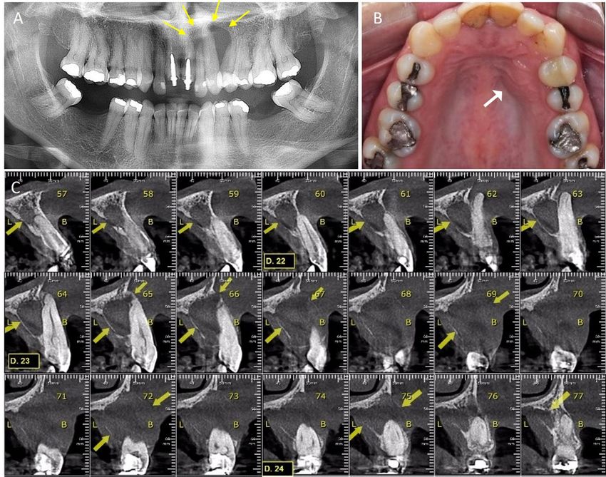
A rare presentation of central granular cell odontogenic tumor
 REPORTE DE CASO / CASE REPORT                                                                               Sander Koth V. y col.

RESÚMEN

El tumor odontogénico de células granulares central (TOCGC) es una neoplasia odontogénica rara con
predilección por la mandíbula de las mujeres de edad mediana. La lesión muestra un crecimiento lento y una
característica radiográfica de una lesión radiolúcida unilocuclar con bordes escleróticos. El objetivo del presente
estudio es reportar un nuevo caso de TOCGC con envolvimiento atípico del maxilar anterior. El examen clínico
mostró aumento de volumen del paladar duro y el área vestibular del canino izquierdo y premolar del maxilar
superior. La tomografía computarizada de haz cónico mostró una lesión unilocular hipodensa bien delimitada
que se extendía del incisivo central izquierdo a la región premolar izquierda. El análisis histopatológico mostró
células eosinofílicas granulares e islas de epitelio odontogénico e inmunotinción positiva para CK14, vimentina,
CD68, Ki67 y S-100, lo que estableció el diagnóstico de TOCGC. El paciente está en seguimiento sin recidiva
durante dieciséis meses.

PALABRAS CLAVE: Neoplasia, neoplasias por tipo histológico, vimentina, tumor odontogénico de células
granulares central, estudio radiográfico.

INTRODUCTION                                                history of trauma in the region two years earlier. The
                                                            patient denied smoking and alcohol drinking habits.
   The central granular cell odontogenic tumor              On clinical exam, a marked swelling was observed
(CGCOT) is a rarely seen neoplasm of odontogenic            on the left side of anterior area of the hard palate and
origin whose histogenesis remains unknown (1,2).            a slight swelling was seen in the vestibular area of
Until 2013, only 38 cases reporting central CGCOT           left canine and pre-molar of maxilla. The overlying
were published (3). On the report of latest update of       mucosa of the region did not present color alteration
the World Health Organization (WHO) classification          nor epithelial dissolution. Panoramic x-ray showed
of head and neck tumors, this entity should represent       a radiolucent unilocular lesion on the left side of
a variant of intraosseous odontogenic fibroma (4).          maxilla involving canine and lateral incisors. Cone
                                                            beam computed tomography of maxilla showed a
   Female patients are more affected than male              hypodense unilocular well delimited lesion extending
patients (3:1) and the average age at diagnosis is 45       from central left incisive to second left pre-molar
years (2,3,5). The neoplasm occurs mostly in the            with 1,9 x 2,0 cm (figure 1). The lesion caused
posterior region of the jaws (2,3,5) and image exams        displacement of teeth and expansion of the cortex. A
frequently shows a radiolucent unilocular lesion with       small area of disruption of cortical margin is observed
a radiopaque border, that may present radiopaque            in palatal cortex.
areas in the interior due to calcification (1,3,6).
                                                               The lesion was surgically removed in view
  The present work aims to report a new case of             of a clinical diagnosis of lateral periodontal
CGCOT with an atypical involvement of anterior              cyst. Histological exam of the excised specimen
maxilla.                                                    showed abundant polygonal cell with eosinophilic
                                                            granular cytoplasm and eccentric placed nuclei on a
   The present study is a descriptive analysis of a         connective tissue. In addition, there was small islands
case. The patient has provided informed consent for         of odontogenic epithelium surrounded by granular
publication.                                                cells. Immunostaining showed positive expression
                                                            odontogenic epithelium for CK14 antigen. Granular
Case report                                                 cells showed positive expression for vimentin and
                                                            CD68 antigen and negative expression for Ki67 and
   A Caucasian female patient, 42 years old, presented      S-100, however, a focal weak expression of Ki67
a swelling in the anterior region of left maxilla           and S-100 suggested dendritic cells (figure 2). A
without associated pain. The patient could not assure       diagnosis of central granular cell odontogenic tumor
when the lesion began to grow, but she reported a           was established.

126                                                                             Rev Estomatol Herediana. 2021 Abr-Jun;31(2): 125-130
A rare presentation of central granular cell odontogenic tumor
REPORTE DE CASO / CASE REPORT Sander Koth V. y col.
Figure 1. Panoramic radiography shows a radiolucent unilocular lesion on the left side of maxilla involving
lateral incisive and canine (arrow, A). Clinical exam shows marked swelling on the left side of anterior area of
the hard palate (arrow, B). Cone beam computed tomography of maxilla showed a hypodense unilocular well
delimited lesion extending from central left incisive to first left pre-molar (arrows, C).
The patient is under follow-up without recurrence of collagenous fibrous tissue with varying islands
after sixteen months (figure 3). or strands of odontogenic epithelium (1,3,6,9,10)
it typically manifests as a well-defined unicystic
DISCUSSION or multilocular radiolucency, although it can be a
mixed-density lesion as well. In our series, there was
Even though some authors believe that this a narrow spectrum of histologic features consisting
disorder represents a variant of central odontogenic of fibrous tissue of altering density and cellularity
fibroma (2,4,7,8), others emphasize that there are with plentiful numbers of large eosinophilic granular
sufficient clinical and histological differences cells, variable amounts of \”inactive-appearing\”
to support that this is another entity (1,3,6). The odontogenic epithelium, and the variable presence
CGCOT presents an older average of age prevalence, of calcified tissue resembling cementum or
mostly occurring during the fifth decade of women, dystrophic calcifications. The ultrastructural and
as reported in this paper, while central odontogenic immunohistochemical findings in this study support
fibroma usually occurs during the second decade of a mesenchymal origin for the granular cells. One
life of men. CGCOT has a predominance of granular recurrence was documented in the current series in
cells with islands of odontogenic epithelium while contrast to no recurrences in the literature. © 2002,
central odontogenic fibroma shows a predominance Mosby, Inc. The author reviews current knowledge
Rev Estomatol Herediana. 2021 Abr-Jun;31(2): 125-130 127A rare presentation of central granular cell odontogenic tumor
REPORTE DE CASO / CASE REPORT Sander Koth V. y col.
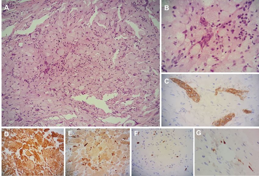
Figure 2. Histological analysis. Hematoxylin and eosin staining shows small islands of odontogenic epithelium
surrounded by polygonal cell with eosinophilic granular cytoplasm and eccentric placed nuclei on a connective
tissue (A, 100x; B, 400x). Immunohistochemical analysis. Odontogenic epithelium shows positive expression
of CK14 (C, 400x). Granular cells show positive expression of vimentin (D, 100x) and CD68 (E, 100x).
Dendritic cells like show positive staining of S-100 (F, 400x) and ki67 (G, 400x).
concerning the central odontogenic fibroma, which at Sarode et al., (3), those are some of the consequences
present is incompletely understood, and reaches the of CGCOT with larger sizes.
following conclusions. 1.
Due to differential diagnosis with central
This study shows a case of CGCOT in which odontogenic fibroma, immunostaining is an
the clinical diagnosis was a lateral periodontal cyst, important tool to diagnose this condition. Granular
as already reported by Cheng et al., (11). However, cells presented positive staining for CD68, vimentin
after histopathological exam, the correct diagnosis and Ck14 while, exclusively cells with dendritic
was established and clinical features of the present morphology were positive for S100 protein, as
case, like sex and age corroborate literature (1,11,12). reported before (5,6).
Unlike most cases reported, this case shows maxilla
involvement. Very few cases in literature reported According to Mesquita et al. (6), positive
occurrence of CGCOT in maxilla (1,5,12–14), staining for CK5, CK19 and CD1 shows that the
and, to our knowledge, only one case described the epithelial islands observed in this neoplasm have
occurrence in anterior area (15), characterizing the an odontogenic origin while granular cells seems
present case with a rare location. to have a mesenchymal origin (6). Sousa et al. (5)
Even though CGCOT usually presents small sizes believe that those granular cells probably represent
and indolent behavior, the present case showed teeth modified fibroblasts, on the other hand, others believe
displacement and cortical expansion associated with that granular cells have a potential for histiocytic
a small area of cortical disruption. According to differentiation (3,11). Silva et al. (13) summarizes this
128 Rev Estomatol Herediana. 2021 Abr-Jun;31(2): 125-130A rare presentation of central granular cell odontogenic tumor
REPORTE DE CASO / CASE REPORT Sander Koth V. y col.
Figure 3. Panoramic radiography shows radiolucent unilocular lesion causing displacement of
teeth (arrow, A). Bone defect and partial teeth realignment are seen one month after surgery (arrow,
B). Complete bone recovery and teeth realignment 12 months after surgery (arrow, C).
by saying that the granular cells are of mesenchymal although it can be a mixed-density lesion as well. In
origin, with a possible histiocytic cell lineage. our series, there was a narrow spectrum of histologic
features consisting of fibrous tissue of altering
The CGCOT has a good prognosis, with slow density and cellularity with plentiful numbers of
growing and well demarcated borders. Conservative large eosinophilic granular cells, variable amounts of
surgical excision has been reported (6,7,11,12,16) with \”inactive-appearing\” odontogenic epithelium, and
only twice recurrences (1,17)it typically manifests as the variable presence of calcified tissue resembling
a well-defined unicystic or multilocular radiolucency, cementum or dystrophic calcifications. The
Rev Estomatol Herediana. 2021 Abr-Jun;31(2): 125-130 129A rare presentation of central granular cell odontogenic tumor
REPORTE DE CASO / CASE REPORT Sander Koth V. y col.
ultrastructural and immunohistochemical findings JE, de Almeida OP. Central granular cell odontogenic
in this study support a mesenchymal origin for the tumor: a histopathologic and immunohistochemical
granular cells. One recurrence was documented in study. Ann Diagn Pathol. 2009; 13(6):405–412.
the current series in contrast to no recurrences in the 7. Meer S, Altini M, Coleman H, Daya N. Central
Granular Cell Odontogenic Tumor:
literature. © 2002, Mosby, Inc. However, Chiang et al.,
Immunohistochemistry and Ultrastructure. Am J
(18) emphasize that if clinical and histopathological Otolaryngol - Head Neck Med Surg. 2004; 25(1):73–
findings show an aggressive behavior, the treatment 78.
should also be more invasive with longer periods of 8. Rühl GH, Akuamoa-Boateng E. Granular cells in
follow-up. odontogenic and non-odontogenic tumours. Virchows
Arch A Pathol Anat Histopathol. 1989;415(5):403–409.
The cause of this odontogenic neoplasm remains 9. Gardner DG. Central odontogenic fibroma current
unknown. Could the trauma suffered by the patient concepts. J Oral Pathol Med. 1996; 25(10):556–561.
at maxilla in the past be the starter of that? The 10. Correa Pontes FS, Lacerda de Souza L, Paula de Paula
limitations of the present study only allow speculation L, de Melo Galvão Neto E, Silva Gonçalves PF, Rebelo
Pontes HA. Central odontogenic fibroma: An updated
regarding this issue. In conclusion, CGCOT is an
systematic review of cases reported in the literature
unusual neoplasm with odontogenic origin not with emphasis on recurrence influencing factors. J
recognized by WHO. The present case reports a new Cranio-Maxillofacial Surg. 2018; 46(10):1753–1757.
case of CGCOT with a rare involvement of anterior 11. Cheng SJ, Wang YP, Chen HM, Chiang CP. Central
maxilla in a Caucasian woman. granular cell odontogenic tumor ofthe mandible. J
Formos Med Assoc. 2013; 112(9):583–585.
Corresponding author: 12. White DK, Chen S-Y, Hartman KS, Miller AS, Gomez
LF. Central granular-cell tumor of the jaws (the so-
Valesca Sander Koth called granular-cell ameloblastic fibroma). Oral
Serviço de Estomatologia – Hospital São Lucas, Surgery, Oral Med Oral Pathol. 1978; 45(3):396–405.
13. Silva BS de F, Yamamoto FP, Cruz e Silva BT, Habib
PUCRS
Souza Aquime JR, Shinohara EH, Pinto D dos S.
Av. Ipiranga, 6690 Sala 231, Porto Alegre, RS 90610- Central Granular Cell Odontogenic Tumor of the
000, Brazil Maxilla. J Craniofac Surg. 2012; 23(2):e117–119.
14. Lotay HS, Kalmar J, DeLeeuw K. Central odontogenic
Declaration of Conflicts of Interests fibroma with features of central granular cell
odontogenic tumor. Oral Surgery, Oral Med Oral Pathol
The authors declare there is no conflict of interest Oral Radiol Endodontology. 2010; 109(2):e63–66.
related to this work. 15. Calvo N, Alonso D, Prieto M, Junquera L. Central
odontogenic fibroma granular cell variant: A case report
REFERENCES and review of the literature. J Oral Maxillofac Surg.
2002; 60(10):1192–1194.
16. Gesek DJ, Adrian JC, Reid EN. Central granular cell
1. Brannon RB, Goode RK, Eversole LR, Carr RF. The
odontogenic tumor. A case report including light
central granular cell odontogenic tumor: Report of 5
microscopy, immunohistochemistry, and literature
new cases. Oral Surg Oral Med Oral Pathol Oral Radiol
review. J Oral Maxillofac Surg. 1995; 53(8):945–9.
Endod. 2002; 94(5):614–621.
17. Piattelli A, Rubini C, Goteri G, Fioroni M, Maiorano E.
2. Carvalho AC, Figueira LCG, Tanabe MN, Martini MZ,
Central granular cell odontogenic tumour: Report of
Shinohara EH. Tumor Odontogênico de Células
the first malignant case and review of the literature.
Granulares. Odonto. 2010; 18(35):96–105.
Oral Oncol. 2003; 39(1):78–82.
3. Sarode SC, Sarode GS, Vaidya K. Central granular cell
18. Chiang CT, Hu KY, Tsai CC. Central granular cell
odontogenic tumor: A systematic review. J Oral Pathol
odontogenic tumor: The first reported case in Oriental
Med. 2014; 43(3):167–176.
people and literature review. J Formos Med Assoc.
4. Takata T, Slootweg P. WHO Classification of Head and
Neck Tumours. 4th ed. Lyons: IARC; 2017. 228. 2014; 113(5):321–325.
5. Sousa SM, Araújo NS, Melhado RM, Araújo VC De.
Central odontogenic granular cell tumor:
Immunohistochemical study of two cases. J Oral
Maxillofac Surg. 1998; 56(6): 787–791. Recibido : 11-12-2020
6. Mesquita ATM, Santos CRR, Gomez RS, Jorge J, León Aceptado: 15-05-2021
130 Rev Estomatol Herediana. 2021 Abr-Jun;31(2): 125-130You can also read