Bacillus subtilis Colonization of Arabidopsis thaliana Roots Induces Multiple Biosynthetic Clusters for Antibiotic Production
←
→
Page content transcription
If your browser does not render page correctly, please read the page content below

ORIGINAL RESEARCH
                                                                                                                                         published: 03 September 2021
                                                                                                                                        doi: 10.3389/fcimb.2021.722778

                                             Bacillus subtilis Colonization of
                                             Arabidopsis thaliana Roots Induces
                                             Multiple Biosynthetic Clusters for
                                             Antibiotic Production
                                             Harsh Maan 1†, Omri Gilhar 1†, Ziv Porat 2* and Ilana Kolodkin-Gal 1*
                                             1 Department of Molecular Genetics, Weizmann Institute of Science, Rehovot, Israel, 2 Flow Cytometry Unit, Life Sciences

                                             Core Facilities, Weizmann Institute of Science, Rehovot, Israel

                                             Beneficial and probiotic bacteria play an important role in conferring immunity of their
                        Edited by:
                                             hosts to a wide range of bacterial, viral, and fungal diseases. Bacillus subtilis is a Gram-
                        Xihui Shen,
 Northwest A and F University, China         positive bacterium that protects the plant from various pathogens due to its capacity to
                       Reviewed by:          produce an extensive repertoire of antibiotics. At the same time, the plant microbiome is a
                   Mikael Lenz Strube,       highly competitive niche, with multiple microbial species competing for space and
     Technical University of Denmark,
                              Denmark
                                             resources, a competition that can be determined by the antagonistic potential of each
                        Talia Karasov,       microbiome member. Therefore, regulating antibiotic production in the rhizosphere is of
 The University of Utah, United States
                                             great importance for the elimination of pathogens and establishing beneficial host-
                    *Correspondence:
                                             associated communities. In this work, we used B. subtilis as a model to investigate the
                                Ziv Porat
               ziv.porat@weizmann.ac.il      role of plant colonization in antibiotic production. Flow cytometry and imaging flow
                      Ilana Kolodkin-Gal     cytometry (IFC) analysis supported the notion that Arabidopsis thaliana specifically
     Ilana.kolodkin-gal@weizmann.ac.il
     †
                                             induced the transcription of the biosynthetic clusters for the non-ribosomal peptides
      These authors have contributed
                 equally to this work
                                             surfactin, bacilysin, plipastatin, and the polyketide bacillaene. IFC was more robust in
                                             quantifying the inducing effects of A. thaliana, considering the overall heterogeneity of the
                    Specialty section:       population. Our results highlight IFC as a useful tool to study the effect of association with
          This article was submitted to
                                             a plant host on bacterial gene expression. Furthermore, the common regulation of multiple
                     Bacteria and Host,
                a section of the journal     biosynthetic clusters for antibiotic production by the plant can be translated to improve the
               Frontiers in Cellular and     performance and competitiveness of beneficial members of the plant microbiome.
                 Infection Microbiology

            Received: 09 June 2021           Keywords: flow cytometry, imaging, antibiotics, Bacillus, plant
         Accepted: 16 August 2021
      Published: 03 September 2021

                                 Citation:
                                             INTRODUCTION
        Maan H, Gilhar O, Porat Z and
 Kolodkin-Gal I (2021) Bacillus subtilis
                                             Rhizobacteria can promote plant growth directly by colonization of the root and exert beneficial
  Colonization of Arabidopsis thaliana
  Roots Induces Multiple Biosynthetic
                                             effects on plant growth and development (Kloepper et al., 2004). These bacteria are often designated
    Clusters for Antibiotic Production.      plant growth-promoting rhizobacteria (PGPR). To date, various PGPR have been isolated,
Front. Cell. Infect. Microbiol. 11:722778.   including various Bacillus species, Burkholderia cepacia, and Pseudomonas fluorescens. These
       doi: 10.3389/fcimb.2021.722778        beneficial rhizobacteria can also confer fitness on their hosts by activating their immune system

Frontiers in Cellular and Infection Microbiology | www.frontiersin.org              1                                    September 2021 | Volume 11 | Article 722778
Maan et al.                                                                                              Quantifying Host Effects on Antibiotic Production

and by antagonizing plant pathogens (Berg et al., 2017; Berg and             primarily controlled by the transition phase regulators Spo0A
Raaijmakers, 2018; Allaband et al., 2019). In addition to the                and CodY and is also affected by DegU, ComA, and ScoC
direct promotion of plant growth, PGPR enhance the efficiency                 (Vargas-Bautista et al., 2014). Interestingly, we recently found
of fertilizers and aid in degrading xenobiotic compounds (Adam               that the plant host can enhance the efficiency of the killing of
et al., 2016; Berg et al., 2017).                                            Serratia plymuthica by B. subtilis by inducing the synthesis of the
    Among growth-promoting strains and biocontrol agents,                    antibiotic bacillaene (Ogran et al., 2019). These preliminary
Bacillus subtilis and subtilis clade members, such as B.                     results raise the question on whether additional antibiotics that
atrophaeus, B. velezensis, and B. mojavensis, are considered                 contribute to rhizocompatibility are induced by Arabidopsis
model organisms (Fan et al., 2017). In particular, the                       thaliana to promote the colonization of preferred symbionts.
antimicrobial activity of B. subtilis has so far been                        However, as the transcriptional regulation of the biosynthetic
demonstrated against bacterial, viral, and fungal soil-borne                 clusters for antibiotics is diverse, this hypothesis needs to be
plant pathogens (Kloepper et al., 2004; Nagorska et al., 2007).              evaluated experimentally.
This activity is mediated largely by antibiotic production:                      To address this question systematically, we considered the
approximately 5% of the B. subtilis genome is dedicated to the               overall effect of the plant host in regulating the transcription
synthesis of antimicrobial molecules by non-ribosomal peptide                from four distinct promoters for B. subtilis antibiotics: surfactin,
synthetases (NRPSs) or polyketide synthases (PKSs) (Stein, 2005;             bacillaene, bacilysin, and plipastatin. As the population within
Ongena and Jacques, 2008; Kinsella et al., 2009; Caulier et al.,             B. subtilis biofilms and root associated communities is
2019). In vitro and in planta studies have indicated the                     heterogeneous (Lopez et al., 2009; Beauregard et al., 2013; Tian
importance of four antibiotics for plant protection: surfactin,              et al., 2021), we monitored the expression in the single-cell level
bacilysin, plipastatin, and bacillaene (Stein, 2005; Hou and                 relying on flow cytometry and imaging flow cytometry (IFC).
Kolodkin-Gal, 2020; Arnaouteli et al., 2021; Ngalimat                        The latter combines the power and speed of traditional flow
et al., 2021).                                                               cytometers with the resolution of a microscope. It therefore
    Surfactin is a small cyclic lipopeptide induced during the               allows for high rate complex morphometric measurements in a
development of genetic competence (Magnuson et al., 1994). The               phenotypically defined way (Zuba-Surma et al., 2007). Our
machinery for surfactin synthesis is encoded within the srfAA–               results indicated that the attachment with the root can
AB–AC–AD operon (Kluge et al., 1988). Surfactin is a powerful                specifically enhance antibiotic production and therefore may
surfactant with antibacterial (Gonzalez et al., 2011) and                    affect the competitiveness of root-associated bacteria compared
antifungal properties (Falardeau et al., 2013). The expression of            with their free-living counterparts.
srfAA-AD operon is induced by the ComQXPA quorum-sensing                         The use of IFC to study gene expression in bacteria was
system at the end of the exponential growth phase. In response to            shown in several studies, as, for example, a study that examined
the competence pheromones, the phosphorylated response                       the promoter activity of various genes in E. coli during the lag
regulator ComA~P activates the transcription of the srf operon               phase (Madar et al., 2013). However, to the best of our
(Roggiani and Dubnau, 1993; Auchtung et al., 2006). A recent                 knowledge, this study is the first time this system has been
study reported that interaction with rice seedlings induces the              used to study plant–bacteria interactions and the impact of such
expression on srfAA in B. subtilis OKB105 (Xie et al., 2015).                interactions on production of all four NRPs/PKS antibiotics.
    Bacilysin is a non-ribosomal dipeptide composed of L-alanine
and amino acid L-anticapsin (Hernandez-Valdes et al., 2020),
which demonstrates antibacterial activity against a wide range of            RESULTS
pathogens (Walker and Abraham, 1970; Chen et al., 2009; Hou
and Kolodkin-Gal, 2020). Its synthesis is controlled mainly by               We wanted to investigate whether the plant can affect the
the bac operon (bacABCDE) (Inaoka et al., 2003; Rajavel                      production of several NRPs/PKS antibiotics and to compare
et al., 2009).                                                               between the different plant influences. Hence, we generated B.
    Fengycin/plipastatin is a lipopeptide comprising 10 amino                subtilis strains harboring transcriptional fusions for PsrfAA-yfp
acid core linked to a b-hydroxy fatty acid and is synthetized by             (surfactin), PpksC-gfp (bacillaene), PbacA-gfp (bacilysin), and PppsA-
five plipastatin synthetases (ppsA, ppsB, ppsC, ppsD, and ppsE)               gfp (plipastatin) (Table 1). The native promoters of these antibiotics
(Tsuge et al., 2007). The ppsA-E operon is repressed by the                  regulate transcription of NRPs/PKS and the rate of transcription is
transition state regulator AbrB during the exponential growth                influenced by binding of specific transcription factors.
phase and is induced in the stationary phase. The pleiotropic                   First, we asked whether the association of B. subtilis with the
regulator degQ gene increases the transcription from the                     plant root impacts the expression of each of the antibiotics.
plipastatin promoter (Rajavel et al., 2009).                                 Hence, using flow cytometry, we examined the expression of
    Bacillaene and dihydrobacillaene (Butcher et al., 2007;                  transcriptional reporters by comparing bacteria cultured in
Straight et al., 2007) are polyketides synthesized by an                     liquid MSgg medium with bacteria attached to plant roots. In
enzymatic complex encoded in the pks gene cluster (Butcher                   order to select the bacteria attached to plant roots, the plant roots
et al., 2007; Straight et al., 2007). The 5′ UTR of pksC was found           were washed to remove non-adherent cells. Our flow cytometry
to be an element for induction of the pks operon. The                        analysis showed that the percentage of cells expressing the
transcription of the biosynthetic clusters for bacillaene is                 fluorescent reporter of each operon significantly increased after

Frontiers in Cellular and Infection Microbiology | www.frontiersin.org   2                               September 2021 | Volume 11 | Article 722778
Maan et al.                                                                                                                       Quantifying Host Effects on Antibiotic Production

TABLE 1 | Common and distinct regulation of NRPS-PKS biosynthetic gene clusters.

NRPs/            Promoter                                                                Regulation (Transcription Factor)
PKS

Surfactin   PsrfAA (Nakano         CodY (Brinsmade, 2017), ComA (Kunst et al., 1994), ComK (Kunst et al., 1994), SigD (Allmansberger, 1997), and SigW (Haldenwang,
            et al., 1991)          1995)
Bacillaene PpksC (Vargas-          ComK (Kunst et al., 1994), ScoC (Kallio et al., 1991), TnrA (Tojo et al., 2005), PerR (Herbig and Helmann, 2001), Fur (Fuangthong et al.,
            Bautista et al.,       2002), CodY (Brinsmade, 2017), SigW (Haldenwang, 1995), SigX (Ogura and Asai, 2016), and DegU (Kunst et al., 1994)
            2014)
Bacilysin   PbacA (Mariappan       ComK (Kunst et al., 1994), AbrB (Strauch et al., 2007), DegU (Kunst et al., 1994), SigH (Saujet et al., 2011), and SigA (Haldenwang,
            et al., 2012)          1995)
Plipastatin PppsA (Yaseen          ComK (Kunst et al., 1994), AbrB (Strauch et al., 2007), Xre (Wood et al., 1990), DegU (Kunst et al., 1994), SigE (Haldenwang, 1995),
            et al., 2016)          and SigA (Haldenwang, 1995).

This table indicates the common and distinct putative transcription factors that potentially bind to the native promoter region of each operon. Transcriptional regulation analysis was
performed using DBTBS Database (https://dbtbs.hgc.jp/) at a threshold: p-value 0.05. References refer to the transcription factors and their diverse roles in B. subtilis physiology.

root attachment in srf (p < 0.0001), pks (p < 0.0005), and bac (p =                               This analysis indeed allowed us to exclude most of the bacterial
0.03) promoters, while it was not significantly increased in pps                               doublets or small aggregates and calculate more accurately both
(p = 0.579). In addition, the mean intensity of cells expressing the                          the percentage of positive GFP cells and bacterial cell length and
promoters increased significantly in pps (p < 0.0001) and pks (p <                             GFP intensity normalized for cell size. This detailed analysis of
0.0001) (Figure 1).                                                                           antibiotic reporters demonstrated that the percentage of
     This conventional flow cytometry allows accurate high-                                    fluorescent positive cells for all four antibiotics increased when
throughput quantification of fluorescence intensities; however,                                 attached to the plant roots (Figure 2). Interestingly, when attached
it is less accurate for bacterial analysis. Higher fluorescence levels                         to plant roots, percentage of cells from PsrfAA-yfp (surfactin)
may be interpreted as higher expression levels, but also can result                           showed an increase of ≈2-fold, PpksC-gfp (bacillaene) showed an
from larger bacterial size or small aggregates (Branda et al., 2001;                          increase of ≈5-fold, PbacA-gfp (bacilysin) showed an increase of
Vlamakis et al., 2008; Chai et al., 2010). Furthermore, the mean                              ≈10-fold, and PppsA-gfp (plipastatin) showed an increase of ≈3-
intensities measured were indicators of the entire fluorescent                                 fold. Such clear differences were not observed in the conventional
positive population rather than an individual bacterial cell.                                 flow cytometry. Furthermore, the mean fluorescent pixel intensity
Therefore, to increase our resolution into the manner by which                                of the cells significantly increased following root attachment in all
antibiotic promoters respond to the attachment of the root, we                                four antibiotic promoters (Figures 3A, B).
used IFC. By collecting large numbers of digital images per                                       Interestingly conventional flow cytometry could not explore
sample and providing a numerical representation of image-based                                differences in the mean fluorescence intensity of cells from
features, the ImageStreamX Mark II combines the per-cell                                      PsrfAA-yfp (surfactin) and PbacA-gfp (bacilysin) when attached
information content provided by standard microscopy with the                                  to roots (Figure 1) compared with cells grown in MSgg medium
statistical significance afforded by large sample sizes common to                              (p = 0.253 and p = 0.442, respectively). However, IFC indicated
traditional flow cytometry.                                                                    that the mean pixel intensity of the fluorescent populations of

                               A                                                              B

 FIGURE 1 | The indicated B. subtilis strain harboring PsrfAA-yfp (surfactin), PpksC-gfp (bacillaene), PbacA-gfp (bacilysin), and PppsA-gfp (plipastatin) reporters was
 analyzed by flow cytometry for (A) positively expressing fluorescent populations. Graphs represent mean ± SD. (B) The mean intensity of the fluorescent populations.
 Box and whisker plot shows median and interquartile range, together with the maximum and minimum values and outlier points. B. subtilis reporter strains were
 grown in either MSgg medium or MSgg medium in the presence of A. thaliana seedlings. Data were collected from 24 h post inoculation, and 100,000 cells were
 counted. Graphs represent results from three independent experiments with n = 3/experiment (total n = 9/group). Statistical analysis was performed using two-way
 ANOVA followed by Tukey’s multiple comparison post hoc testing. p < 0.05 was considered statistically significant.

Frontiers in Cellular and Infection Microbiology | www.frontiersin.org                    3                                        September 2021 | Volume 11 | Article 722778
Maan et al.                                                                                                              Quantifying Host Effects on Antibiotic Production

                                        A                                                  B

                                        C                                                  D

 FIGURE 2 | The indicated B. subtilis strain harboring (A) PsrfAA-yfp (surfactin), (B) PpksC-gfp (bacillaene), (C) PbacA-gfp (bacilysin), and (D) PppsA-gfp (plipastatin)
 reporters was analyzed by imaging flow cytometry for positively expressing fluorescent populations. B. subtilis reporter strains were grown in either MSgg medium or
 MSgg medium in the presence of A. thaliana seedlings. Data were collected from 24 h post inoculation, and 100,000 cells were counted. Graphs represent results
 from three independent experiments with n = 3/experiment (total n = 9/group). The percentage of fluorescent positive cells was measured by imaging flow cytometry
 and analyzed with IDEAS 6.3. Statistical analysis was performed using unpaired t-test with Welch correction. p < 0.05 was considered statistically significant. Graphs
 represent mean ± SD.

these two reporters was significantly higher (p = 0.005 and p =                          Overall, these results suggest that IFC is a sensitive and
0.011, respectively), showing a higher sensitivity of the method.                       accurate technique to study weakly expressed genes in bacterial
    The quantification performed with IFC also yielded a higher                          cells. Importantly, both flow cytometry and IFC agreed that all
percentage of fluorescence positive cells as compared with the                           four antibiotics are induced by the plant.
traditional flow cytometry in all four promoters in the presence                             Next, we examined if the plant and its secretions specifically
of plant root. This may be explained by the sensitivity and ability                     regulate antibiotic production or induce all genes in B. subtilis due
to acquire individual pixels by IFC, allowing the detection of low                      to non-specific effect of its growth, as the plant may affect the
fluorescence intensity signals of small objects that might be                            synthesis or stability of GFP regardless of its promoter. For this
within the electronic noise of conventional flow cytometers, as                          purpose, we measured the expression of transcriptional reporter of
well as elimination of debris and aggregates. This results in better                    the b-lactamase PenP. b-lactamases are enzymes that account for
identification and separation of the bacterial population from the                       an additional layer of defense as they hydrolyze the b-lactam ring of
background noise and often provides more robust results.                                b-lactams, thus inactivating the antibiotic before it reaches its target,

Frontiers in Cellular and Infection Microbiology | www.frontiersin.org              4                                     September 2021 | Volume 11 | Article 722778
Maan et al.                                                                                                                     Quantifying Host Effects on Antibiotic Production

                            A

                            B                                                                                               C

 FIGURE 3 | The indicated B. subtilis strain harboring PsrfAA-yfp (surfactin), PpksC-gfp (bacillaene), PbacA-gfp (bacilysin), and PppsA-gfp (plipastatin) reporters was analyzed
 by imaging flow cytometry for (A) mean pixel intensity of the fluorescent positive populations. Bacteria expressing the fluorescent reporters were cultured in the absence
 or presence of A. thaliana seedlings. Box and whisker plot shows median and interquartile range, together with the maximum and minimum values and outlier points.
 (B) Representative bright-field and fluorescent images related to expression of reporters in MSgg and on A. thaliana roots. Scale bar = 7 µm. (C) Comparison between
 cell length of all four reporters when grown in MSgg medium and cell length of the same reporters grown on A. thaliana roots. Graphs represent mean ± SD. Data were
 collected from 24 h post inoculation, and 100,000 cells were counted. The mean pixel intensity and cell length of fluorescent positive cells were measured by imaging
 flow cytometry and analyzed with IDEAS 6.3. Graphs represent results from three independent experiments with n = 3/experiment (total n = 9/group). Statistical analysis
 was performed using unpaired t-test with Welch correction. p < 0.05 was considered statistically significant.

the PBPs (penicillin binding proteins) (Therrien and Levesque,                             reporter allows us to monitor gene expression in real time by
2000). The active b-lactamase of B. subtilis (Takagi et al., 1993;                         monitoring light production in a plate reader with an
Bucher et al., 2019) was not induced but rather modestly decreased                         illuminometer. When grown on liquid defined medium, wild-
(p = 0.015) in the presence of the plant (Supplementary Figure S1).                        type cells expressed luciferase from srfAA and pksC promoters
In addition, the expression of the core metabolic enzyme lactate                           robustly. However, while root secretions did not alter the kinetics
dehydrogenase ldh was also unaffected by the root (Supplementary                           of the expression, they were sufficient to significantly increase the
Figure S1). Therefore, the effect of the plant is not through post-                        intensity of the luciferase emission (Figure 4). Therefore, the
translational effects on the GFP reporter, and at least two enzymes                        measurement of the inducing effect on the level of the population
unrelated to the biosynthesis of NRP/polyketide antibiotics are not                        agreed with our single-cell analysis and confirmed an induction
induced by the plant.                                                                      by A. thaliana and also suggested that this induction is mediated
    To study whether attachment to plant roots also influences                              by a secreted factor that can activate the distinct promoters for
bacterial cell length, we grouped all measurements into two                                bacillaene and surfactin synthesis (Table 1). Consistently, using
groups, bacteria grown in MSgg medium (control) and bacteria                               confocal scanning laser microscopy, we could clearly confirm the
attached to plant roots. Interestingly, when we compared the cell                          expression of the pks promoter and, to some extent, surfactin on
length among the bacteria from all the reporters attached to plant                         cells attached with A. thaliana roots (Figure 5). This experiment
roots to that of MSgg, the data in the MSgg group showed a                                 also exemplified the adherence of the bacteria to the plant root
higher degree of dispersion (SD =1.76) as compared to the                                  after 24 h of co-culture, which was also the time point used to
bacteria attached to the plant roots (SD = 0.90). An F-test                                quantify the expression from the four promoters of the antibiotic
further confirmed that the variances were significantly different                            biosynthetic clusters by flow cytometry and IFC.
between two groups (F-test, p-value = 0.0001) (Figure 3C).
    To confirm that the impact of the plant root on antibiotic
production could be due to a secreted factor, we monitored the                             DISCUSSION
expression from PsrfAA and PpksC fused to a luciferase reporter in
the presence and absence of root secretions. The use of the                                Since B. subtilis was first described by Ferdinand Cohn in the late
unstable luciferase (an enzyme that degrades rapidly and                                   1800s, it was shown to specialize in the production of secondary
therefore is not accumulated) (McLoon et al., 2011) as a                                   metabolites (Steinke et al., 2021). Many of the biosynthetic

Frontiers in Cellular and Infection Microbiology | www.frontiersin.org                 5                                        September 2021 | Volume 11 | Article 722778
Maan et al.                                                                                                                   Quantifying Host Effects on Antibiotic Production

                                         A                                                 B

 FIGURE 4 | A. thaliana secretions increase the expression of (A) PsrfAA-lux (surfactin) and (B) PpksC-lux (bacilllane) in B. subtilis cells. B. subtilis strains expressing
 luciferase under the control of the srf and pks promoters were cultured in liquid MSgg medium or liquid MSgg medium supplemented with A. thaliana secretions
 (15% v/v). Luminescence was monitored for 24 h using a microplate reader. Graphs represent results from three independent experiments. Error bars represent ±
 SEM. Luciferase activity was normalized to avoid artifacts related to differential cell numbers as RLU/OD.

 FIGURE 5 | Visualizing the expression of PpksC-gfp (bacillaene) and PsrfAA-gfp (surfactin) on A. thaliana roots. Bacteria expressing GFP under the control of the pks
 and srf promoters were cultured in the presence of A. thaliana seedlings in MSgg medium. After 24 h, the bacteria colonizing the roots were photographed with a
 confocal microscope.

Frontiers in Cellular and Infection Microbiology | www.frontiersin.org                 6                                      September 2021 | Volume 11 | Article 722778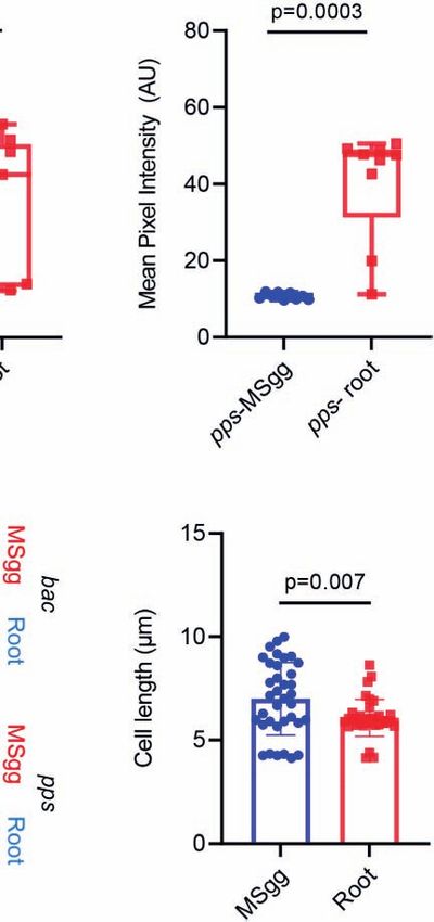
Maan et al.                                                                                                                 Quantifying Host Effects on Antibiotic Production

pathways for these metabolites are conserved either across the                            of cells, with IFC to analyze the population at the single-cell level,
entire Bacillus genus or within specific phylogenetic clades.                              and luciferase-based reporters to screen for potential activators.
Surfactin, bacillaene, bacilysin, and plipastatin have essentially                        While flow cytometry can detect the trends efficiently, statistical
been observed within the subtilis clade (Aleti et al., 2015; Hou                          significance is frequently not achieved for antibiotic producers
and Kolodkin-Gal, 2020). Therefore, the different environmental                           while performing multiple comparisons. These cases include
niches inhabited by members of the B. subtilis clade may select                           weakly expressing promoters and heterogeneous populations.
for conservation of metabolites with distinct (or potentially                                 Our findings that all four biosynthetic clusters were induced
redundant) beneficial functions.                                                           by the root strongly indicate co-evolution of the regulation of
    Here, we found that the production of non-ribosomal                                   biosynthetic clusters for antibiotic production. The complexity of
peptides and polyketides was specifically activated during                                 these antibiotic–host interactions suggests that the plant host
symbiotic interaction with A. thaliana. Our results                                       actively promotes the establishment of the most beneficial
demonstrated that direct interactions with the root increased                             bacterial community. Our findings provide a simple example
the expression of four different biosynthetic clusters with distinct                      of high-order interactions that shape microbiomes; the host
promoters encoding for antibiotics with significance for B.                                modulates antibiotic production in the desired bacterial
subtilis competitiveness, and the secretions were sufficient to                            colonizers, providing the colonizers a clear advantage over less
induce surfactin and bacillaene expression. The four promoters                            beneficial potential residents.
of the biosynthetic clusters differ in their sequence and
regulations (Table 1). Therefore, it is not intuitive that they
will be co-induced together following attachment to the root.                             MATERIALS AND METHODS
    Indeed, various studies reported that plant metabolites induce
the expression of NRPs and antibiotic production genes. We                                Strains and Media
previously demonstrated that the interaction with the plant                               All strains used in this study are in Table 2. The strains were
increases the capacity of B. subtilis to compete with Serattia                            grown in LB broth (Difco) or MSgg medium (Branda et al., 2001)
plymuthica, and our current results further indicate that the root                        [5 mM potassium phosphate, 100 mM MOPS (pH 7), 2 mM
is an active regulator of the competitive interactions occurring on                       MgCl2, 50 µM MnCl2, 50 µM FeCl3, 700 µM CaCl2, 1 µM ZnCl2,
its roots (Ogran et al., 2019). An increase in bacterial                                  2 µM thiamine, 0.5% glycerol, 0.5% glutamate, 50 µg/ml
competitiveness due to increased antibiotic production may be                             threonine, tryptophan, and phenylalanine) (Branda et al.,
a conserved feature of rhizobacteria: plants enhance the killing                          2001]. Solid LB medium contained 1.5% bacto agar (Difco).
efficiency of Xanthomonas citri by Paenibacillus polymyxa SC2
(Liu et al., 2021), wheat extract induces the expression of                               Plant Growth Conditions
biosynthetic genes for antibiotic production in Pseudomonas                               Seeds of A. thaliana Col-0 were surface-sterilized and seeded on
genotypes (Rieusset et al., 2021), and barley induces the                                 petri dishes containing Murashige and Skoog medium (4.4 g/L),
antifungal genes of Pseudomonas fluorescens (Jousset et al.,                               pH 5.7, supplied with 0.5% (w/v) plant agar (Duchefa) and 0.5%
2011). Our methodology here offers a practical approach to                                sucrose (Sigma-Aldrich), and then stratified at 4°C for 2 days.
study the effect of plant metabolites on heterogeneous                                    The seeds were further transferred to a growth chamber (MRC)
communities, even when expressed by a small subpopulation                                 at 23°C in a 12-h light/12-h dark regime.

TABLE 2 | Strains used for this work.

Strain                                                                   Description                                                                 Source or reference

B. subtilis       Wild type                                                                                                                         (Branda et al., 2001)
PpksC-lux         B. subtilis sacA::PpksC-lux (Cm r), promoter of bacillaene operon tagged to the luciferase reporter integrated in the             (Ogran et al., 2019)
                  neutral SacA locus
PsrfAA-lux        B. subtilis sacA::PsrfAA-lux (Cm r), promoter of surfactin operon tagged to the luciferase reporter integrated in the neutral     (Maan et al., 2021)
                  sacA locus
PsrfAA-yfp        B. subtilis sacA:: PsrfAA-yfp (Sp r), promoter of surfactin operon tagged to the YFP reporter integrated in the neutral           Avigdor Eldar Lab, TAU,
                  amyE locus                                                                                                                        Israel
PpksC-gfp         B. subtilis amyE:: PsrfAA-yfp (Cm r), promoter of bacillaene operon tagged to the GFP reporter integrated in the neutral           (Ogran et al., 2019)
                  amyE locus
PbacA-gfp         B. subtilis amyE:: PbacA-gfp (Cm r), promoter of bacilysin operon tagged to the GFP reporter integrated in the neutral            (Maan et al., 2021)
                  amyE locus
PppsA-gfp         B. subtilis amyE:: PppsA-gfp (Cm r), promoter of plipastatin operon tagged to the GFP reporter integrated in the neutral          (Maan et al., 2021)
                  amyE locus
PpenP-gfp         B. subtilis amyE:: PpenP-gfp (Sp r), promoter of plipastatin operon tagged to the GFP reporter integrated in the neutral          (Bucher et al., 2019)
                  amyE locus
Pldh-gfp          B. subtilis amyE:: Pldh-gfp                                                                                                       (Hassanov et al., 2018)
    r                               r
Cm , chloramphenicol resistance; Sp , spectinomycin resistance.

Frontiers in Cellular and Infection Microbiology | www.frontiersin.org                7                                      September 2021 | Volume 11 | Article 722778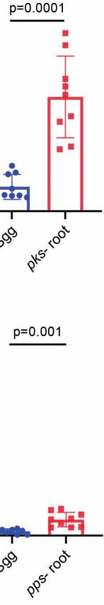
Maan et al.                                                                                             Quantifying Host Effects on Antibiotic Production

Extraction of Plant Secretions                                               image by detecting large changes of pixel values in the image).
Plant secretions were retrieved from 14-day-old A. thaliana                  Cells expressing GFP were selected using the Intensity (the sum
seedlings cultured in 6 ml of liquid MSgg of each well of a six-             of the background subtracted pixel values within the image) and
well microplate (Thermo Scientific). Eight seedlings were put in              Max Pixel values (the largest value of the background-subtracted
each well. The plant secretions were collected after 4 days, filtered         pixels) of the GFP channel (Ch02). GFP expression was
with a 0.22-µm filter, and stored at 4°C for further use.                     quantified using the Mean Pixel feature (the mean of the
                                                                             background-subtracted pixels contained in the input mask).
Luminescence Experiments                                                     The size of bacteria was quantified using the Length feature
Luminescence reporters were grown in either MSgg medium or                   (measures the longest part of an object, in microns) of the bright-
MSgg medium containing plant secretions. Experiments were                    field image.
carried using a flat bottom 96-well plate with white opaque walls
(Corning). Measurements were performed every 30 min at 30°C                  Statistical Analysis
for a period for 24 h, using a microplate reader (Synergy 2;                 All experiments were performed three separate and independent
BioTek, Winooski, VT, USA). Luciferase activity was normalized               times in triplicates. Datasets were compared using a standard two-
to avoid artifacts related to differential cells numbers as                  way ANOVA, followed by Tukey’s multiple comparison post hoc
RLU/OD.                                                                      testing, or a pairwise comparison using unpaired t-test with Welch’s
                                                                             correction in order to correct for groups with significantly unequal
Confocal Microscopy                                                          variances. Error bars represented ± SD, unless stated otherwise. p <
Plants cultured with bacteria were washed in PBS and mounted                 0.05 was considered statistically significant.
on a microscope slide and covered with a poly-L-Lysine 31                        Statistical analyses were performed with GraphPad Prism 9.0
(Sigma)-treated coverslip. Cells were visualized and                         (GraphPad Software, Inc., San Diego, CA).
photographed using a laser scanning confocal microscope
(Zeiss LSM 780) equipped with a high-resolution microscopy
Axiocam camera, as required. Data were captured using Zen                    DATA AVAILABILITY STATEMENT
black software (Zeiss, Oberkochen, Germany).
                                                                             The original contributions presented in the study are included in
Flow Cytometry                                                               the article/Supplementary Material. Further inquiries can be
Indicated strains used in the experiments were inoculated in 1.5             directed to the corresponding authors.
ml of liquid MSgg without seedlings (control) and MSgg with 14-
day-old A. thaliana seedlings in a 24-well plate (Thermo
Scientific); each well contained eight seedlings. The set was
incubated for 24 h in a growth chamber (MRC) at 23°C in a                    AUTHOR CONTRIBUTIONS
12-h light/12-h dark regime. After incubation, the seedlings were            HM, OG, IK-G, and ZP designed the experiments. HM, OG, and
removed from the liquid medium and washed in PBS for the
                                                                             ZP performed the experiments. HM, ZP, and IK-G analyzed the
purpose of removing non-adherent bacteria. Samples were
                                                                             data. IK-G and ZP wrote the manuscript. All authors contributed
transferred to Eppendorf tube in 500 µl of PBS and vortexed
                                                                             to the article and approved the submitted version.
for 1 min, for the purpose of detaching the bacteria from the root.
Samples were measured using a BD LSR II flow cytometer (BD
Biosciences), using laser excitation of 488 nm, coupled with 505
LP and 525/50 sequential filters. For each sample, 100,000 cells              FUNDING
were counted and samples were analyzed using Diva 8 software
(BD Biosciences).                                                            The IK-G laboratory is supported by the Israel Science
                                                                             Foundation grant number 119/16 and ISF-JSPS 184/20 and
Imaging Flow Cytometry                                                       Israel Ministry of Science—Tashtiot (Infrastructures)—123402
Samples were prepared as for the flow cytometer. Data were                    in Life Sciences and Biomedical Sciences. IK-G is supported by
acquired by ImageStreamX Mark II (AMNIS, part of Luminex                     an internal grant from the Estate of Albert Engleman provided by
corp., Austin Tx) using a 60× lens (NA = 0.9). Lasers used were              the Angel–Faivovich Fund for Ecological Research and by a
488 nm (200 mW) for GFP excitation and 785 nm (5 mW) for                     research grant from the Benoziyo Endowment Fund for the
side scatter measurement. During acquisition, bacterial cells were           Advancement of Science. IK-G is a recipient of the Rowland and
gated according to their area (in square microns) and side scatter,          Sylvia Career Development Chair.
which excluded the calibration beads (that run in the instrument
along with the sample). For each sample, 100,000 events were
collected. Data were analyzed using IDEAS 6.3 (AMNIS). Single                SUPPLEMENTARY MATERIAL
event bacteria were selected according to their area (in square
microns) and aspect ratio (width divided by the length of a best-            The Supplementary Material for this article can be found online
fit ellipse). Focused events were selected by the Gradient RMS                at: https://www.frontiersin.org/articles/10.3389/fcimb.2021.
and Contrast features (measures the sharpness quality of an                  722778/full#supplementary-material

Frontiers in Cellular and Infection Microbiology | www.frontiersin.org   8                               September 2021 | Volume 11 | Article 722778
Maan et al. Quantifying Host Effects on Antibiotic Production
Supplementary Figure S1 | Indicated reporter strains for PpenP-gfp and Pldh-gfp MSgg medium or in MSgg medium in presence of A. thaliana seedlings. Data were
was analyzed by flow cytometry for (A) positively expressing fluorescent populations. collected from 24 h post inoculation, 100,000 cells were counted. Graphs represent
Graphs represent mean ± SD or (B) the mean intensity of the fluorescent populations. results from two independent experiments with n = 2/experiment (total n = 6/group).
Box and whisker plot shows median and interquartile range, together with the Statistical analysis was performed using Two-way ANOVA followed by Tukey’s
maximum and minimum values and outlier points. Reporter stains were either grown in multiple comparison post hoc testing. p < 0.05 was considered statistically significant.
Fuangthong, M., Herbig, A. F., Bsat, N., and Helmann, J. D. (2002). Regulation of
REFERENCES the Bacillus Subtilis Fur and perR Genes by PerR: Not All Members of the PerR
Adam, E., Groenenboom, A. E., Kurm, V., Rajewska, M., Schmidt, R., Tyc, O., et al. Regulon Are Peroxide Inducible. J. Bacteriol. 184, 3276–3286. doi: 10.1128/
(2016). Controlling the Microbiome: Microhabitat Adjustments for Successful JB.184.12.3276-3286.2002
Biocontrol Strategies in Soil and Human Gut. Front. Microbiol. 7, 1079. doi: Gonzalez, D. J., Haste, N. M., Hollands, A., Fleming, T. C., Hamby, M., Pogliano,
10.3389/fmicb.2016.01079 K., et al. (2011). Microbial Competition Between Bacillus Subtilis and
Aleti, G., Sessitsch, A., and Brader, G. (2015). Genome Mining: Prediction of Staphylococcus Aureus Monitored by Imaging Mass Spectrometry.
Lipopeptides and Polyketides From Bacillus and Related Firmicutes. Comput. Microbiology 157, 2485–2492. doi: 10.1099/mic.0.048736-0
Struct. Biotechnol. J. 13, 192–203. doi: 10.1016/j.csbj.2015.03.003 Haldenwang, W. G. (1995). The Sigma Factors of Bacillus Subtilis. Microbiol. Rev.
Allaband, C., McDonald, D., Vazquez-Baeza, Y., Minich, J. J., Tripathi, A., 59, 1–30. doi: 10.1128/mr.59.1.1-30.1995
Brenner, D. A., et al. (2019). Microbiome 101: Studying, Analyzing, and Hassanov, T., Karunker, I., Steinberg, N., Erez, A., and Kolodkin-Gal, I. (2018).
Interpreting Gut Microbiome Data for Clinicians. Clin. Gastroenterol. Novel Antibiofilm Chemotherapies Target Nitrogen From Glutamate and
Hepatol. 17, 218–230. doi: 10.1016/j.cgh.2018.09.017 Glutamine. Sci. Rep. 8, 7097. doi: 10.1038/s41598-018-25401-z
Allmansberger, R. (1997). Temporal Regulation of sigD From Bacillus Subtilis Herbig, A. F., and Helmann, J. D. (2001). Roles of Metal Ions and Hydrogen
Depends on a Minor Promoter in Front of the Gene. J. Bacteriol. 179, 6531– Peroxide in Modulating the Interaction of the Bacillus Subtilis PerR Peroxide
6535. doi: 10.1128/jb.179.20.6531-6535.1997 Regulon Repressor With Operator DNA. Mol. Microbiol. 41, 849–859. doi:
Arnaouteli, S., Bamford, N. C., Stanley-Wall, N. R., and Kovacs, A. T. (2021). 10.1046/j.1365-2958.2001.02543.x
Bacillus Subtilis Biofilm Formation and Social Interactions. Nat. Rev. Hernandez-Valdes, J. A., Zhou, L., de Vries, M. P., and Kuipers, O. P. (2020).
Microbiol. 19 (9), 600–614. doi: 10.1038/s41579-021-00540-9 Impact of Spatial Proximity on Territoriality Among Human Skin Bacteria.
Auchtung, J. M., Lee, C. A., and Grossman, A. D. (2006). Modulation of the ComA- NPJ Biofilms Microbiomes 6 (1), 30. doi: 10.1038/s41522-020-00140-0
Dependent Quorum Response in Bacillus Subtilis by Multiple Rap Proteins and Hou, Q., and Kolodkin-Gal, I. (2020). Harvesting the Complex Pathways of
Phr Peptides. J. Bacteriol. 188, 5273–5285. doi: 10.1128/JB.00300-06 Antibiotic Production and Resistance of Soil Bacilli for Optimizing Plant
Beauregard, P. B., Chai, Y., Vlamakis, H., Losick, R., and Kolter, R. (2013). Bacillus Microbiome. FEMS Microbiol. Ecol. 96 (9), fiaa142. doi: 10.1093/femsec/fiaa142
Subtilis Biofilm Induction by Plant Polysaccharides. Proc. Natl. Acad. Sci. Inaoka, T., Takahashi, K., Ohnishi-Kameyama, M., Yoshida, M., and Ochi, K.
U. S. A. 110, E1621–E1630. doi: 10.1073/pnas.1218984110 (2003). Guanine Nucleotides Guanosine 5’-Diphosphate 3’-Diphosphate
Berg, G., Koberl, M., Rybakova, D., Muller, H., Grosch, R., and Smalla, K. (2017). and GTP Co-Operatively Regulate the Production of an Antibiotic Bacilysin
Plant Microbial Diversity Is Suggested as the Key to Future Biocontrol and in Bacillus Subtilis. J. Biol. Chem. 278, 2169–2176. doi: 10.1074/jbc.
Health Trends. FEMS Microbiol. Ecol. 93 (5). doi: 10.1093/femsec/fix050 M208722200
Berg, G., and Raaijmakers, J. M. (2018). Saving Seed Microbiomes. ISME J. 12, Jousset, A., Rochat, L., Lanoue, A., Bonkowski, M., Keel, C., and Scheu, S. (2011).
1167–1170. doi: 10.1038/s41396-017-0028-2 Plants Respond to Pathogen Infection by Enhancing the Antifungal Gene
Branda, S. S., Gonzalez-Pastor, J. E., Ben-Yehuda, S., Losick, R., and Kolter, R. Expression of Root-Associated Bacteria. Mol. Plant Microbe Interact. 24, 352–
(2001). Fruiting Body Formation by Bacillus Subtilis. Proc. Natl. Acad. Sci. 358. doi: 10.1094/MPMI-09-10-0208
U. S. A. 98, 11621–11626. doi: 10.1073/pnas.191384198 Kallio, P. T., Fagelson, J. E., Hoch, J. A., and Strauch, M. A. (1991). The Transition-
Brinsmade, S. R. (2017). CodY, a Master Integrator of Metabolism and Virulence in State Regulator Hpr of Bacillus-Subtilis Is a DNA-Binding Protein. J. Biol.
Gram-Positive Bacteria. Curr. Genet. 63, 417–425. doi: 10.1007/s00294-016-0656-5 Chem. 266, 13411–13417. doi: 10.1016/S0021-9258(18)98855-1
Bucher, T., Keren-Paz, A., Hausser, J., Olender, T., Cytryn, E., and Kolodkin-Gal, I. Kinsella, K., Schulthess, C. P., Morris, T. F., and Stuart, J. D. (2009). Rapid
(2019). An Active Beta-Lactamase Is a Part of an Orchestrated Cell Wall Stress Quantification of Bacillus Subtilis Antibiotics in the Rhizosphere. Soil Biol.
Resistance Network of Bacillus Subtilis and Related Rhizosphere Species. Biochem. 41, 374–379. doi: 10.1016/j.soilbio.2008.11.019
Environ. Microbiol. 21 (3), 1068–1085. doi: 10.1111/1462-2920.14526 Kloepper, J. W., Ryu, C. M., and Zhang, S. (2004). Induced Systemic Resistance
Butcher, R. A., Schroeder, F. C., Fischbach, M. A., Straight, P. D., Kolter, R., Walsh, and Promotion of Plant Growth by Bacillus Spp. Phytopathology 94, 1259–
C. T., et al. (2007). The Identification of Bacillaene, the Product of the PksX 1266. doi: 10.1094/PHYTO.2004.94.11.1259
Megacomplex in Bacillus Subtilis. Proc. Natl. Acad. Sci. U. S. A. 104, 1506– Kluge, B., Vater, J., Salnikow, J., and Eckart, K. (1988). Studies on the Biosynthesis
1509. doi: 10.1073/pnas.0610503104 of Surfactin, a Lipopeptide Antibiotic From Bacillus-Subtilis Atcc-21332. FEBS
Caulier, S., Nannan, C., Gillis, A., Licciardi, F., Bragard, C., and Mahillon, J. (2019). Lett. 231, 107–110. doi: 10.1016/0014-5793(88)80712-9
Overview of the Antimicrobial Compounds Produced by Members of the Kunst, F., Msadek, T., Bignon, J., and Rapoport, G. (1994). The DegS/DegU and
Bacillus Subtilis Group. Front. Microbiol. 10, 302. doi: 10.3389/fmicb.2019.00302 ComP/ComA Two-Component Systems Are Part of a Network Controlling
Chai, Y., Norman, T., Kolter, R., and Losick, R. (2010). An Epigenetic Switch Degradative Enzyme Synthesis and Competence in Bacillus Subtilis. Res.
Governing Daughter Cell Separation in Bacillus Subtilis. Genes Dev. 24, 754– Microbiol. 145, 393–402. doi: 10.1016/0923-2508(94)90087-6
765. doi: 10.1101/gad.1915010 Liu, H., Li, Y., Ge, K., Du, B., Liu, K., Wang, C., et al. (2021). Interactional
Chen, X. H., Scholz, R., Borriss, M., Junge, H., Mogel, G., Kunz, S., et al. (2009). Mechanisms of Paenibacillus Polymyxa SC2 and Pepper (Capsicum Annuum
Difficidin and Bacilysin Produced by Plant-Associated Bacillus Amyloliquefaciens L.) Suggested by Transcriptomics. BMC Microbiol. 21, 70. doi: 10.1186/s12866-
Are Efficient in Controlling Fire Blight Disease. J. Biotechnol. 140, 38–44. 021-02132-2
doi: 10.1016/j.jbiotec.2008.10.015 Lopez, D., Vlamakis, H., and Kolter, R. (2009). Generation of Multiple Cell Types
Falardeau, J., Wise, C., Novitsky, L., and Avis, T. J. (2013). Ecological and in Bacillus Subtilis. FEMS Microbiol. Rev. 33, 152–163. doi: 10.1111/j.1574-
Mechanistic Insights Into the Direct and Indirect Antimicrobial Properties 6976.2008.00148.x
of Bacillus Subtilis Lipopeptides on Plant Pathogens. J. Chem. Ecol. 39, 869– Maan, H., Friedman, J., and Kolodkin-Gal, I. (2021). Resolving the Conflict
878. doi: 10.1007/s10886-013-0319-7 Between Antibiotic Production and Rapid Growth by Recognition of
Fan, B., Blom, J., Klenk, H. P., and Borriss, R. (2017). Bacillus Amyloliquefaciens, Peptidoglycan of Susceptible Competitors bioRxiv 2021.2002.2007.430110.
Bacillus Velezensis, and Bacillus Siamensis Form an” Operational Group B. Madar, D., Dekel, E., Bren, A., Zimmer, A., Porat, Z., and Alon, U. (2013).
Amyloliquefaciens” Within the B-Subtilis Species Complex. Front. Microbiol. 8, Promoter Activity Dynamics in the Lag Phase of Escherichia Coli. BMC Syst.
22. doi: 10.3389/fmicb.2017.00022 Biol. 7, 136. doi: 10.1186/1752-0509-7-136
Frontiers in Cellular and Infection Microbiology | www.frontiersin.org 9 September 2021 | Volume 11 | Article 722778Maan et al. Quantifying Host Effects on Antibiotic Production
Magnuson, R., Solomon, J., and Grossman, A. D. (1994). Biochemical and Genetic Takagi, M., Ohta, T., Johki, S., and Imanaka, T. (1993). Characterization of the
Characterization of a Competence Pheromone From B. Subtilis. Cell 77, 207– Membrane Sensor PenJ for Beta-Lactam Antibiotics From Bacillus
216. doi: 10.1016/0092-8674(94)90313-1 Licheniformis by Amino Acid Substitution. FEMS Microbiol. Lett. 110, 127–
Mariappan, A., Makarewicz, O., Chen, X. H., and Borriss, R. (2012). Two- 131. doi: 10.1111/j.1574-6968.1993.tb06306.x
Component Response Regulator DegU Controls the Expression of Bacilysin Therrien, C., and Levesque, R. C. (2000). Molecular Basis of Antibiotic Resistance
in Plant-Growth-Promoting Bacterium Bacillus Amyloliquefaciens FZB42. and Beta-Lactamase Inhibition by Mechanism-Based Inactivators: Perspectives
J. Mol. Microbiol. Biotechnol. 22, 114–125. doi: 10.1159/000338804 and Future Directions. FEMS Microbiol. Rev. 24, 251–262. doi: 10.1016/S0168-
McLoon, A. L., Kolodkin-Gal, I., Rubinstein, S. M., Kolter, R., and Losick, R. 6445(99)00039-X
(2011). Spatial Regulation of Histidine Kinases Governing Biofilm Formation Tian, T., Sun, B., Shi, H., Gao, T., He, Y., Li, Y., et al. (2021). Sucrose Triggers a
in Bacillus Subtilis. J. Bacteriol. 193, 679–685. doi: 10.1128/JB.01186-10 Novel Signaling Cascade Promoting Bacillus Subtilis Rhizosphere
Nagorska, K., Bikowski, M., and Obuchowski, M. (2007). Multicellular Behaviour Colonization. ISME J. 15, 2723–2737. doi: 10.1038/s41396-021-00966-2
and Production of a Wide Variety of Toxic Substances Support Usage of Tojo, S., Satomura, T., Morisaki, K., Deutscher, J., Hirooka, K., and Fujita, Y.
Bacillus Subtilis as a Powerful Biocontrol Agent. Acta Biochim. Pol. 54, 495– (2005). Elaborate Transcription Regulation of the Bacillus Subtilis Ilv-Leu
508. doi: 10.18388/abp.2007_3224 Operon Involved in the Biosynthesis of Branched-Chain Amino Acids
Nakano, M. M., Xia, L. A., and Zuber, P. (1991). Transcription Initiation Region of Through Global Regulators of CcpA, CodY and TnrA. Mol. Microbiol. 56,
the Srfa Operon, Which Is Controlled by the Comp-Coma Signal Transduction 1560–1573. doi: 10.1111/j.1365-2958.2005.04635.x
System in Bacillus-Subtilis. J. Bacteriol. 173, 5487–5493. doi: 10.1128/ Tsuge, K., Matsui, K., and Itaya, M. (2007). Production of the non-Ribosomal
jb.173.17.5487-5493.1991 Peptide Plipastatin in Bacillus Subtilis Regulated by Three Relevant Gene
Ngalimat, M. S., Yahaya, R. S. R., Baharudin, M. M. A., Yaminudin, S. M., Karim, Blocks Assembled in a Single Movable DNA Segment. J. Biotechnol. 129, 592–
M., Ahmad, S. A., et al. (2021). A Review on the Biotechnological Applications 603. doi: 10.1016/j.jbiotec.2007.01.033
of the Operational Group Bacillus Amyloliquefaciens. Microorganisms 9 (3), Vargas-Bautista, C., Rahlwes, K., and Straight, P. (2014). Bacterial Competition
614. doi: 10.3390/microorganisms9030614 Reveals Differential Regulation of the Pks Genes by Bacillus Subtilis.
Ogran, A., Yardeni, E. H., Keren-Paz, A., Bucher, T., Jain, R., Gilhar, O., et al. J. Bacteriol. 196, 717–728. doi: 10.1128/JB.01022-13
(2019). The Plant Host Induces Antibiotic Production to Select the Most Vlamakis, H., Aguilar, C., Losick, R., and Kolter, R. (2008). Control of Cell Fate by
Beneficial Colonizers. Appl. Environ. Microbiol 85 (13). doi: 10.1128/ the Formation of an Architecturally Complex Bacterial Community. Genes
AEM.00512-19 Dev. 22, 945–953. doi: 10.1101/gad.1645008
Ogura, M., and Asai, K. (2016). Glucose Induces ECF Sigma Factor Genes, sigX Walker, J. E., and Abraham, E. P. (1970). The Structure of Bacilysin and
and Sigm, Independent of Cognate Anti-Sigma Factors Through Acetylation of Other Products of Bacillus Subtilis. Biochem. J. 118, 563–570. doi: 10.1042/
CshA in Bacillus Subtilis. Front. Microbiol. 7, 1918. doi: 10.3389/ bj1180563
fmicb.2016.01918 Wood, H. E., Devine, K. M., and McConnell, D. J. (1990). Characterisation of a
Ongena, M., and Jacques, P. (2008). Bacillus Lipopeptides: Versatile Weapons for Repressor Gene (Xre) and a Temperature-Sensitive Allele From the Bacillus
Plant Disease Biocontrol. Trends Microbiol. 16, 115–125. doi: 10.1016/ Subtilis Prophage, PBSX. Gene 96, 83–88. doi: 10.1016/0378-1119(90)90344-Q
j.tim.2007.12.009 Xie, S., Wu, H., Chen, L., Zang, H., Xie, Y., and Gao, X. (2015). Transcriptome
Rajavel, M., Mitra, A., and Gopal, B. (2009). Role of Bacillus Subtilis BacB in the Profiling of Bacillus Subtilis OKB105 in Response to Rice Seedlings. BMC
Synthesis of Bacilysin. J. Biol. Chem. 284, 31882–31892. doi: 10.1074/ Microbiol. 15, 21. doi: 10.1186/s12866-015-0353-4
jbc.M109.014522 Yaseen, Y., Gancel, F., Drider, D., Bechet, M., and Jacques, P. (2016). Influence of
Rieusset, L., Rey, M., Gerin, F., Wisniewski-Dye, F., Prigent-Combaret, C., and Promoters on the Production of Fengycin in Bacillus Spp. Res. Microbiol. 167,
Comte, G. (2021). A Cross-Metabolomic Approach Shows That Wheat 272–281. doi: 10.1016/j.resmic.2016.01.008
Interferes With Fluorescent Pseudomonas Physiology Through Its Root Zuba-Surma, E. K., Kucia, M., Abdel-Latif, A., Lillard, J. W.Jr., and Ratajczak, M.
Metabolites. Metabolites 11 (2), 84. doi: 10.3390/metabo11020084 Z. (2007). The ImageStream System: A Key Step to a New Era in Imaging. Folia
Roggiani, M., and Dubnau, D. (1993). ComA, a Phosphorylated Response Histochem. Cytobiol. 45, 279–290.
Regulator Protein of Bacillus Subtilis, Binds to the Promoter Region of srfA.
J. Bacteriol. 175, 3182–3187. doi: 10.1128/jb.175.10.3182-3187.1993 Conflict of Interest: The authors declare that the research was conducted in the
Saujet, L., Monot, M., Dupuy, B., Soutourina, O., and Martin-Verstraete, I. (2011). absence of any commercial or financial relationships that could be construed as a
The Key Sigma Factor of Transition Phase, SigH, Controls Sporulation, potential conflict of interest.
Metabolism, and Virulence Factor Expression in Clostridium Difficile.
J. Bacteriol. 193, 3186–3196. doi: 10.1128/JB.00272-11 Publisher’s Note: All claims expressed in this article are solely those of the authors
Stein, T. (2005). Bacillus Subtilis Antibiotics: Structures, Syntheses and Specific and do not necessarily represent those of their affiliated organizations, or those of
Functions. Mol. Microbiol. 56, 845–857. doi: 10.1111/j.1365-2958.2005.04587.x the publisher, the editors and the reviewers. Any product that may be evaluated in
Steinke, K., Mohite, O. S., Weber, T., and Kovacs, A. T. (2021). Phylogenetic this article, or claim that may be made by its manufacturer, is not guaranteed or
Distribution of Secondary Metabolites in the Bacillus Subtilis Species Complex. endorsed by the publisher.
mSystems 6 (2), e00057-21. doi: 10.1128/mSystems.00057-21
Straight, P. D., Fischbach, M. A., Walsh, C. T., Rudner, D. Z., and Kolter, R. (2007). Copyright © 2021 Maan, Gilhar, Porat and Kolodkin-Gal. This is an open-access
A Singular Enzymatic Megacomplex From Bacillus Subtilis. Proc. Natl. Acad. article distributed under the terms of the Creative Commons Attribution License
Sci. U. S. A. 104, 305–310. doi: 10.1073/pnas.0609073103 (CC BY). The use, distribution or reproduction in other forums is permitted, provided
Strauch, M. A., Bobay, B. G., Cavanagh, J., Yao, F., Wilson, A., and Le Breton, Y. the original author(s) and the copyright owner(s) are credited and that the original
(2007). Abh and AbrB Control of Bacillus Subtilis Antimicrobial Gene publication in this journal is cited, in accordance with accepted academic practice. No
Expression. J. Bacteriol. 189, 7720–7732. doi: 10.1128/JB.01081-07 use, distribution or reproduction is permitted which does not comply with these terms.
Frontiers in Cellular and Infection Microbiology | www.frontiersin.org 10 September 2021 | Volume 11 | Article 722778You can also read