Novel MYO1D Missense Variant Identified Through Whole Exome Sequencing and Computational Biology Analysis Expands the Spectrum of Causal Genes of ...
←
→
Page content transcription
If your browser does not render page correctly, please read the page content below
ORIGINAL RESEARCH
                                                                                                                                              published: 13 September 2021
                                                                                                                                             doi: 10.3389/fmed.2021.724826

                                              Novel MYO1D Missense Variant
                                              Identified Through Whole Exome
                                              Sequencing and Computational
                         Edited by:
                                              Biology Analysis Expands the
                  Thirumal Kumar D.,
        Meenakshi Academy of Higher
                                              Spectrum of Causal Genes of
        Education and Research, India

                       Reviewed by:
                                              Laterality Defects
                    Ibrahim Jelaidan,
     King Faisal Cardiac Center, King         Rabab Said Alsafwani 1† , Khalidah K. Nasser 1,2† , Thoraia Shinawi 1 ,
  Abdulaziz Medical City, Saudi Arabia        Babajan Banaganapalli 2,3 , Hanan Abdelhalim ElSokary 2 , Zhaher F. Zaher 4,5 ,
              Prashantha Karunakar,           Noor Ahmad Shaik 2,3,6 , Gaser Abdelmohsen 4,7 , Jumana Yousuf Al-Aama 2,3 ,
                 PES University, India        Adam J. Shapiro 8 , Osman O. Al-Radi 9*, Ramu Elango 2,3* and Turki Alahmadi 4,10*
                    *Correspondence:          1
                                               Department of Medical Laboratory Technology, Faculty of Applied Medical Sciences, King Abdulaziz University, Jeddah,
                           Ramu Elango
                                              Saudi Arabia, 2 Princess Al-Jawhara Center of Excellence in Research of Hereditary Disorders, King Abdulaziz University,
                    relango@kau.edu.sa
                                              Jeddah, Saudi Arabia, 3 Department of Genetic Medicine, Faculty of Medicine, King Abdulaziz University, Jeddah, Saudi
                      Osman O. Al-Radi
                                              Arabia, 4 Department of Pediatrics, Faculty of Medicine, King Abdulaziz University, Jeddah, Saudi Arabia, 5 Pediatric Cardiac
                       oradi@kau.edu.sa
                                              Center of Excellence, King Abdulaziz University Hospital, King Abdulaziz University, Jeddah, Saudi Arabia, 6 Department of
                          Turki Alahmadi
                                              Genetics, Al Borg Medical Laboratories, Jeddah, Saudi Arabia, 7 Pediatric Cardiology Division, Department of Pediatrics,
                tsalahmadi@kau.edu.sa
                                              Cairo University, Kasr Al Ainy Faculty of Medicine, Cairo, Egypt, 8 Division of Pediatric Respiratory Medicine, McGill University
     † These   authors have contributed       Health Centre Research Institute, Montreal Children’s Hospital, Montreal, QC, Canada, 9 Department of Surgery Faculty of
                    equally to this work      Medicine, King Abdulaziz University, Jeddah, Saudi Arabia, 10 Pediatric Department, Faculty of Medicine in Rabigh, King
                                              Abdulaziz University, Jeddah, Saudi Arabia

                    Specialty section:
          This article was submitted to       Laterality defects (LDs) or asymmetrically positioned organs are a group of rare
                    Precision Medicine,
                a section of the journal      developmental disorders caused by environmental and/or genetic factors. However, the
                   Frontiers in Medicine      exact molecular pathophysiology of LD is not yet fully characterised. In this context,
            Received: 14 June 2021            studying Arab population presents an ideal opportunity to discover the novel molecular
         Accepted: 10 August 2021
                                              basis of diseases owing to the high rate of consanguinity and genetic disorders.
      Published: 13 September 2021
                                              Therefore, in the present study, we studied the molecular basis of LD in Arab patients,
                              Citation:
  Alsafwani RS, Nasser KK, Shinawi T,         using next-generation sequencing method. We discovered an extremely rare novel
        Banaganapalli B, ElSokary HA,         missense variant in MYO1D gene (Pro765Ser) presenting with visceral heterotaxy and left
 Zaher ZF, Shaik NA, Abdelmohsen G,
Al-Aama JY, Shapiro AJ, O. Al-Radi O,
                                              isomerism with polysplenia syndrome. The proband in this index family has inherited this
Elango R and Alahmadi T (2021) Novel          homozygous variant from her heterozygous parents following the autosomal recessive
    MYO1D Missense Variant Identified
                                              pattern. This is the first report to show MYO1D genetic variant causing left–right
   Through Whole Exome Sequencing
   and Computational Biology Analysis         axis defects in humans, besides previous known evidence from zebrafish, frog and
     Expands the Spectrum of Causal           Drosophila models. Moreover, our multilevel bioinformatics-based structural (protein
           Genes of Laterality Defects.
               Front. Med. 8:724826.
                                              variant structural modelling, divergence, and stability) analysis has suggested that Ser765
     doi: 10.3389/fmed.2021.724826            causes minor structural drifts and stability changes, potentially affecting the biophysical

Frontiers in Medicine | www.frontiersin.org                                           1                                          September 2021 | Volume 8 | Article 724826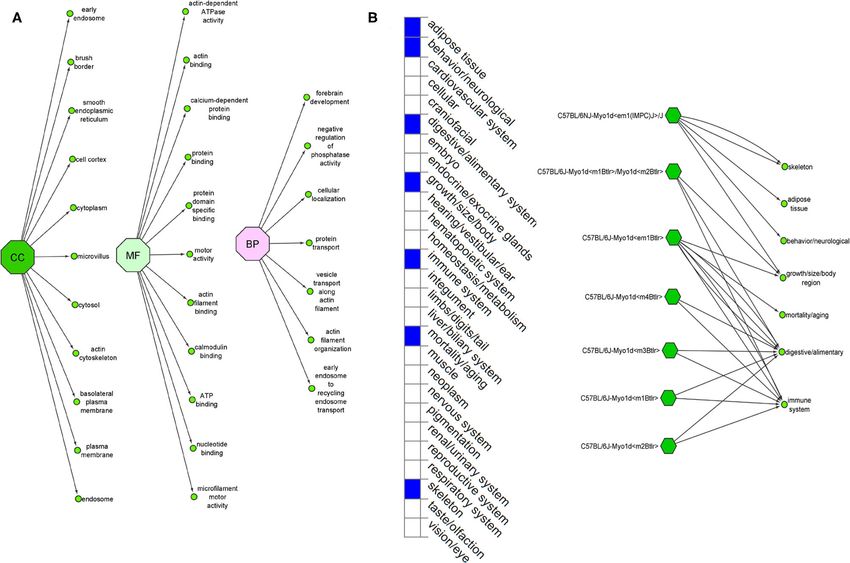
Alsafwani et al.                                                                                                              Novel MYO1D Gene Variant in LD

                                              and functional properties of MYO1D protein like calmodulin binding and microfilament
                                              motor activities. Functional bioinformatics analysis has shown that MYO1D is ubiquitously
                                              expressed across several human tissues and is reported to induce severe phenotypes
                                              in knockout mouse models. In conclusion, our findings show the expanded genetic
                                              spectrum of LD, which could potentially pave way for the novel drug target identification
                                              and development of personalised medicine for high-risk families.

                                              Keywords: laterality defects, whole exome sequencing, microfilament, gene expression, variant

INTRODUCTION                                                                        due to its potential in expanding the genetic spectrum of the
                                                                                    disease. However, literature searches reveal sparse data on the
Laterality defects (LDs) are a group of developmental diseases                      Arab LD patients. We hypothesise that genetically investigating
that affect internal organ positioning in the body. In general,                     LD patients from a consanguineous Arab society will offer
human LDs can be divided into three categories: (1) situs solitus                   new insights into disease pathogenesis by identifying novel
(SS) with normally expected organ arrangement; (2) situs inversus                   genes or novel variants in known genes, as demonstrated in
(SI) characterised by complete mirror image of organs; and (3)                      other complex diseases (20). Therefore, the objective of the
situs ambiguus (SA) with organ arrangement falling along a                          present study was to identify the genetic cause of LD in
spectrum of various anomalies between SS and SI, including                          Arab patients, using WES and multilevel bioinformatics-based
congenital heart defects (CHDs). Within SA, a subgroup of                           structural (protein variant structural modelling, divergence, and
patients presents a severe and complex form of congenital heart                     stability) and functional (gene expression and knockout mouse
disease, which is commonly known as heterotaxy (1). Defective                       model) analysis approaches.
left–right (LR) patterning of internal organs is associated with
multiple congenital diseases affecting the cardiovascular system,
                                                                                    MATERIALS AND METHODS
kidneys, liver, and biliary tract (2, 3). According to the National
Birth Defects Prevention Study (4), the estimated prevalence                        The ethical approval for the present study was obtained
of LD is 1.1 per 10,000 in the United States. Despite the rare                      from the institutional ethics committee of King Abdulaziz
likelihood of LD, its incidence is excepted to be higher among                      University Hospital (KAUH), Jeddah, Saudi Arabia. Informed
the Arab population due to their high rate of consanguinity and                     consent forms were collected from both adult parents and
genetic disorders (5).                                                              their children (parental consent and children’s assent for those
    The aetiology of LD is complex and includes both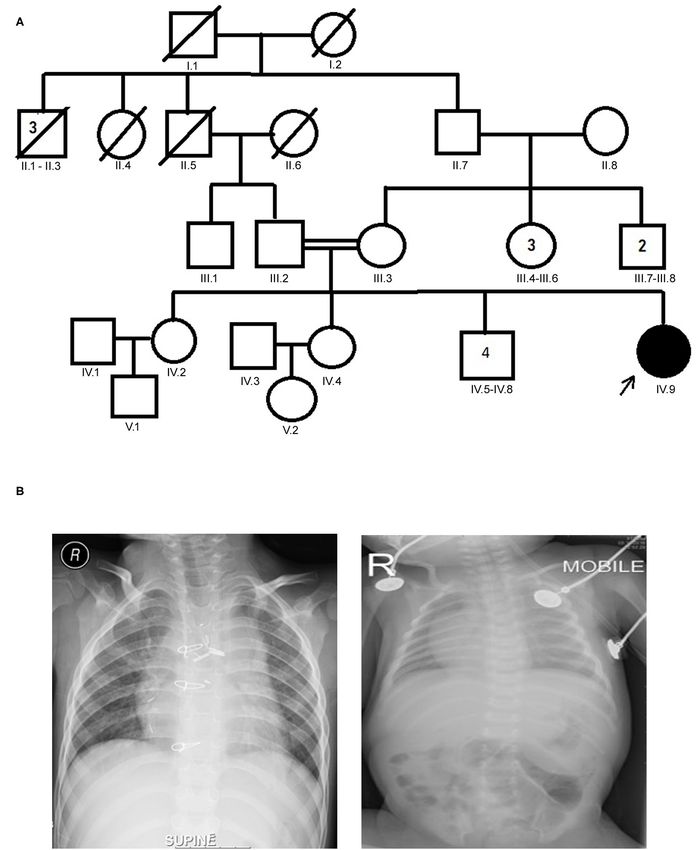
Alsafwani et al.                                                                                                                       Novel MYO1D Gene Variant in LD

 FIGURE 1 | (A) Family pedigree chart of laterality defect (LD) Arab family. Proband is indicated by the arrow. The affected proband (shaded circle) is homozygous of
 c.2293C>T mutation in MYO1D gene. Both parents were consanguineous (double horizontal lines) and heterozygous carriers of the identified mutation. (B) Chest
 X-rays showing left isomerism (heterotaxy).

Frontiers in Medicine | www.frontiersin.org                                        3                                        September 2021 | Volume 8 | Article 724826
Alsafwani et al.                                                                                                                     Novel MYO1D Gene Variant in LD

TABLE 1 | List of LD potential candidate variants that showed autosomal recessive inheritance pattern.

        Gene name        Chrom    ref    Alt     Effect     HGVS.c          HGVS.p           dbSNP151_ID       1000G_AF         ExAC_AF        gnomAD_exomes_AF

(1)     ZC3H12A          chr1     G      T       Missense   c.99G>T         p.Arg33Ser       rs116208741       0.00139776       0.002405       0.002499959
(2)     ZC3H12A          chr1     C      T       Missense   c.95C>T         p.Pro32Leu       rs115805535       0.00119808       0.002117       0.002117
(3)     TMEM18A          chr7     C      T       Missense   c.116G>A        p.Gly39Glu       rs183593116       0.00219649       0.004772       0.00515989
(4)     CPNE7            chr16    C      T       Missense   c.898C>T        p.Pro300Ser      rs150443459       0.000399361      0.0002966      0.0003931566
(5)     ASXL2            chr2     C      T       Missense   c.1489G>A       p.Ala497Thr      rs192716734       0.00139776       0.003805       0.004480134
(6)     MYO1D            chr17    G      A       Missense   c.2293C>T       p.Pro765Ser      rs7209106         0.0043           0.002759       0.002424917
(7)     EPB42            chr15    C      T       Missense   c.1477G>A       p.Gly493Ser      rs148871144       0.000199681      0.0007578      0.0007836991
(8)     FAM220A          chr7     G      A       Missense   c.437C>T        p.Pro146Leu      rs75910050        0.00139776       0.0009884      0.0009261591
(9)     OR10A2           chr11    C      A       Missense   c.741C>A        p.Phe247Leu      rs150322658       0.00119808       0.0014         0.001421724

LD, laterality defect.

TABLE 2 | Computational pathogenicity prediction scores of the LD candidate variants.

S. no              Variant              Gene                CADD           FATHMM             MetaLR            MutationTaster             PROVEAN            REVEL

(1)                rs115805535          ZC3H12A             16.06          0.22678            0.0431            0.08975                    0.30964            0.027
(2)                rs116208741          ZC3H12A             –              –                  –                 –                          –                  –
(3)                rs150443459          CPNE7               –              –                  –                 –                          –                  –
(4)                rs192716734          ASXL2               14.94          0.18248            0.025             0.25126                    0.24026            0.079
(5)                rs148871144          EPB42               0.135          0.78537            0.1928            0.08975                    0.23156            0.121
(6)                rs75910050           FAM220A             0.524          0.06931            0.0102            0.08975                    0.58248            0.049
(7)                rs150322658          OR10A2              –              –                  –                 –                          –                  –
(8)                rs183593116          TMEM184A            9.855          0.43279            0.0355            0.08975                    0.07008            0.039
(9)                rs7209106            MYO1D               23.2           0.88298            0.6557            0.81001                    0.80682            0.553

LD, laterality defect.
CADD: >25 = damaging; 0.5 = damaging;  0.5 = damaging; 0.5 = damaging; 0.5 = damaging; 0.5 = damaging;  30 score) were included. All variants with a                          matrices (PSSMs). The output file demonstrates the links between
minor allele frequency (MAF)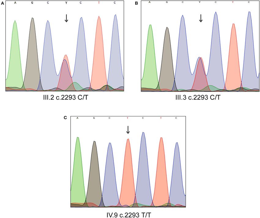
Alsafwani et al.                                                                                                              Novel MYO1D Gene Variant in LD

 FIGURE 2 | Sanger sequencing analysis of MYO1D gene. (A) Affected proband, homozygous for the variant c.2293C>T. (B,C) Heterozygous carriers of mother and
 father.

of the native protein was then used to construct the mutated                     LD candidate gene. This database provides the expression profile
version of candidate protein, which was then energy minimised                    of the query gene or protein based on primary antibody staining
and then analysed for structural deformities like amino acid or                  data in a series of immunohistochemistry pictures of clinical
whole structure level deviations using YASARA software (24).                     specimens. The functional enrichment analysis of the potential
The impact of candidate variant on the stability of protein                      LD candidate gene was done using gene ontology (GO) webtool
structure was estimated using DUET webserver, which contains                     hosted in Ensembl web browser. Moreover, Mouse Genome
Protein Data Bank (PDB) structures of query proteins to predict                  Informatics (MGI) database (http://www.informatics.jax.org/)
the Gibbs free energy (G) values (25).                                           was used to better understand the functional role of potential LD
                                                                                 gene on phenotype characteristics of knockout mouse models.
                                                                                 The MGI resource provides a comprehensive set of data, tools,
RNA Expression, Gene Ontology, and Mouse Gene                                    and analysis designed specifically for use in mouse laboratory
Knockout Model                                                                   model. It accepts input data in the form of a gene symbol and
The Human Protein Atlas (HPA) (https://www.proteinatlas.org/)                    provides output corresponding to the physiological condition of
database was used to determine the RNA expression status of the                  knockout mice.

Frontiers in Medicine | www.frontiersin.org                                  5                                     September 2021 | Volume 8 | Article 724826
Alsafwani et al. Novel MYO1D Gene Variant in LD
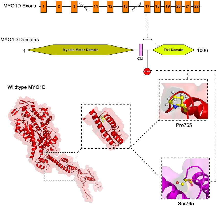
FIGURE 3 | The exonic, functional domain and 3D structural annotation of MYO1D (Pro765Ser) variant.
RESULTS inferior vena cava with absent supra-renal segment, and
azygos continuation (a rare congenital abnormality often
Clinical Assessment combined with cardiovascular and visceral malformations).
The proband aged 4 years 6 months at the time of clinical At the age of 10 months, the proband underwent thorough
diagnosis was born to an apparently healthy consanguineous palliative cardiac procedures in the form of ductal stenting in
parents of Arab origin (Figure 1A). The proband exhibited a the neonatal period followed by Kawashima cavo-pulmonary
spectrum of phenotypes including visceral heterotaxy (abnormal shunt (a palliative surgical procedure performed in cases of left
arrangements of thoracoabdominal organs) (Figure 1B), isomerism and azygos continuation of the inferior vena cava, and
congenital cyanotic heart disease in the form of single ventricle common atrioventricular valve with or without regurgitation
physiology, left isomerism with polysplenia syndrome, double and pulmonary stenosis) in addition to left pulmonary artery
inlet atrioventricular connection (a heart defect that affects (LPA) balloon dilatation procedure. At 3 years of age, Fontan
the valves and chambers), pulmonary atresia, interrupted completion was performed via incorporation of hepatic veins to
Frontiers in Medicine | www.frontiersin.org 6 September 2021 | Volume 8 | Article 724826Alsafwani et al. Novel MYO1D Gene Variant in LD
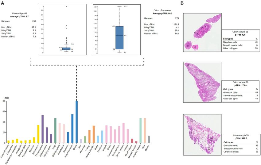
FIGURE 4 | Protein Atlas expression analysis of MYO1D. (A) Bar graph represents the MYO1D expression in sigmoid and transverse colon samples. (B)
Histopathological examinations of colonic samples from three different patients showing the MYO1D protein expression.
pulmonary artery correcting thereby blood flow from the lower reported as homozygous for this variant in the gnomAD.
body parts directly to the lungs. But their clinical details are not provided in the gnomAD
database. In the index family studied here, both parents were
heterozygous and do not have any symptoms associated with
Genetic Analysis LD, confirming the autosomal recessive inheritance pattern as
Whole-Exome Sequencing Variant Filtering and Novel we initially deduced from their pedigree analysis. Moreover,
Gene Identification more than 80% (5/6; 83.34) of the computational prediction
The sequencing of the index case generated approximately 98,000 methods like CADD, FATHMM, MetaLR, Mutation Taster,
variants, including 12,150 synonymous variants, 13,000 missense PROVEAN, and REVEL have attributed pathogenicity scores to
variants, and 11,500 indels. Variant filtration was based on this MYO1D (p.Pro765Ser) variant (Table 2). Functional biology
its rare frequency, deleterious potential, autosomal recessive data available from model organisms like Drosophila, zebrafish,
mode of inheritance, and functional relevance to disease (LD, and frog have proved the functional role of MYO1D gene
primary ciliary dyskinesia (PCD), congenital heart disease, and in LDs.
heterotaxy). Nine genetic variants were identified as potential
candidates (Table 1). Among these variants, only one missense
variant (rs7209106: NM_015194.2:c.2293C>T; p.Pro765Ser) in Sanger Sequencing Validation
MYO1D novel gene has survived our variant filtration criteria. Sanger sequencing analysis confirmed that the LD patient is
This allele is absent in local databases like GME (Greater Middle homozygous for c.2293C>T variant in MYO1D gene (1V.9,
East) (http://igm.ucsd.edu/gme/), DALIA (Disease Alleles in Figures 1A, 2), whereas the mother and father were heterozygous
Arabs) (http://clingen.igib.res.in/dalia/index), and Saudi Human carriers (111.2, 111.3, Figures 1A, 2). This variant was absent in
Genome Program (SHGP) (https://shgp.kacst.edu.sa/index.en. apparently healthy siblings and were homozygous for the T allele
html#home).The MAF of this variant in international databases (1V.2, 1V.4, and 1V.5–8; Figure 1A). Two additional clinically
like 1,000 Genomes and gnomAD databases is 0.005 and diagnosed LD families were screened for this variant, and none
0.002, respectively. Although it has an allele frequency of carries this mutation, suggesting that MYO1D (p.Pro765Ser)
0.013 in the African population, only eight individuals are variant is a rare private mutation in this family.
Frontiers in Medicine | www.frontiersin.org 7 September 2021 | Volume 8 | Article 724826Alsafwani et al. Novel MYO1D Gene Variant in LD
FIGURE 5 | (A) Ensembl function annotations of MYO1D. (B) Mouse MYO1D knockout analysis and phenotypic changes.
Computational Functional Analysis and template proteins (Figure 3). The stereochemical evaluation
Variant Mapping on MYOID1 Protein Domain of the energy-minimised MYO1D protein model revealed that
The mapping of conserved amino acid domains is a vital step 96.2% of the amino acids are in the allowed portion of the
in deducing the association between the nucleotide sequence, protein, whereas 3.8% are in the non-allowed region. As per the
protein structure, and function of disease-causing proteins. The above outlined processes, the native MYO1D model was used as
CDD analysis showed that MYO1D protein is made up of three a template to create a mutant variant by manually substituting
domains, namely, motor (11–682 amino acids), IQ calmodulin- proline for serine at the 765th position. PyMOL was used to
binding motif (699–719 amino acids), and Myosin TH1 (803– depict native and mutant proteins.
1,000 amino acids) domains. The Pro765Ser variant is located
between the Myosin TH1 and calmodulin-binding domains Structural Deviation and Stability Findings
(Figure 3). We have used YASARA tool to analyze Cα-atom coordinates
of native and mutant MYO1D 3D structures to evaluate
MYO1D 3D Model Construction their structural drifts (in terms of RMSD) at residue and
The PDB database search revealed the availability of partial 707 whole structure levels. RMSD value is used to quantitatively
aa (between 10 and 717 of 1,006 AA long MYO1D protein) measure the structural similarity between two atomic coordinates
X-ray crystal protein model (4L79) with 2.3-Å resolution. when superimposed on each other. The impact of substitution
Hence, the remaining 306 aa long chain was simulated with mutations on amino acid structures can be calculated when
iterative threading assembly modification (I-TASSER) webserver there is a divergence at the polypeptide chain level. We noticed
following an ab initio approach. From the I-TASSER output, minor structural drifts in MYO1D structure only at 765th residue
the best MYO1D model was chosen based on its polypeptide position due to the RMSD value difference (2.28) induced by
prediction quality scores like confidence score (C = −1.52), the substitution of proline with serine (Figure 3). The DUET
template modelling (TM = 0.53 ± 0.15), and root-mean-square analysis of the MYO1D (P765S) variant predicted Gibbs free
deviation (RMSD) (12.7 ± 4.3 Å) scores. These quality metrics energy (11G) alterations shifting the energy equilibrium to
indicate the very good structural similarity between the query negative value, i.e., −0.959 kcal/mol, suggesting that the queried
Frontiers in Medicine | www.frontiersin.org 8 September 2021 | Volume 8 | Article 724826Frontiers in Medicine | www.frontiersin.org
Alsafwani et al.
TABLE 3 | Phenotypes and genetic data of LD and/or PCD among Arabs.
Nationality Situs inversus PCD Dextrocardia Heterotaxy of Recurrent chest Chronic rhinitis Bronchiectasis Chronic or Gene Reference
(SI) abdominal infections recurrent otitis
organs media
Yemeni + – + Visceral – – – – MYO1D Present
heterotaxy, study
polysplenia
syndrome
Saudi (6 patients) +6/6 – NR NR – NR NR NR DNAH5, PKD1L1, (26)
DNAAF5/CYP21A2,
DNAI1
Arab + NR + NR NR NR NR NR GDF1 (27)
Saudi + NR + Abdominal NR NR NR NR NR (28)
ultrasound
revealed that the
liver and
gallbladder were
located in the left
hypochondrium,
spleen on the right
side
Arab + NR + + NR NR NR NR HES7 (29)
Saudi (75 patients) + +73/75 NR NR +15/75 NR NR NR CCDC151, (26)
9
CCDC39,
CCDC40,
DNAH11,
GOLGA3, RSPH9,
CCNO, RSPH9,
ITCH, MCIDAS,
RSPH4A, DNAH5,
CEP164, GOLGA3
Morocco + + + + + + + – NR (30)
Saudi + + + NR NR + + + DNAH1 (31)
Kuwaiti + + NR NR + NR NR DNAH5 (32),
Saudi NR NR NR NR CCDC151 (33)
September 2021 | Volume 8 | Article 724826
+ + + +
Palestine NR + + NR NR NR + + LRRC6 (34)
Saudi NR + NR NR NR NR + NR RSPH9 (35)
Novel MYO1D Gene Variant in LD
UAE NR + NR NR + NR + NR RSPH9 (36)
UAE – + NR NR + + + + RSPH9 (37)
Saudi + + NR NR NR NR + NR 19q13.3 (27)
Saudi + + – Situs inversus of + NR + – NR (38)
cardiac shadow
and gastric air
bubble
+, presence of symptoms; –, absence of symptoms; NR, not reported; PCD, primary ciliary dyskinesia.Alsafwani et al. Novel MYO1D Gene Variant in LD
variant is potentially deleterious to the protein stability owing to Various studies suggested the role of MYO1D gene in laterality
its destabilising behaviour. disease in Drosophila, zebrafish, and frog (43–47). MYO1D has
a role in organ asymmetry in Drosophila, which lacks cilia
RNA Expression Analysis and nodal pathway while developing LD by using polar cell
The HPA shows the positive expression status of MYO1D gene in polarity (PCP), MYO1D, and HOX gene Abd-B (48, 49). Another
different tissues and organs of the human body like the colon, study demonstrated the function of MYO1D in Xenopus laevis
lungs, and thyroid gland. In particular, the highest expression and influence the orientation of the cilia on the LR organiser
was seen in the digestive system with the colon, where the 274 (LRO) through planar cell polarity pathway as implicated in
transverse colon samples showed a maximum of 221.5 protein Drosophila (45). In zebrafish, MYO1D plays a fundamental
transcripts per million (pTPM) and 233 sigmoid colon samples role in the LR organisation (43, 50). Aside from the above
showed a maximum of 97.8 pTPM. The RNA-Seq analysis initial reports, our understanding of MYO1D function in the
of immunohistochemistry tissue specimens from three control context of human LR patterning remains largely unexplored.
specimens showed that glandular cells showed the highest pTPM The situs inversus (SI) phenotype is reported in approximately
status of MYO1D gene when compared with smooth muscle cells 50% of PCD cases with congenital cardiac defects (51, 52).
and other cell types in the colon (Figure 4). Approximately 3–7% of LD patients have CHDs (53). Many
studies (54, 55) in a variety of species failed to identify a unifying
Gene Ontology and Gene Knockout Analysis mechanism for LR patterning. However, recent studies (43, 50)
The Ensembl GO analysis of MYO1D gene showed its provided the first evidence of a shared origin of laterality in both
involvement in 43 GO terms, including those connected to arthropods and chordates through MYO1D gene. The clinical
biological processes (seven GO terms), molecular functions features are consistent with previous observations in zebrafish
(11 GO terms), and cellular component (25 GO terms) and Drosophila, indicating that MYO1D has an important role
(Supplementary Table 1). All the annotations collectively in LR patterning during embryogenesis. Moreover, asymmetric
highlight that MYO1D is localised in the cytoplasm (cellular clustering of cilia was disrupted in ependymal cells of MYO1D
component) and plays an important role in actin filament KO rat models, consistent with LD (56).
organisation (biological process) as well as microfilament motor MYO1D gene knockout in different mouse strains (seven
activity function (molecular function). Supplementary Table 2 different strains) is shown to demonstrate a variety of phenotypes
shows details of disease phenotypes corresponding to the like decreased body fat amount (adipose tissue), decreased
MYO1D genetic background in different knockout mice models. startle reflex (behaviour/neurological), increased susceptibility to
colitis (digestive/alimentary), decreased bone mineral content
DISCUSSION (skeleton), and increased susceptibility to weight loss (growth
size/body weight and immune system and mortality or ageing)
Recent evolution and easy accessibility of next-generation (Figure 5). Our study is the first one to report the association of
sequencing resulted in accurate molecular diagnosis of variety of defective MYO1D to LD in humans, confirming that the function
genetic diseases from around the globe (39, 40). Owing to the is evolutionarily conserved from Drosophila, to zebrafish, to
genetic heterogeneity, identification of specific molecular cause frog, to humans. Thus, MYO1D gene can be considered as the
of LD is very challenging in up to 80% of the cases. Majority new causal gene for LD in humans. Though the Drosophila
of known LD causative genes are structural proteins of the cilia and zebrafish models clearly showed the visceral heterotaxy,
and are known for their involvement in NODAL/TGFβ or SHH mouse KO phenotypes were surprisingly not showing any LD-
signalling pathways. During embryonic development, these genes related phenotypes.
play an important role in symmetrical LR positioning of the Extensive computational analysis of the protein structure
organs (10). and function adds the supporting evidence for MYO1D in LD.
MYO1D gene consists of 27 exons mapped to chromosome MYO1D protein is 1,006 aa long with a molecular weight of
17q11.2. Myosin heavy chain class 1 is a member of the myosin 116 kDa. It consists of a large, highly conserved Myocin Motor
superfamily, playing essential roles in cytoskeletal structure, Domain (671 aa), short calmodulin-binding motif (20 aa), and
mechanical signal-transduction membrane dynamics (41), and a basic C-terminal tail homology-1 (TH1) domain (197 aa).
endosome processing (42). In the present study, we identified The amino acid residue level structural deviation observed with
the first case of a homozygous missense (c.2293C>T) mutation the variant serine (Pro765Ser) in MYO1D is likely to disturb
in MYO1D gene causing LD in the proband of an Arab the primary, secondary, tertiary, and quaternary structural
consanguineous family. Further screening of LD participants features in the protein. Numerous studies have shown the strong
from two additional Arab families did not reveal any mutations correlation between deviations in residue level RMSD score and
in this gene. In Middle Eastern Arab databases (with more structural properties for the disease-causing variants (23, 57–59).
than 10,000 exome data combined) such as SHGP, GMC, and Disease causative pathogenic mutations have often changed the
DALIA, not a single case was recorded for this variant. Also, energy equilibrium, which is required to maintain the protein
this variant was extremely rare (Alsafwani et al. Novel MYO1D Gene Variant in LD
binding, actin-dependent ATPase activity, calcium-dependent data in the public domain and (b) as per the local Institutional
protein binding, and microfilament motor activities (61). Ethics committee approval and Saudi national policy on genomic
LD is a complex disease, and its clinical phenotype data sharing in the public domain outside the country. Requests
presentations often overlap with PCD symptoms. A recent to access the data should be directed to RE or TS.
study from Saudi Arabia reported the overlapping clinical
symptoms between PCD and LD patients (26). This report ETHICS STATEMENT
investigated a total of 81 patients, including 58 patients with
sinopulmonary infections (SPIs), 15 patients with combined The Ethical approval for the present study was obtained from
LD with SPIs, and six patients with LD alone. They reported institutional Ethics Committee of King Abdulaziz University
mutations in the known PCD genes as follows: RSPH9, Hospital (KAUH), Jeddah, Saudi Arabia and informed consent
CCNO, DNAAF5, RSPH4A, MCIDAS, and CCDC40 gene forms were collected from both adult parents and their children
mutations in PCD patients with SPIs; CCDC151, DNAH11, (guardian consent for Language evaluation).
CCDC40, DNAH5, and CCDC39 gene mutations in LD
patients with SPIs; PKD1L1 and DNAAF5 gene mutations CONSENT TO PARTICIPATE
in LD patients; and RSPH9 and MCIDAS gene mutations
in neonatal respiratory distress. Additionally, they have also Informed consent was obtained from all subjects involved in
identified gene mutations in ITCH and CEP164 in two the study.
patients, demonstrating ITCH-related syndrome and Bardet–
Biedl syndrome. Sparrow et al. (29) reported HES7 as a AUTHOR CONTRIBUTIONS
cause of spondylocostal dysostosis with SI and dextrocardia.
Molecular diagnosis of LD and PCD in Arab patients has TA, RE, and KN: conceptualisation. KN, RA, BB, HE, and RE:
revealed a spectrum of mutations in many genes with methodology. BB: software and visualisation. KN, RA, BB, NS,
variable clinical presentations (35), which is summarised in and RE: formal analysis. RA, KN, TS, and RE: investigation.
Table 3. KN, TS, and BB: resources. RA, KN, TS, BB, ZZ, GA, NS,
JA-A, AS, OA-R, RE, and TA: writing—original draft preparation.
CONCLUSION KN, TS, NS, and RE: writing—review and editing. KN, BB,
and RE: supervision. TS: project administration and funding
In conclusion, we discovered missense mutation in MYO1D acquisition. All authors contributed to the article and approved
gene (c.2293C>T) in an Arab patient presenting with visceral the submitted version.
heterotaxy and left isomerism with polysplenia syndrome by
using higher-throughput WES technology. This is the first report FUNDING
to establish the relationship between MYO1D variants and LD,
supporting the previous findings in Drosophila zebrafish, and This research work was funded by Institutional Fund Projects
frog. This exciting finding may support the critical role of under grant no. (IFPRC-132-290-2020). Therefore, authors
MYO1D gene for LR patterning in humans. This study has gratefully acknowledge technical and financial support from the
some sincere limitations, as this is the first case identified with Ministry of Education and King Abdulaziz University, Jeddah,
MYO1D mutation potentially contributing to LD phenotypes, Saudi Arabia.
and there are no reported cases with MYO1D variants to
compare our data with. Therefore, testing MYO1D variants for ACKNOWLEDGMENTS
LD patients in large cohort studies is recommended to verify
our findings. Future functional studies are also recommended to The authors gratefully acknowledge technical and financial
investigate the specific molecular role and therapeutic prospects support from the Ministry of Education and King Abdulaziz
of targeting MYO1D genetic variants in patients demonstrating University, Jeddah, Saudi Arabia.
LD phenotypes.
SUPPLEMENTARY MATERIAL
DATA AVAILABILITY STATEMENT
The Supplementary Material for this article can be found
The datasets presented in this article are not readily available online at: https://www.frontiersin.org/articles/10.3389/fmed.
because (a) participants’ refusal to store or distribute the genomic 2021.724826/full#supplementary-material
REFERENCES dyskinesia: insights into situs ambiguus and heterotaxy. Chest. (2014)
146:1176–86. doi: 10.1378/chest.13-1704
1. Shapiro AJ, Davis SD, Ferkol T, Dell SD, Rosenfeld M, Olivier KN, et 2. Kosaki K, Casey B. Genetics of human left-right axis malformations. Semin
al. Laterality defects other than situs inversus totalis in primary ciliary Cell Dev Biol. (1998) 9:89–99. doi: 10.1006/scdb.1997.0187
Frontiers in Medicine | www.frontiersin.org 11 September 2021 | Volume 8 | Article 724826Alsafwani et al. Novel MYO1D Gene Variant in LD
3. Lopez KN, Li AH, Hanchard N, Azamian M, Lalani S, Dickerson hypercholesterolemia causative PCSK9 functional domain mutations reveals
H, et al. Whole exome sequencing in congenital heart disease reveals binding affinity alterations with LDLR. Int J Pept Res Ther. (2021) 27:719–
variants in left-right patterning genes previously associated with heterotaxy 33. doi: 10.1007/s10989-020-10121-8
syndrome and primary ciliary dyskinesia. J Am Coll Cardiol. (2015) 65(10 24. Shaik NA, Bokhari HA, Masoodi TA, Shetty PJ, Ajabnoor GMA, Elango R, et
Suppl.):A490. doi: 10.1016/S0735-1097(15)60490-9 al. Molecular modelling and dynamics of CA2 missense mutations causative
4. Lin AE, Krikov S, Riehle-Colarusso T, Frías JL, Belmont J, Anderka M, et al. to carbonic anhydrase 2 deficiency syndrome. J Biomol Struct Dyn. (2020)
Laterality defects in the national birth defects prevention study (1998-2007): 38:4067–80. doi: 10.1080/07391102.2019.1671899
birth prevalence and descriptive epidemiology. Am J Med Genet A. (2014) 25. Pires DE, Ascher DB, Blundell TL. DUET: a server for predicting effects of
164A:2581–91. doi: 10.1002/ajmg.a.36695 mutations on protein stability using an integrated computational approach.
5. Best S, Shoemark A, Rubbo B, Patel MP, Fassad MR, Dixon M, et al. Nucleic Acids Res. (2014) 42:W314–319. doi: 10.1093/nar/gku411
Risk factors for situs defects and congenital heart disease in primary ciliary 26. Shamseldin HE, Al Mogarri I, Alqwaiee MM, Alharbi AS,
dyskinesia. Thorax. (2019) 74:203–5. doi: 10.1136/thoraxjnl-2018-212104 Baqais K, Alsaadi M, et al. An exome-first approach to aid in
6. Kuehl KS, Loffredo C. Risk factors for heart disease associated with abnormal the diagnosis of primary ciliary dyskinesia. Hum Genet. (2020)
sidedness. Teratology. (2002) 66:242–8. doi: 10.1002/tera.10099 139:1273–83. doi: 10.1007/s00439-020-02170-2
7. Kuehl KS, Loffredo CA. Population-based study of l-transposition of the great 27. Marek-Yagel D, Bolkier Y, Barel O, Vardi A, Mishali D, Katz U, et al. A founder
arteries: possible associations with environmental factors. Birth Defects Res A truncating variant in GDF1 causes autosomal-recessive right isomerism and
Clin Mol Teratol. (2003) 67:162–7. doi: 10.1002/bdra.10015 associated congenital heart defects in multiplex Arab kindreds. Am J Med
8. Sutherland MJ, Ware SM. Disorders of left-right asymmetry: Genet A. (2020) 182:987–93. doi: 10.1002/ajmg.a.61509
heterotaxy and situs inversus. Am J Med Genet C. (2009) 28. Saleh A. Dextrocardia with situs inversus totalis in a boy: a case report. Int J
151C:307–17. doi: 10.1002/ajmg.c.30228 Contemp Pediatr. (2016) 1096–11098. doi: 10.18203/2349-3291.ijcp20162398
9. Deng H, Xia H, Deng S. Genetic basis of human left-right asymmetry 29. Sparrow DB, Faqeih EA, Sallout B, Alswaid A, Ababneh F, Al-Sayed M,
disorders. Expert Rev Mol Med. (2015) 16:e19. doi: 10.1017/erm.2014.22 et al. Mutation of HES7 in a large extended family with spondylocostal
10. Li AH, Hanchard NA, Azamian M, D’alessandro LCA, Coban- dysostosis and dextrocardia with situs inversus. Am J Med Genet A. (2013)
Akdemir Z, Lopez KN, et al. Genetic architecture of laterality defects 161A:2244–9. doi: 10.1002/ajmg.a.36073
revealed by whole exome sequencing. Eur J Hum Genet. (2019) 30. Raoufi M, Sator H, Lahma J, El Ayoubi A, Nitassi S, Oujilal A, et al. A
27:563–73. doi: 10.1038/s41431-018-0307-z case of Kartagener syndrome with rhinolalia clausa. Pan Afr Med J. (2016)
11. Ware SM, Jefferies JL. New genetic insights into congenital heart disease. J 23:159. doi: 10.11604/pamj.2016.23.159.8664
Clin Exp Cardiol. (2012) S8:003. doi: 10.4172/2155-9880.S8-003 31. Imtiaz F, Allam R, Ramzan K, Al-Sayed M. Variation in DNAH1
12. Collignon J, Varlet I, Robertson EJ. Relationship between asymmetric nodal may contribute to primary ciliary dyskinesia. BMC Med Genet. (2015)
expression and the direction of embryonic turning. Nature. (1996) 381:155– 16:14. doi: 10.1186/s12881-015-0162-5
8. doi: 10.1038/381155a0 32. Marafie MJ, Al Suliman IS, Redha AM, Alshati AM. Primary ciliary
13. Olson EN, Srivastava D. Molecular pathways controlling heart development. dyskinesia: Kartagener syndrome in a family with a novel DNAH5 gene
Science. (1996) 272:671–6. doi: 10.1126/science.272.5262.671 mutation and variable phenotypes. Egypt J Med Hum Genet. (2015) 16:95–
14. Iratni R, Yan YT, Chen C, Ding J, Zhang Y, Price SM, et al. Inhibition of excess 9. doi: 10.1016/j.ejmhg.2014.08.001
nodal signaling during mouse gastrulation by the transcriptional corepressor 33. Hjeij R, Onoufriadis A, Watson CM, Slagle CE, Klena NT, Dougherty GW,
DRAP1. Science. (2002) 298:1996–9. doi: 10.1126/science.1073405 et al. CCDC151 mutations cause primary ciliary dyskinesia by disruption of
15. Otto EA, Schermer B, Obara T, O’toole JF, Hiller KS, Mueller AM, et the outer dynein arm docking complex formation. Am J Hum Genet. (2014)
al. Mutations in INVS encoding inversin cause nephronophthisis type 2, 95:257–74. doi: 10.1016/j.ajhg.2014.08.005
linking renal cystic disease to the function of primary cilia and left-right axis 34. Horani A, Ferkol TW, Shoseyov D, Wasserman MG, Oren YS, Kerem B, et al.
determination. Nat Genet. (2003) 34:413–20. doi: 10.1038/ng1217 LRRC6 mutation causes primary ciliary dyskinesia with dynein arm defects.
16. Qian F, Germino FJ, Cai Y, Zhang X, Somlo S, Germino GG. PKD1 interacts PLoS One. (2013) 8:e59436. doi: 10.1371/journal.pone.0059436
with PKD2 through a probable coiled-coil domain. Nat Genet. (1997) 16:179– 35. Alsaadi MM, Gaunt TR, Boustred CR, Guthrie PA, Liu X, Lenzi L, et al. From
83. doi: 10.1038/ng0697-179 a single whole exome read to notions of clinical screening: primary ciliary
17. Yoshiba S, Shiratori H, Kuo IY, Kawasumi A, Shinohara K, dyskinesia and RSPH9 p.Lys268del in the Arabian Peninsula. Ann Hum Genet.
Nonaka S, et al. Cilia at the node of mouse embryos sense (2012) 76:211–20. doi: 10.1111/j.1469-1809.2012.00704.x
fluid flow for left-right determination via Pkd2. Science. (2012) 36. Reish O, Slatkin M, Chapman-Shimshoni D, Elizur A, Chioza B, Castleman
338:226–31. doi: 10.1126/science.1222538 V, et al. Founder mutation(s) in the RSPH9 gene leading to primary ciliary
18. Calvet JP. Ciliary signaling goes down the tubes. Nat Genet. (2003) 33:113– dyskinesia in two inbred Bedouin families. Ann Hum Genet. (2010) 74:117–
4. doi: 10.1038/ng1078 25. doi: 10.1111/j.1469-1809.2009.00559.x
19. Grimes DT, Keynton JL, Buenavista MT, Jin X, Patel SH, Kyosuke 37. Castleman VH, Romio L, Chodhari R, Hirst RA, De Castro SC, Parker KA, et
S, et al. Genetic analysis reveals a hierarchy of interactions al. Mutations in radial spoke head protein genes RSPH9 and RSPH4A cause
between polycystin-encoding genes and genes controlling cilia primary ciliary dyskinesia with central-microtubular-pair abnormalities. Am
function during left-right determination. PLoS Genet. (2016) J Hum Genet. (2009) 84:197–209. doi: 10.1016/j.ajhg.2009.01.011
12:e1006070. doi: 10.1371/journal.pgen.1006070 38. Bashi S, Khan MA, Guirjis A, Joharjy IA, Abid MA. Immotile-
20. Saadah OI, Banaganapalli B, Kamal NM, Sahly AN, Alsufyani HA, cilia syndrome with azoospermia: a case report and review of the
Mohammed A, et al. Identification of a rare exon 19 skipping mutation in literature. Br J Dis Chest. (1988) 82:194–6. doi: 10.1016/0007-0971(88)9
ALMS1 gene in alström syndrome patients from two unrelated saudi families. 0043-5
Front Pediatr. (2021) 9:652011. doi: 10.3389/fped.2021.652011 39. Bokhari HA, Shaik NA, Banaganapalli B, Nasser KK, Ageel HI, Al Shamrani
21. Marchler-Bauer A, Lu S, Anderson JB, Chitsaz F, Derbyshire MK, AS, et al. Whole exome sequencing of a Saudi family and systems biology
Deweese-Scott C, et al. CDD: a Conserved Domain Database for the analysis identifies CPED1 as a putative causative gene to Celiac Disease. Saudi
functional annotation of proteins. Nucleic Acids Res. (2010) 39:D225– J Biol Sci. (2020) 27:1494–502. doi: 10.1016/j.sjbs.2020.04.011
D9. doi: 10.1093/nar/gkq1189 40. Gaboon NEA, Banaganapalli B, Nasser K, Razeeth M, Alsaedi MS, Rashidi
22. Ahmed Awan Z, Bima A, Rashidi OM, Jamil K, Khan IA, Almukadi HS, et OM, et al. Exome sequencing and metabolomic analysis of a chronic
al. Low resolution protein mapping and KB-R7943 drug-protein molecular kidney disease and hearing loss patient family revealed RMND1 mutation
interaction analysis of long-QT syndrome linked KCNH2 mutations. All Life. induced sphingolipid metabolism defects. Saudi J Biol Sci. (2020) 27:324–
(2020) 13:183–93. doi: 10.1080/26895293.2020.1737249 34. doi: 10.1016/j.sjbs.2019.10.001
23. Awan ZA, Bahattab R, Kutbi HI, Noor AOJ, Al-Nasser MS, Shaik 41. Huber LA, Fialka I, Paiha K, Hunziker W, Sacks DB, Bähler M, et al.
NA, et al. Structural and molecular interaction studies on familial Both calmodulin and the unconventional myosin Myr4 regulate membrane
Frontiers in Medicine | www.frontiersin.org 12 September 2021 | Volume 8 | Article 724826Alsafwani et al. Novel MYO1D Gene Variant in LD
trafficking along the recycling pathway of MDCK cells. Traffic. (2000) 1:494– 56. Hegan PS, Ostertag E, Geurts AM, Mooseker MS. Myosin Id is required
503. doi: 10.1034/j.1600-0854.2000.010607.x for planar cell polarity in ciliated tracheal and ependymal epithelial cells.
42. De La Cruz EM, Ostap EM. Relating biochemistry and function Cytoskeleton (Hoboken). (2015) 72:503–16. doi: 10.1002/cm.21259
in the myosin superfamily. Curr Opin Cell Biol. (2004) 16:61– 57. Nasser KK, Banaganapalli B, Shinawi T, Elango R, Shaik NA. Molecular
7. doi: 10.1016/j.ceb.2003.11.011 profiling of lamellar ichthyosis pathogenic missense mutations on the
43. Juan T, Geminard C, Coutelis JB, Cerezo D, Poles S, Noselli S, et al. Myosin1D structural and stability aspects of TGM1 protein. J Biomol Struct Dyn. (2020)
is an evolutionarily conserved regulator of animal left-right asymmetry. Nat 1–11. doi: 10.1080/07391102.2020.1782770
Commun. (2018) 9:1942. doi: 10.1038/s41467-018-04284-8 58. Ibrahim AZ, Thirumal Kumar D, Abunada T, Younes S, George Priya
44. Lebreton G, Geminard C, Lapraz F, Pyrpassopoulos S, Cerezo D, Speder P, et Doss C, Zaki OK, et al. Investigating the structural impacts of a
al. Molecular to organismal chirality is induced by the conserved myosin 1D. novel missense variant identified with whole exome sequencing in an
Science. (2018) 362:949–52. doi: 10.1126/science.aat8642 Egyptian patient with propionic acidemia. Mol Genet Metab Rep. (2020)
45. Tingler M, Kurz S, Maerker M, Ott T, Fuhl F, Schweickert A, et al. A conserved 25:100645. doi: 10.1016/j.ymgmr.2020.100645
role of the unconventional myosin 1D in laterality determination. Curr Biol. 59. Thirumal Kumar D, Udhaya Kumar S, Nishaat Laeeque AS, Apurva
(2018) 28:810–16.e813. doi: 10.1016/j.cub.2018.01.075 Abhay S, Bithia R, Magesh R, et al. Computational model to analyze and
46. Yuan S, Brueckner M. Left-right asymmetry: myosin 1D at the center. Curr characterize the functional mutations of NOD2 protein causing inflammatory
Biol. (2018) 28:R567–R9. doi: 10.1016/j.cub.2018.03.01 disorder - Blau syndrome. Adv Protein Chem Struct Biol. (2020) 120:379–
47. Blum M, Ott T. Mechanical strain, novel genes and evolutionary insights: 408. doi: 10.1016/bs.apcsb.2019.11.005
news from the frog left-right organizer. Curr Opin Genet Dev. (2019) 56:8– 60. Shaik NA, Nasser KK, Alruwaili MM, Alallasi SR, Elango R, Banaganapalli B.
14. doi: 10.1016/j.gde.2019.05.005 Molecular modelling and dynamic simulations of sequestosome 1 (SQSTM1)
48. Spéder P, Ádám G, Noselli S. Type ID unconventional myosin missense mutations linked to Paget disease of bone. J Biomol Struct Dyn.
controls left-right asymmetry in Drosophila. Nature. (2006) (2021) 39:2873–84. doi: 10.1080/07391102.2020.1758212
440:803–7. doi: 10.1038/nature04623 61. Ko Y-S, Bae JA, Kim KY, Kim SJ, Sun EG, Lee KH, et al. MYO1D binds with
49. González-Morales N, Géminard C, Lebreton G, Cerezo D, Coutelis J-B, kinase domain of the EGFR family to anchor them to plasma membrane
Noselli S. The atypical cadherin dachsous controls left-right asymmetry in before their activation and contributes carcinogenesis. Oncogene. (2019)
Drosophila. Dev Cell. (2015) 33:675–89. doi: 10.1016/j.devcel.2015.04.026 38:7416–32. doi: 10.1038/s41388-019-0954-8
50. Blum M, Ott T. Animal left-right asymmetry. Curr Biol. (2018) 28:R301–
R4. doi: 10.1016/j.cub.2018.02.073 Conflict of Interest: The authors declare that the research was conducted in the
51. Mirra V, Werner C, Santamaria F. Primary ciliary dyskinesia: an update on absence of any commercial or financial relationships that could be construed as a
clinical aspects, genetics, diagnosis, and future treatment strategies. Front potential conflict of interest.
Pediatr. (2017) 5:135. doi: 10.3389/fped.2017.00135
52. Boon M, Smits A, Cuppens H, Jaspers M, Proesmans M, Dupont LJ, et
Publisher’s Note: All claims expressed in this article are solely those of the authors
al. Primary ciliary dyskinesia: critical evaluation of clinical symptoms and
diagnosis in patients with normal and abnormal ultrastructure. Orphanet J and do not necessarily represent those of their affiliated organizations, or those of
Rare Dis. (2014) 9:11. doi: 10.1186/1750-1172-9-11 the publisher, the editors and the reviewers. Any product that may be evaluated in
53. Escobar-Diaz MC, Friedman K, Salem Y, Marx GR, Kalish BT, Lafranchi this article, or claim that may be made by its manufacturer, is not guaranteed or
T, et al. Perinatal and infant outcomes of prenatal diagnosis of heterotaxy endorsed by the publisher.
syndrome (asplenia and polysplenia). Am J Cardiol. (2014) 114:612–
7. doi: 10.1016/j.amjcard.2014.05.042 Copyright © 2021 Alsafwani, Nasser, Shinawi, Banaganapalli, ElSokary, Zaher,
54. Blum M, Feistel K, Thumberger T, Schweickert A. The evolution and Shaik, Abdelmohsen, Al-Aama, Shapiro, O. Al-Radi, Elango and Alahmadi. This
conservation of left-right patterning mechanisms. Development. (2014) is an open-access article distributed under the terms of the Creative Commons
141:1603–13. doi: 10.1242/dev.100560 Attribution License (CC BY). The use, distribution or reproduction in other forums
55. Coutelis JB, González-Morales N, Géminard C, Noselli S. Diversity is permitted, provided the original author(s) and the copyright owner(s) are credited
and convergence in the mechanisms establishing L/R asymmetry and that the original publication in this journal is cited, in accordance with accepted
in metazoa. EMBO Rep. (2014) 15:926–37. doi: 10.15252/embr.2014 academic practice. No use, distribution or reproduction is permitted which does not
38972 comply with these terms.
Frontiers in Medicine | www.frontiersin.org 13 September 2021 | Volume 8 | Article 724826You can also read