Cardioprotective Effect of Nec-1 in Rats Subjected to MI/R: Downregulation of Autophagy-Like Cell Death - Hindawi.com
←
→
Page content transcription
If your browser does not render page correctly, please read the page content below
Hindawi
Cardiovascular erapeutics
Volume 2021, Article ID 9956814, 8 pages
https://doi.org/10.1155/2021/9956814
Research Article
Cardioprotective Effect of Nec-1 in Rats Subjected to MI/R:
Downregulation of Autophagy-Like Cell Death
Liang Wang ,1 Xuebai Lv,2 Jue Tian,3,4 Xiaoliang Wang,3 Ye Wu,3,5,6
and Hui Rong Liu 3,5,6
1
Department of Cardiology, Peking University International Hospital, Beijing 030001, China
2
Third Medical Center, The General Hospital of the People's Liberation Army, Beijing 102206, China
3
Department of Physiology, Shanxi Medical University, Taiyuan 030001, China
4
Department of Pathophysiology, Ningxia Medical University, Yinchuan, Ningxia 750004, China
5
Department of Pathophysiology, School of Basic Medical Sciences, Capital Medical University, Beijing 100029, China
6
Beijing Key Laboratory of Metabolic Disorders Related Cardiovascular Disease, Capital Medical University, Beijing 100069, China
Correspondence should be addressed to Liang Wang; wangliang_1891@126.com and Hui Rong Liu; liu_huirong@21cn.com
Liang Wang and Xuebai Lv contributed equally to this work.
Received 19 March 2021; Revised 20 May 2021; Accepted 26 June 2021; Published 13 July 2021
Academic Editor: Hangang Yu
Copyright © 2021 Liang Wang et al. This is an open access article distributed under the Creative Commons Attribution License,
which permits unrestricted use, distribution, and reproduction in any medium, provided the original work is properly cited.
Objective. Necrostatin-1 (Nec-1), an inhibitor of necroptosis, has been reported to protect against myocardial ischemia-reperfusion
(MI/R) injury. However, the contribution of the potential antinecroptotic effect of Nec-1 on its infarct limitation and cardiac
function improvement effects after MI/R has not been investigated. Methods. The present study investigated the effect of Nec-1
on myocardial infarct size, necroptosis, and cardiac functional recovery in rats subjected to myocardial ischemia-reperfusion
(MI/R 30 min/12, 24, 48, and 72 h). Results. The study showed that Nec-1 might reduce myocardial cell death and maintain
myoarchitectonic integrity, consequently inhibiting the reactive fibrosis process in rats in myocardial ischemia/late reperfusion.
Moreover, the administration of Nec-1 (0.6 mg/kg) at the onset of reperfusion significantly reduced the release of creatine kinase
and downregulation of autophagy within 24 h after reperfusion, and there was a significantly positive correlation between them.
Conclusion. These results suggest that antinecroptosis treatment may improve the clinical outcomes of patients with ischemic
heart disease.
1. Introduction Growing evidence from animal experiments and clinical
observations indicates that myocardial infarction after ische-
Ischemic heart disease is the most severe cause of death in mia and reperfusion is caused by necrosis, a traditional cell
developed countries [1]. Early reperfusion after coronary death pathway, and apoptosis, a gene-controlled programmed
obstruction is the most effective means of limiting ischemic cell death. Although the contribution of necrosis to infarct size
myocardial injury. However, abundant evidence suggests is considerable [8, 9], apoptosis as programmed cell death is
reperfusion may cause additional cell death called reperfu- generally considered to be more controllable than necrosis.
sion injury [2, 3]. It has long been recognized that the loss Recently, numerous studies have substantiated that apoptosis
of cardiomyocytes, an increase in replacement, and reactive is not an exclusively programmed cell death [10–13]. For
fibrosis resulting in scar formation lead to cardiac dysfunc- example, autophagic cell death as a necrosis-like cell death
tion after myocardial ischemia-reperfusion (MI/R) [4, 5]. has been confirmed as a novel programmed cell death (termed
Blocking the signal transduction leading to cell death signif- type II programmed cell death) [11, 12, 14].
icantly reduces myocardial infarct size and improves myo- Type II programmed cell death is characterized by a mas-
cardial functional recovery after reperfusion [4, 6, 7]. sive cytosolic autophagosome, which can be suppressed by
2                                                                                                   Cardiovascular Therapeutics

downregulating the formation of autophagosomes [15].               2.2. Myocardial I/R Protocol. Male Sprague-Dawley rats
Autophagy was considered a self-salvaged cell mechanism            (200–285 g) were anesthetized with 0.3 g/kg i.p. of 10% chlor-
in response to the serious condition, which has a protective       aldurate and ventilated with a small animal respirator. A limb
role within a certain threshold. Exceeding the threshold will      Lead II electrocardiogram (ECG) was recorded. Myocardial
result in cell death [16, 17]. However, recent studies have        ischemia (MI) was produced by temporarily exteriorizing
found that autophagic cell death induced by MI/R could             the heart through a left thoracic incision and placing a 6-0
not be inhibited by blocking the autophagy pathway [18–            silk suture slipknot around the left anterior descending coro-
20]. Therefore, this research considered that an upstream          nary artery (LAD). After 30 min of MI, the slipknot was
mechanism of autophagy might be a critical event in mediat-        released, and the myocardium was reperfused (R) for 12,
ing cell death during MI/R, but a lack of suitable tools limited   24, 48, and 72 h. Rats were randomized to receive either the
further investigation.                                             vehicle (0.05% DMSO) or Necrostatin-1 (0.6 mg/kg, Sigma-
    Degterev et al. discovered a small tryptophan-based mol-       Aldrich, Inc., USA) via the caudal vein at the onset of reper-
ecule called Necrostatin-1 (Nec-1), which can specifically          fusion. Sham-operated control rats (sham MI/R) underwent
inhibit a nonapoptosis pathway from necrosis-termed                the same surgical procedures except that the suture placed
necroptosis and protect the cerebral cortex against ische-         under the left coronary artery was not tied. At the end of
mia/reperfusion (I/R) injury. Furthermore, they showed that        the R period, the heart was quickly excised, and the I/R car-
necroptosis was a delayed process, and autophagy could be          diac tissue was isolated and processed according to the proce-
induced by necroptosis in cerebral I/R injury [13]. Some           dures described below.
studies have discovered that neurons and cardiomyocytes,
as terminally differentiated cells, are more sensitive to           2.3. Determination of Cardiac Histology. At 72 h after reper-
autophagy than other cell types [21, 22]. Therefore, the           fusion, myocardial samples were fixed in 10% formalin and
effects of Nec-1 on the heart and brain may be similar and          embedded in paraffin and transversely sectioned off (5 μm).
could potentially have a significant therapeutic value to           Masson’s trichrome stain (Sigma-Aldrich, Inc., USA) was
MI/R injury. It has recently been demonstrated that Nec-1          used to trace the areas of the myocardial infarct zone and
had cardioprotective effects in 2 and 4 h reperfusions after        the extent of collagen deposition and organization of collagen
30 min of myocardial ischemia [23, 24]. These results              fibrils at 72 h after reperfusion.
strongly suggest that necroptosis occurring in ischemic/re-            After reperfusion, samples at 48 h were fixed in 2.5% glu-
perfused cardiomyocytes may cause cardiac cell death and           taraldehyde in 0.1 M phosphate buffer (pH 7.4) for 2 h at 4°C.
that Nec-1 may exert its infarct reduction effect partially         The specimens were then rinsed in buffer and postfixed with
through an antiautophagic effect. However, to date, the con-        1% osmium tetroxide in the phosphate buffer for 2 h at 4°C.
tribution of the potential antinecroptotic effect of Nec-1 on       After being dehydrated with a graded series of dehydrating
its infarct limitation and cardiac function improvement            acetone, the specimens were infiltrated with Epoxy resin
effects after myocardial I/R has not been directly investigated.    618. Ultrathin sections (50 μm thickness) were cut using the
    Accordingly, the aims of the present experiment were (1)       LKB Ultramicrotome IV after polymerization at 60°C,
to investigate whether administration of Nec-1, a specific          stained with uranyl acetate and lead citrate solution, and
inhibitor of necroptosis, may reduce autophagy-like cell           observed using a 100-CX transmission electron microscope.
death induced by MI/R and thus contribute to its infarct           The relevant calculation formula was myocardial infarction
reduction and myocardial functional recovery after reperfu-         = LV infarct zone/LV area × 100%.
sion in vivo, and, if so, (2) to determine the influence of
myoarchitectonic necessary changes on antinecroptosis              2.4. Determination of Cardiac Function Injury. MI/R-
effects associated with the administration of Nec-1 in the set-     induced cardiac dysfunction was monitored 10 min before
ting of myocardial ischemia and reperfusion.                       ischemia and 0 min, 12, 24, 48, and 72 h after MI/R. The left
                                                                   ventricular pressure (LVD), including left ventricular sys-
2. Materials and Methods                                           tolic, diastolic, and end-diastolic pressures (LVSP, LVDP,
                                                                   and LVEDP, respectively), was digitally processed via a
2.1. Animal Experiment Protocol. The experiments were per-         hemodynamic analyzing system (PowerLab Hardware,
formed in adherence to the Guide for the Care and Use of           ADInstruments). Heart rates (HR) and maximal positive
Laboratory Animals protocol, published by the Ministry of          and negative values of the instantaneous first derivative of
the People’s Republic of China (issued June 3, 2004) and           LVP (+dp/dtmax and −dp/dtmax, respectively) were derived
approved by the Institutional Committee on Animal Care             from computer algorithms. The postischemic recovery of
(Ethical Number: 2017-025 (BMR)). The Sprague-Dawley               cardiac function was expressed as a percentage of the preis-
rats used in the present study were obtained from the Bill         chemic value.
Animal Farm (Mianyang, Sichuan province, China). The rats
were housed in pathogen-free conditions at approximately           2.5. Serum Creatine Kinase Level Assay. Blood samples were
20°C and were exposed to a light cycle of 12 h light and 12        collected from the abdominal aorta at different time points
h dark. They were fed rat chow and water ad libitum                (12, 24, 48, and 72 h) after reperfusion from rats treated with
throughout the study period. An acclimation period of at           the vehicle and Nec-1 and centrifuged at 1,000 × g for 10 min.
least one week was provided before initiating the experimen-       The serum was stored at −70°C until used. According to the
tal protocol.                                                      supplier’s instructions, serum concentrations of creatine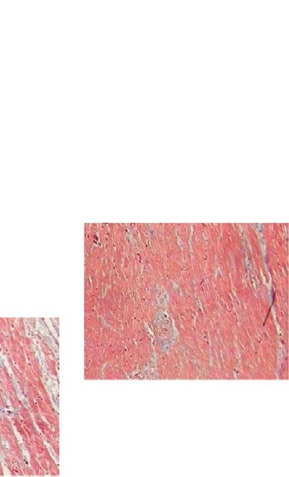
Cardiovascular Therapeutics 3
Nec-1
Vehicle
(a) (b)
18 ⁎⁎
Myocardial Infarction (% LVA)
15
12
9
6
3
0
Vechicle Necrostatin-1
(c)
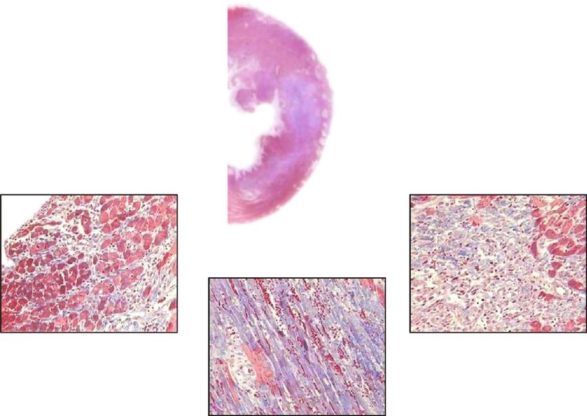
Figure 1: Nec-1 protects adult rats from myocardial ischemia/reperfusion injury. (a, b) Representative Masson’s trichrome staining in adult
rat hearts subjected to ischemia for 30 min and reperfusion for 72 h, respectively, treated with DMSO or Nec-1 at the onset of reperfusion.
Reactive fibrosis for myocardial injury is stained in blue and expands from the endocardium to the epicardium. In contrast with the
control, the administration of Necrostatin-1 can obviously reduce the extent of myocardial injury from the endocardium to the
epicardium. (c) Graph shows myocardial infarct size measured by Masson’s trichrome staining at 72 h of reperfusion. All data were
expressed as mean ± SD; n = 10 for the vehicle group and n = 9 for the Nec-1 group. Scale bar, 1 mm and 40 μm. ∗∗ P < 0:01 Nec-1 group
vs. vehicle group. LVA: left ventricular area.
kinase (CK) were determined using a diagnostic kit (Jian- 2.7. Statistics. Results are expressed as a mean ± standard
cheng, Nanjing, China). deviation. The statistics were calculated using a two-group t
-test (unpaired, one- or two-sided). The correlation between
2.6. Western Blotting Assay. The expression of MAP-LC3β the two variables was analyzed by single linear regression
(microtubule-associated protein light chain 3β, LC3β) was analysis with SPSS 15.0 statistics software. Values of P <
examined by western blot. Proteins from a tissue homogenate 0:05 were considered to be statistically significant.
were separated by SDS-PAGE and electroblotted onto nitro-
cellulose. The blots were blocked overnight in TBS with 3% 3. Results
BSA (bovine serum albumin) and then probed with rabbit
anti-LC3β antiserum (1 : 3,000, Santa Cruz sc-28266) at 4°C 3.1. Effect of Nec-1 in Late Reperfusion after Myocardial
overnight while agitated. The blots were washed once in Ischemia. The histomorphological changes and myocardial
TBST (TBS with 0.05% Tween 20) and twice for 5 min in infarct size at 72 h after reperfusion were examined to deter-
TBS followed by incubation for 1 h at room temperature with mine the effects of Nec-1 in rats subjected to myocardial
HRP-conjugated Goat Anti-Rabbit IgG Antibody (1 : 3,000, ischemia and late reperfusion. In anesthetized S-D rats sub-
Zhongshan Jinqiao, China). The blots were washed three jected to I/R protocol, the Nec-1 (0.6 mg/kg) that was admin-
times for 5 min in TBST and developed with a chemilumines- istered at the onset of reperfusion significantly reduced
cence detection kit (Pierce). The immunoblotting was scanned reactive myocardial fibrosis from the endocardium to the epi-
with an HP scanner, and the blot densities were analyzed with cardium and the infarct size from 15:1 ± 1:7% (vehicle
Image-Pro Plus 5.0 software. The relevant calculation formula group) to 5:3 ± 2% (P < 0:01; Figure 1) by Masson’s tri-
was myocardial infarction = LV infarct zone/LV area × 100%. chrome stains [5].4 Cardiovascular Therapeutics
Vehicle Nec-1
(a) (b)
Figure 2: Ultrastructure of the myocardial infarct area under a transmission electron microscope at 48 h reperfusion after ischemia (x8000).
(a) Electron micrographs of hearts subjected to I 30 min/R 48 h and treated with DMSO show massive vacuoles in myocardial tissue. (b)
Electron micrographs of hearts subjected to I 30 min/R 48 h and treated with Nec-1+DMSO show a few autophagosomes, but the
myocardial ultrastructure has more integrity than the vehicle group. Arrows indicate autophagosomes. n = 10 for the vehicle group and n
= 9 for the Nec-1 group.
Through observing LV tissue at 48 h after reperfusion ulated compared to the control group. Treatment with Nec-1
with a transmission electron microscope, it was further found resulted in a marked reduction in expression levels of LC3β
that myocardial ultrastructure in the vehicle group had been at 12 and 24 h after reperfusion but had no significant
severely damaged; cardiomyocytes were disintegrated and changes at 48 and 72 h after reperfusion, compared with the
had formed massive vacuoles but almost no autophago- relative vehicle group (Figures 4(a) and 4(b)). And interest-
somes. Inversely, treatment with Nec-1 can maintain myo- ingly, expression levels of LC3β in the Nec-1 group were sig-
cardial ultrastructural integrity and markedly reduce the nificantly upregulated compared with the vehicle group at 48
loss of cardiomyocytes. However, there were a few autopha- h after reperfusion. This result may explain why the number
gosomes with diffuse distribution in some cardiomyocytes of autophagosomes observed under the transmission electron
(Figure 2). Thus, Nec-1 can not only salvage left ventricular microscope at 48 h after reperfusion in the Nec-1 group was
(LV) tissue from ultimate death but also inhibit LV remodel- higher than in the relative vehicle group.
ing processes by reducing myocardial cell death.
3.4. The Results of CK Activity after Treatment with Nec-1. As
3.2. Cardiac Functional Assessment at Different Time Points an essential indicator of myocardial damage, serum CK levels
after I/R In Vivo. Consistent with previous studies, myocardial were monitored at different time points after I/R. Treatment
ischemia and reperfusion resulted in a significant decrease in with Nec-1 significantly reduced serum CK levels at 12 and
cardiac function (Figure 3). As shown in Figure 3, treatment 24 h after reperfusion, compared with the relative vehicle
with Nec-1 markedly improved LVDP, LVEDP, −dp/dtmax, group (722 ± 111 and 929 ± 271 U/L versus 1,433 ± 124 and
and +dp/dtmax (Figures 3(a)–3(d)). Specifically, the LVDP 1,438 ± 174 U/L, P < 0:01 and P < 0:05, respectively), while
and LVEDP (each at 12 and 48 h after reperfusion) of rats CK levels in the control group were 232 ± 34 U/L
treated with Nec-1 at the onset of reperfusion were signifi- (Figure 4(c)). However, treatment with Nec-1 could not
cantly lower than those of rats treated with the vehicle diminish serum CK levels at 48 and 72 h after reperfusion;
(Figures 3(a) and 3(b)); −dp/dtmax and +dp/dtmax (each inversely, serum CK levels of the Nec-1 group at 48 h after
at 12 h after reperfusion) of rats treated with Nec-1 at the reperfusion had markedly increased, compared with those
onset of reperfusion were significantly higher than those of of the relative vehicle group (559 ± 175 versus 1,238 ± 158,
rats treated with the vehicle (Figures 3(c) and 3(d)). Notably, P < 0:01).
augmented LVDP and LVEDP in the vehicle group likely Because the trend of immunoblotting showed a striking
result from alterations in myocardial compliance due to the similarity to the serum CK levels, in which Nec-1 reduced
larger area of myocardial infarction. Treatment with Nec-1 the value less than 12 h after reperfusion and delayed the
can markedly diminish the alteration. peak to 48 h after reperfusion, it was decided to investigate
the correlation between them. The results showed that,
3.3. The Changes of LC3β at Different Time Points in the WB although these parameters of MI/R existed with interindivid-
Assay. Autophagy is a cellular degradation process responsi- ual variation, the expression of MAP-LC3β was significantly
ble for the turnover of unnecessary or dysfunctional organ- correlated with the serum CK levels (r = 0:89, P < 0:001,
elles and cytoplasmic proteins. In the process, cytoplasmic Figure 4(d)). These results demonstrated that the necroptosis
proteins or dysfunctional organelles are sequestrated in a induced by MI/R delayed autophagy-like cell death and
double-membrane-bound vesicle, termed autophagosome, played a vital role in MI/R.
delivered to the lysosome by fusion as an autolysosome and
then degraded [25]. LC3β was localized in the autophago- 4. Discussion
some or autolysosome membrane and identified as an excel-
lent marker of autophagic activity [13, 18–20, 26]. Through This study demonstrated that Nec-1 (0.6 mg/kg) could main-
determining myocardial levels of LC3β at different time tain myocardial ultrastructural integrity and markedly
points after I/R in vivo, we found that the expression levels reduce myocardial infarct size and reactive fibrosis at late
of LC3β at all time points after I/R were significantly upreg- reperfusion after MI. Additionally, LVDP and LVEDP, asCardiovascular Therapeutics 5
1.5 ⁎⁎
1
⁎
LVDP (kPa)
0.5
0
–0.5
–1
Baseline I30 min R12 h R24 h R48 h R72 h
Vehicle
Nec-1
(a)
4
⁎⁎
3 ⁎
LVEDP (kPa)
2
1
0
Baseline I30 min R12 h R24 h R48 h R72 h
Vehicle
Nec-1
(b)
800
700
600
−dp/dtmax (kPa/s)
⁎⁎
500
400
300
200
Baseline I30 min R12 h R24 h R48 h R72 h
Vehicle
Nec-1
(c)
Figure 3: Continued.6 Cardiovascular Therapeutics
1000
800
+dp/dtmax (kPa/s)
600
⁎
400
200
Baseline I30 min R12 h R24 h R48 h R72 h
Vehicle
Nec-1
(d)
Figure 3: Assessment of cardiac function in vehicle and Nec-1-treated rats at different time points (12 h, 24 h, 48 h, and 72 h) during MI/R: (a)
LVDP; (b) LVEDP; (c) −dp/dtmax; (d) +dp/dtmax in different groups. n = 8 mice for baseline; for vehicle group: I30min n = 10, R12h n = 10,
R24h n = 10, R48h n = 10, R72h n = 10; for Nec-1 group: I30min n = 10, R12h n = 10, R24h n = 10, R48h n = 10, R72h n = 10. All data were
expressed as mean ± SD. ##P < 0:01, baseline vs. vehicle group (I30 min). ∗ P < 0:05 or ∗∗ P < 0:01, Nec-1 group vs. vehicle group
(I30min/R12h, 24 h, 48 h, 72 h).
essential determinants of LV remodeling, were also signifi- poptosis pathway characterized by autophagy, and considered
cantly lower in the Nec-1 group than in the vehicle group. autophagy as a downstream consequence of necroptosis
Thus, we confirmed that Nec-1 (0.6 mg/kg) could protect rather than a contributing factor to necroptotic cell death in
the myocardium against I/R injury in rats by reducing the cerebral I/R injury [18]. Because both necroptosis and
loss of myocardial cells and LV remodeling. Moreover, it also autophagy are mechanisms of delayed ischemic brain injury,
found that Nec-1 at higher concentrations (1.8 mg/kg) would we observed the alterations of LC3β expression levels and
increase the mortality rate of rats subjected to chronic myo- serum CK levels, respectively, at 12, 24, 48, and 72 h reperfu-
cardial ischemia (data not shown). The results indicated that sion after MI, and found that the administration of Nec-1 at
Nec-1 was cardioprotective at lower concentrations but had the onset of reperfusion significantly reduced the release of
harmful actions with increased concentrations, which creatine kinase and downregulation of autophagy at 12 and
appears to coincide with the report of C. C. Smith et al. [24]. 24 h after reperfusion, but the effects were not sustained to
Another interesting work was to determine the feature of 48 and 72 h after reperfusion. A correlation exists between
necroptosis in MI/R. Autophagy is a highly conserved cellu- the release of creatine kinase and the expression of autoph-
lar mechanism of protein recycling that may lead to pro- agy. Administering Nec-1 to coincide with reperfusion could
grammed cell death (type II programmed cell death) [11, decrease and delay the peak to 48 h after reperfusion. These
12, 14, 27]. Therefore, autophagy plays a dual role as a cell- data are similar to those of Zhao et al., who demonstrated
survival pathway and as an intrinsic cell death mechanism that infarct size increased to a peak at 24 h of reperfusion
under some circumstances. As a terminally differentiated with no further increase at 48 and 72 h of reperfusion [29],
cell, the cardiomyocyte is thought to be more sensitive to consistent with the peak of autophagic activity. This evidence
autophagy than other cell types. Some studies have shown provides strong support for our hypothesis that necroptosis
that autophagy is a protective mechanism in chronic ische- is not only an autophagy-like cell death but is also an essen-
mia [20, 21], and inhibiting autophagy resulted in a signifi- tial contribution to delayed reperfusion injury after myocar-
cant aggravation of cardiomyocytes in I/R injury. Inversely, dial ischemia.
enhancing autophagy has an underlying protective response This study confirmed that the myocardial protective
against I/R injury in heart cells [20]. Recently, the effect of effect of Necrostatin-1 in rat myocardial I/R injury is related
autophagy in mediating cell survival and death in MI/R to autophagy. However, the effective dose of the drug and the
injury is still controversial [26]. It has been considered that related pharmacological mechanism are not clear. To further
the upstream mechanism of autophagy could be a critical determine the effect of Nec-1, additional research can explore
event in mediating cell survival and death during ischemia the lowest effective dose, the best therapeutic dose and side
and reperfusion in the heart. However, autophagy is consid- effects of the drug, and the molecular biological mechanisms
ered as the end of many paths [27, 28]; thus, it is difficult to related to Nec-1.
judge which pathway will be essential. In summary, this research demonstrated that the adminis-
In 2005, L. Yan et al. discovered a small molecule called tration of Nec-1 inhibited an autophagy-like cell death that
Necrostatin-1 (Nec-1), which can inhibit necroptosis, a nona- may play an important role during delayed reperfusion injuryCardiovascular Therapeutics 7
2 ##
⁎⁎
1.6
⁎⁎
LC3II/actin
12 h 24 h 48 h 72 h 1.2
Ischemia 30 min/reperfusion + + + + + + + +
Nec-1 + – + – + – + – 0.8 ⁎⁎
LC3 0.4
Actin
0
Control R12 h R24 h R48 h R72 h
MI/R+DMSO+Nec-1
MI/R+DMSO
(a) (b)
2000
⁎⁎ 1700
⁎⁎
1500
CK activity (U/L)
1300
CK activity (U/L)
1000
⁎⁎ 900 r = 0.89, p < 0.001
500 500
0 100
Control R12 h R24 h R48 h R72 h 0.2 0.4 0.6 0.8 1 1.2 1.4 1.6
LC3 II/actin
MI/R+DMSO
MI/R+Nec-1
MI/R+DMSO+Nec-1
MI/R+Vehicle
(c) (d)
Figure 4: Detection and analysis of correlation between expression of MAP-LC3β in infarct zone and serum CK levels at different time points
(12 h, 24 h, 48 h, and 72 h) during MI/R. (a) Representative immunoblot of LC3β protein in infarct zone (the tissue of the infarct zone is pale)
of vehicle and Nec-1-treated rats at different time points (12 h, 24 h, 48 h, and 72 h) during MI/R. (b) Expression of LC3β protein in the infarct
zone of vehicle and Nec-1-treated rats at different time points (12 h, 24 h, 48 h, and 72 h) during MI/R. Each column represents the ratio of the
gray value of LC3β to IOD (actin). ∗∗ P < 0:01, Nec-1 group vs. vehicle group. ##P < 0:01, Nec-1 group (R12h) vs. Nec-1 group (R24h). (c)
Measurement of serum CK levels on rats subjected to 12 h, 24 h, 48 h, and 72 h reperfusion after myocardial ischemia for 30 min. Comparing
the difference between the Nec-1 group and the vehicle group at different time points. Each column represents mean ± SD. ∗∗ P < 0:01, Nec-1
group vs. vehicle group. (d) Relationship between LC3β immunoreactivity (% control) and serum CK levels (U/L) at different time points
(12 h, 24 h, 48 h, and 72 h) during MI/R. Analysis of correlation shows that LC3β immunoreactivity was highly correlated with serum CK
levels (r = 0:89, P < 0:001). All data were expressed as mean ± SD, n = 8 mice for baseline; for the vehicle group: I30min n = 10, R12h n = 10,
R24h n = 10, R48h n = 10, R72h n = 10; for the Nec-1 group: I30min n = 10, R12h n = 10, R24h n = 10, R48h n = 10, R72h n = 10.
after myocardial ischemia, and it significantly improved car- Conflicts of Interest
diac function after myocardial ischemia and reperfusion, sug-
gesting that antinecroptosis treatment may improve the The authors declare that they have no conflicts of interest.
clinical outcomes of patients with ischemic heart disease.
However, the mechanism of autophagy-like cell death needs
to be investigated further. Thus, in the following study, Nec- Authors’ Contributions
1 of binding targets will be identified, and other therapeutic
strategies explored to reduce myocardial cell death responsible Liang Wang and Xuebai Lv contributed equally to this work.
for myocardial I/R injury.
Data Availability Acknowledgments
The datasets used and analyzed during the current study are This work was supported by the grants from the National
available from the corresponding author on reasonable Natural Science Foundation of China (Nos. 30572084,
request. 30670835, and 30700276).8 Cardiovascular Therapeutics
References [18] L. Yan, D. E. Vatner, S. J. Kim et al., “Autophagy in chronically
ischemic myocardium,” PNAS, vol. 102, no. 39, pp. 13807–
[1] F. Poustchi, H. Amani, Z. Ahmadian et al., “Combination ther- 13812, 2005.
apy of killing diseases by injectable hydrogels: from concept to [19] L. Yan, J. Sadoshima, D. E. Vatner, and S. F. Vatner, “Autoph-
medical applications,” Advanced Healthcare Materials, vol. 10, agy: a novel protective mechanism in chronic ischemia,” Cell
no. 3, article e2001571, 2021. Cycle, vol. 5, no. 11, pp. 1175–1177, 2006.
[2] D. M. Yellon and D. J. Hausenloy, “Myocardial reperfusion [20] A. Hamacher-Brady, N. R. Brady, and R. A. Gottlieb, “Enhanc-
injury,” New England Journal of Medicine, vol. 357, no. 11, ing macroautophagy protects against ischemia/reperfusion
pp. 1121–1135, 2007. injury in cardiac myocytes,” The Journal of Biological Chemis-
[3] H. M. Piper, Y. Abdallah, and C. Schäfer, “The first minutes of try, vol. 281, no. 40, pp. 29776–29787, 2006.
reperfusion: a window of opportunity for cardioprotection,” [21] K. E. Larsen and D. Sulzer, “Autophagy in neurons: a review,”
Cardiovascular Research, vol. 61, no. 3, pp. 365–371, 2004. Histology and Histopathology, vol. 17, no. 3, pp. 897–908, 2002.
[4] X. Wang, F. Liang, X. Jiao et al., “Diverse effects of L-arginine [22] H. D. Sybers, J. Ingwall, and M. DeLuca, “Autophagy in car-
on cardiac function of rats subjected to myocardial ischemia diac myocytes,” Recent Advances in Studies on Cardiac Struc-
and reperfusion in vivo,” Acta Biochimica et Biophysica Sinica, ture and Metabolism, vol. 12, pp. 453–463, 1976.
vol. 39, no. 3, pp. 201–207, 2007.
[23] B. H. Chua, J. P. Gao, and C. C. Chua, “Necrostatin-1 is a novel
[5] E. Toyota, Y. Kawaguchi, Y. Ogasawara et al., “Novel rat model protector of myocardial infarction,” Circulation, vol. 114,
of ischemic cardiomyopathy induced by repetitive myocardial p. 212, 2006.
ischemia/reperfusion injury while conscious,” Circulation
[24] C. C. Smith, S. M. Davidson, S. Y. Lim, J. C. Simpkin, J. S.
Journal, vol. 71, no. 5, pp. 788–795, 2007.
Hothersall, and D. M. Yellon, “Necrostatin: a potentially novel
[6] A. Haunstetter and S. Izumo, “Toward anti-apoptosis as a new cardioprotective agent?,” Cardiovascular Drugs and Therapy,
treatment modality,” Circulation Research, vol. 86, no. 4, vol. 21, no. 4, pp. 227–233, 2007.
pp. 371–376, 2000.
[25] D. J. Klionsky and S. D. Emr, “Autophagy as a regulated path-
[7] F. Gao, L. Tao, W. Yan et al., “Early anti-apoptosis treatment way of cellular degradation,” Science, vol. 290, no. 5497,
reduces myocardial infarct size after a prolonged reperfusion,” pp. 1717–1721, 2000.
Apoptosis, vol. 9, no. 5, pp. 553–559, 2004.
[26] H. Takagi, Y. Matsui, and J. Sadoshima, “The role of autoph-
[8] M. Clarke, M. Bennett, and T. Littlewood, “Cell death in the agy in mediating cell survival and death during ischemia and
cardiovascular system,” Heart, vol. 93, no. 6, pp. 659–664, reperfusion in the heart,” Antioxidants & Redox Signaling,
2007. vol. 9, no. 9, pp. 1373–1382, 2007.
[9] J. D. McCully, H. Wakiyama, Y. J. Hsieh, M. Jones, and [27] A. M. Cuervo, “Autophagy: many paths to the same end,”
S. Levitsky, “Differential contribution of necrosis and apopto- Molecular and Cellular Biochemistry, vol. 263, no. 1/2,
sis in myocardial ischemia-reperfusion injury,” American pp. 55–72, 2004.
Journal of Physiology. Heart and Circulatory Physiology,
[28] K. Asanuma, I. Tanida, I. Shirato et al., “MAP-LC3, a promis-
vol. 286, no. 5, pp. H1923–H1935, 2004.
ing autophagosomal marker, is processed during the differen-
[10] P. Vandenabeele, T. Vanden Berghe, and N. Festjens, “Caspase tiation and recovery of podocytes from PAN nephrosis,” The
inhibitors promote alternative cell death pathways,” Science's FASEB Journal, vol. 17, no. 9, pp. 1165–1167, 2003.
STKE, vol. 2006, no. 358, p. pe44, 2006.
[29] Z. Q. Zhao, M. Nakamura, N. P. Wang et al., “Dynamic pro-
[11] L. Yu, A. Alva, H. Su et al., “Regulation of an ATG7-beclin 1 gression of contractile and endothelial dysfunction and infarct
program of autophagic cell death by caspase-8,” Science, extension in the late phase of reperfusion,” The Journal of Sur-
vol. 304, no. 5676, pp. 1500–1502, 2004. gical Research, vol. 94, no. 2, pp. 133–144, 2000.
[12] L. Yu, F. Wan, S. Dutta et al., “Autophagic programmed cell
death by selective catalase degradation,” PNAS, vol. 103,
no. 13, pp. 4952–4957, 2006.
[13] A. Degterev, Z. Huang, M. Boyce et al., “Chemical inhibitor of
nonapoptotic cell death with therapeutic potential for ischemic
brain injury,” Nature Chemical Biology, vol. 1, no. 2, pp. 112–
119, 2005.
[14] M. W. Knaapen, M. J. Davies, M. de Bie, A. J. Haven,
W. Martinet, and M. M. Kockx, “Apoptotic versus autophagic
cell death in heart failure,” Cardiovascular Research, vol. 51,
no. 2, pp. 304–312, 2001.
[15] Y. Tsujimoto and S. Shimizu, “Another way to die: autophagic
programmed cell death,” Cell Death and Differentiation,
vol. 12, no. S2, pp. 1528–1534, 2005.
[16] Y. Uchiyama, M. Shibata, M. Koike, K. Yoshimura, and
M. Sasaki, “Autophagy-physiology and pathophysiology,” His-
tochemistry and Cell Biology, vol. 129, no. 4, pp. 407–420, 2008.
[17] W. Bursch, “The autophagosomal-lysosomal compartment in
programmed cell death,” Cell Death and Differentiation,
vol. 8, no. 6, pp. 569–581, 2001.You can also read