Cleidocranial dysplasia syndrome with epilepsy: a case report - BMC Pediatrics
←
→
Page content transcription
If your browser does not render page correctly, please read the page content below
Ma et al. BMC Pediatrics     (2019) 19:97
https://doi.org/10.1186/s12887-019-1472-0

 CASE REPORT                                                                                                                                     Open Access

Cleidocranial dysplasia syndrome with
epilepsy: a case report
Yimei Ma1,3 , Fumin Zhao2,3 and Dan Yu1,3*

  Abstract
  Background: Cleidocranial dysplasia is a rare autosomal dominant disorder resulting in skeletal and dental
  abnormalities due to the disturbance in ossification of the bones. The prevalence of CCD is one in a million of live
  births, and epileptic seizures are rarer in this disease.
  Case presentation: Herein, we present a case of a 10-year-old girl, who not only suffered with cleidocranial
  dysplasia, but experienced frequent seizures. We initiated an anti-epileptic treatment for this patient with dose
  adjustments to her weight of levetiracetam (10 mg/kg, bid) for 3 months. The epileptic seizures were controlled, but
  the intelligence level and control of epilepsy need to be followed up for a longer duration.
  Conclusions: In clinical practice, if a patient has unusual facies, typical clavicle defect, skull bone enlargement, and
  unclosed anterior fontanelle, we should consider the possibility of cleidocranial dysplasia, genetic detection are
  helpful to make a confirmed diagnosis. In such cases, early diagnosis and treatment is important to correct
  deformities and improve the quality of life of patients.
  Keywords: Cleidocranial dysplasia, RUNX2

Background                                                                            tract and ear canal [3, 4]. The incidence of the disease is
Cleidocranial dysplasia is a rare autosomal dominant                                  very low, and combined with seizures are rarer. In this
hereditary skeletal disease (MIM number is 600211). A                                 paper, the clinical data of a patient with CCD accompan-
few of these cases were familial and most were sporadic.                              ied by epileptic seizure were reported. Laboratory, imaging
There was no significant difference in the incidence be-                              studies and related literature were reviewed to discuss the
tween males and females, and the clinical incidence was                               clinical manifestations and genetic characteristics of the
1:1000000 [1]. The main manifestations of CCD are sys-                                disease to improve the clinician’s understanding and
temic skeletal and teeth dysplasia. The typical symptoms                              knowledge in regard to this specific congenital disorder.
include: incomplete or absent development of one or
both clavicles, the formation of pseudo joints, shoulders                             Case presentation
drooping, shoulder joint hypermobility; delayed closure                               A 10 years and 6 months old girl, was admitted to hos-
or unclosed anterior fontanelle, widening of cranial                                  pital due to “epileptic seizures for five months”, which
sutures, cranial ectasia; retention of deciduous teeth,                               manifested as an involuntary nodding movement accom-
delayed sprouting of permanent teeth, malformation of                                 panied by loss of consciousness, with no fever, limb stiff-
roots and tooth cysts with supernumerary teeth [2]. Sys-                              ness, cyanosis, salivation and incontinence. During the
temic manifestations include: pigeon chest or conical                                 early period of illness, these symptoms lasted for about
shape of chest, smaller scapula, widened pubic symphy-                                10 s - with a frequency of about 2 episodes per day.
sis gap, Short stature, repeated infection of respiratory                             Then it gradually increased to 30 s to 1 min before spon-
                                                                                      taneous cessation, with a frequency of about 4–5 times a
* Correspondence: yd540@126.com
1
 Department of Pediatrics, West China Second University Hospital, Sichuan             day. The patient was treated with “carbamazepine and
University, Chengdu, Sichuan 610041, People’s Republic of China                       vitamin B6” but there was no obvious improvement in
3
 Key Laboratory of Birth Defects and Related Diseases of Women and                    symptoms or progression of illness. Physical examination
Children (Sichuan University), Ministry of Education, Chengdu 610041,
Sichuan, China                                                                        done at time of admission: T 36.8 °C, P 89 beats /min, R
Full list of author information is available at the end of the article                19 beats / min, BP 109/68 mmHg. Weight 27 kg, height
                                        © The Author(s). 2019 Open Access This article is distributed under the terms of the Creative Commons Attribution 4.0
                                        International License (http://creativecommons.org/licenses/by/4.0/), which permits unrestricted use, distribution, and
                                        reproduction in any medium, provided you give appropriate credit to the original author(s) and the source, provide a link to
                                        the Creative Commons license, and indicate if changes were made. The Creative Commons Public Domain Dedication waiver
                                        (http://creativecommons.org/publicdomain/zero/1.0/) applies to the data made available in this article, unless otherwise stated.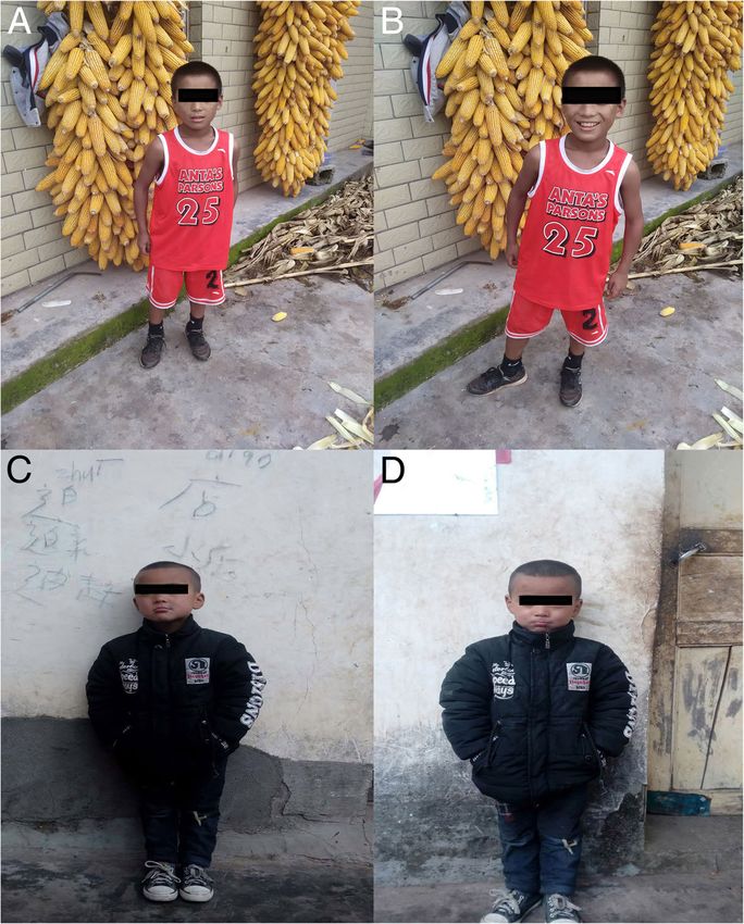
Ma et al. BMC Pediatrics     (2019) 19:97                                                                                              Page 2 of 6

125 cm. The patient is positive for special type of facies-                answer simple questions but cannot add or subtract
witha flat nose, wide-set eyes, micrognathia, deciduous                    within 10. The funnel chest was found a week after birth,
and misaligned teeth, and 9 maxillary and mandibular                       and the anterior fontanelle was not closed at 2 years old,
teeth. Head circumference is 52 cm.The anterior fonta-                     none of them were treated. The child has two brothers,
nelle is open, approximately 4 × 4 cm size, and soft on                    both healthy, with no obvious abnormalities in develop-
palpation. The sagittal and coronal sutures are unclosed.                  ment (Fig. 2). laboratory examination: blood routine:
The width of the sagittal suture is about 6 cm. The width                  WBC 4.6 × 10⁹/L, N 34.9%, HB 134 g/L, PLT 281 × 1012/L;
of the coronal suture is about 0.5 cm. They are all soft                   Blood ammonia: 40 umol/L; Vitamin D determination:
and flat, without tenderness. No résistance was felt in                    12.8 ng/ml; Kidney and kidney function, electrolytes,
the neck. Defects can be observed in the right clavicle,                   blood gas analysis, glucose, pyruvate, β - hydroxybutyrate,
bilateral shoulders can reach the midline. The thoracic                    calcium/phosphate/alkaline phosphatase, urination and
cavity hollows and changes like a funnel. A brown patch                    defecation were both normal. Electrocardiogram is normal
which is approximately 2.5 × 1.0 cm in size is visible on                  overall. X-ray examination showed: Asymmetry in Bilat-
the left wrist and a light brown patch of approximately                    eral clavicles, a third of the right lateral clavicle is absent.
3.5 × 4.0 cm is visible on the right wrist. The double knee                The cranium is higher, the cranial suture are widened,
valgus is deformed in an “X” shape with no limitation of                   and the anterior fontanelle was not closed. The bone
activity. There were no abnormalities in the spine and                     structure of the cranial plate is not completely devel-
joints, and the muscle strength and muscle tones of the                    oped. There are many suture bones in the skull. (Fig. 3).
limbs were normal. The pathological reflex were nega-                      Video EEG: Abnormal: multi-focal sharp wave, sharp
tive (Fig. 1). The child raised his head at 3 months old,                  and slow wave were frequent issued (Fig. 4). Skull clav-
crawled and rolled over at 7 months old, walked alone                      icle dysplasia syndrome RUNX2 gene sequencing sug-
and called daddy and Mommy at 1 year old. At present,                      gested RUNX2 c.947delA p. (His316fs) heterozygous,
she is in the third grade of elementary school, she can                    pathogenic frameshift mutations (Fig. 5).

 Fig. 1 Physical examination showed obvious abnormal development in patient. (a: unusual facies; b: right shoulder towards the midline; c & d:
 funnel chest)

Ma et al. BMC Pediatrics     (2019) 19:97                                                                                                Page 3 of 6

 Fig. 2 No obvious abnormalities in the development of siblings. Brother. (a & b: elder brother; c & d: younger brother)

 Fig. 3 Plain radiographs showing multiple skeletal abnormalities. a: chest radiograph showing bilateral asymmetry clavicles with partial absence
 of the right; b & c: anteroposterior and lateral cephalograms showing the broad sagittal and coronal sutures, delayed closure of the anterior
 fontanel, numerous wormian bones, hypoplastic maxilla and unerupted permanent teeth

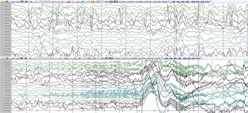
Ma et al. BMC Pediatrics     (2019) 19:97                                                                                               Page 4 of 6

 Fig. 4 Electroencephalogram: abnormal school-age electroencephalogram: multi-focal cusp, sharp and slow wave were frequently issued., The right
 central, top, and mid-temporal regions show pointed and spiked rhythms repeatedly released, which is considered as complex partial seizures

 Fig. 5 Sequencing of the RUNX2 gene: c.947delA p. (His316fs) . This disease is an autosomal dominant inheritance. The child has a heterozygous
 mutation whose parents are normal. This mutation is a frameshift mutation (the RUNX2 translation protein has a disorder in the coding of the
 amino acid residue His at position 316). This mutation is expected to cause the encoded protein to be truncated and lose its normal function.
 (a: reference; b: patient; c: the father; d:the mother)

Ma et al. BMC Pediatrics   (2019) 19:97                                                                                             Page 5 of 6

Discussion and conclusions                                      were case reports which had been published over 10 years
CCD is also known as Marie and Sainton ‘s disease,              ago. B. M. Tress’s research found that among the patients
Schlotzhauer-Marie-Sainton syndrome, osteogenesis               with epilepsy, there were 3 cases in which this was sec-
imperfecta, which was first described by Pierre Marie           ondary to an abscess, 2 with cerebral angiomas and 2 with
and Paul Sainton in 1898 [5]. RUNX2 gene is a CCD               telangiectasia [15]. Oyer, CE reported a 15-day-old female
gene that is located on chromosome 6p21 and is a 130            neonate with CCD. Her postnatal course was character-
kb length protein. It encodes the transcription factors re-     ized by seizures and recognition of hydrocephalus during
quired for chondrocyte proteins and dentin, influences          the first day of life [16].
the differentiation and maturation of osteoblasts, and            This patient had no family history of epilepsy, but cra-
affects intramembranous and endochondral ossification           nial MRI suggested sequela of brain injury. So the eti-
[4, 6–8]. In our case, the sequencing of the RUNX2 gene         ology of epilepsy maybe brain damage. Consistent with
indicate c.947delA p. (His316fs) mutation in patient while      the literature, the occurrence of epilepsy may be related
her parents’ are normal. This mutation is a frameshift mu-      to brain structural abnormalities and damage. But the
tation (the RUNX2 translation protein has a disorder in         number of cases are limited, further research is necessary
the coding of the amino acid residue His at position 316).      in the future. After the patient was admitted to the
This mutation is expected to cause the encoded protein to       hospital, oral levetiracetam (10 mg/kg, bid) was used to
be truncated and lose its normal function. In addition to       control seizure and she received supportive treatment
the typical dystrophy of the cranial clavicle and various       [17, 18]. The intelligence level and control of epilepsy
vertebral and dental abnormalities, recurrent ear infec-        need to be followed up for a longer period of time [19–21].
tions and hearing loss are common in CCD due to max-              In clinical practice, if one patient has unusual facies, typ-
illofacial hypoplasia and osteogenic deficiencies [3]; most     ical clavicle defect, skull bone enlargement, and unclosed
patients have chest malformations, some severe cases can        anterior fontanelle, we should consider the possibility of
lead to an early respiratory distress in infants [4]; some      cleidocranial dysplasia, in such cases, early diagnosis and
case also indicate that CCD patient are more likely to          treatment is important to correct deformities and improve
suffer from hematological disease [9–11]. Osteopenia is         the quality of life of patients.
present in most patients due to osteogenesis and ossifica-
                                                                Abbreviations
tion [5]; CCD patients are often of short stature, their        BP: Blood pressure; CCD: Cleidocranial dysplasia; P: Pulse; R: Respiratory;
birth length is usually normal, but their height is below       T: Temperature
the two percentiles between 4 and 8 years old. The final
average height of the male is 165 cm (±8 cm) and the fe-        Acknowledgements
                                                                The authors thank the patient’s parents for providing permission to use the
male is 156 cm (±10 cm) [12]. The motor development             information of their children, and also thank Dr. Mowshica Rajah for the
may be of slightly delayed onset (age of walking), but in       English revise.
most cases, mental development is normal [13].
                                                                Author’ contributions
   This patient had a typical clavicle defect, skull bone en-   YMM, DY and FMZ analyzed and interpreted the patient data. YMM wrote
largement, and unclosed anterior fontanelle, with funnel        the draft of this article, and DY and FMZ revised this article. All authors read
chest which conforms to the typical CCD performance             and approved the final manuscript.
and the results of genetic sequencing, confirmed this diag-     Funding
nosis. Several neurological disorders associated with CCD       The study was supported by the Science and Technology Department of
have been reported such as mental retardation, spastic          Sichuan Province (NO. 2018SZ0123) for data collection and language polish.
paresis etc. [14]. Seizures associated with this disease are
                                                                Competing interest
very rare. The patient was diagnosed with epilepsy at 5         Each author declares that they have no competing interests, and certifies
years of age but was not on regular treatment. Most of the      that he has no commercial association (e.g. consultancies, stock ownership,
CCD patients had normal intelligence, but the mental de-        equity interest, patent/licensing arrangement, etc.) that might pose a conflict
                                                                of interest in connection with the submitted article.
velopment of this patient was significantly backward com-
pared with children of the same age. As a student of the        Ethics approval and consent to participate
third grade of elementary school, she cannot read and           This study was approved by the Ethics Committee of the West China Second
                                                                University Hospital and written informed consent was obtained from parents
cannot perform simple addition and subtraction in less          of the child.
than 10. Her cranial magnetic resonance imaging (MRI)
showed bilateral parietooccipital atrophy and periventricu-     Consent for publication
                                                                The study obtained informed written consent from the parents of the study
lar leukomalacia. According to her medical history, the         participants in order to publish their clinical details and clinical images.
mental development delay may be related to the brain
damage and poor control of epilepsy. Analysis of the
                                                                Publisher’s Note
Literature on CCD patients with epilepsy, we found that         Springer Nature remains neutral with regard to jurisdictional claims in
there were few related literatures. Most of the articles        published maps and institutional affiliations.
Ma et al. BMC Pediatrics (2019) 19:97 Page 6 of 6
Author details 20. Wakamoto H, Nagao H, Hayashi M, Morimoto T. Long-term medical,
1
Department of Pediatrics, West China Second University Hospital, Sichuan educational, and social prognoses of childhood-onset epilepsy: a
University, Chengdu, Sichuan 610041, People’s Republic of China. population-based study in a rural district of Japan. Brain Dev. 2000;
2
Department of Radiology, West China Second University Hospital, Sichuan 22(4):246–55.
University, Chengdu 610041, Sichuan, China. 3Key Laboratory of Birth Defects 21. Aldenkamp AP, Weber B, Overweg-Plandsoen WC, Reijs R, van Mil S.
and Related Diseases of Women and Children (Sichuan University), Ministry Educational underachievement in children with epilepsy: a model to predict
of Education, Chengdu 610041, Sichuan, China. the effects of epilepsy on educational achievement. J Child Neurol. 2005;
20(3):175–80.
Received: 1 November 2018 Accepted: 27 March 2019
References
1. Machol K, Mendoza-Londono R, Lee B. Cleidocranial Dysplasia Spectrum
Disorder. In: Adam MP, Ardinger HH, Pagon RA, Wallace SE, Bean LJH,
Stephens K, Amemiya A, editors. GeneReviews®. Seattle (WA); 1993.
2. Callea M, Bellacchio E, Di Stazio M, Fattori F, Bertini E, Yavuz I, Clarich G,
Gunay A. A case of cleidocranial dysplasia with peculiar dental features:
pathogenetic role of the RUNX2 mutation and long term follow-up. Oral
Health Dent Manage. 2014;13(2):548–51.
3. Cooper SC, Flaitz CM, Johnston DA, Lee B, Hecht JT. A natural history of
cleidocranial dysplasia. Am J Med Genet. 2001;104(1):1–6.
4. Mundlos S. Cleidocranial dysplasia: clinical and molecular genetics. J Med
Genet. 1999;36(3):177–82.
5. The classic: Marie P., and Sainton P.: Sur la dysostose cleido-cranienne
herediataire, Rev. neurol. 6:835, 1898. On hereditary cleido-cranial dysostosis.
Clin Orthop Relat Res 1968, 58:5–7.
6. Mundlos S, Otto F, Mundlos C, Mulliken JB, Aylsworth AS, Albright S,
Lindhout D, Cole WG, Henn W, Knoll JH, et al. Mutations involving the
transcription factor CBFA1 cause cleidocranial dysplasia. Cell. 1997;89(5):
773–9.
7. El-Gharbawy AH, Peeden JN Jr, Lachman RS, Graham JM Jr, Moore SR,
Rimoin DL. Severe cleidocranial dysplasia and hypophosphatasia in a child
with microdeletion of the C-terminal region of RUNX2. Am J Med Genet A.
2010;152A(1):169–74.
8. Gelb BD, Cooper E, Shevell M, Desnick RJ. Genetic mapping of the
cleidocranial dysplasia (CCD) locus on chromosome band 6p21 to include a
microdeletion. Am J Med Genet. 1995;58(2):200–5.
9. Callea M, Bellacchio E, Fattori F, Bertini E, Callea F, Cammarata-Scalisi F.
Acute myeloid leukemia in a 3 years old child with cleidocranial dysplasia.
Leuk Lymphoma. 2016;57(9):2189–91.
10. Kelkar PS, Ten RM. Association of humoral immunodeficiency, cleidocranial
dysplasia, and von Willebrand's disease in a family cluster. J Allergy Clin
Immunol. 2001;107(4):742.
11. Alexander WN, Ferguson RL. Beta thalassemia minor and cleidocranial
dysplasia: a rare combination of genetic abnormalities in one family. Oral
Surg Oral Med Oral Pathol. 1980;49(5):413–8.
12. Dincsoy Bir F, Dinckan N, Guven Y, Bas F, Altunoglu U, Kuvvetli SS,
Poyrazoglu S, Toksoy G, Kayserili H, Uyguner ZO. Cleidocranial dysplasia:
clinical, endocrinologic and molecular findings in 15 patients from 11
families. Eur J Med Genet. 2017;60(3):163–8.
13. Bharti K, Goswami M. Cleidocranial dysplasia: a report of two cases with
brief review. Intractable Rare Dis Res. 2016;5(2):117–20.
14. Nakahara I, Nozaki K, Ishikawa J. [a case of cleidocranial dysostosis
associated with arachnoid cyst]. No shinkei geka. Neurol Surg. 1987;15(11):
1241–6.
15. Tress BM, Mosely IF. Cleidocranial dysostosis, hereditary haemorrhagic
telangiectasia and epilepsy: a rare association. Neuroradiology. 1977;
12(4):233–6.
16. Oyer CE, Tatevosyants NG, Cortez SC, Hornstein A, Wallach M.
Cleidocranial dysplasia with neonatal death due to central nervous
system injury in utero: case report and literature review. Pediatr Dev
Pathol. 1998;1(4):314–8.
17. Weijenberg A, Brouwer OF, Callenbach PM. Levetiracetam
monotherapy in children with epilepsy: a systematic review. CNS
drugs. 2015;29(5):371–82.
18. Tekgul H, Gencpinar P, Cavusoglu D, Dundar NO. The efficacy, tolerability
and safety of levetiracetam therapy in a pediatric population. Seizure. 2016;
36:16–21.
19. Bailet LL, Turk WR. The impact of childhood epilepsy on neurocognitive and
behavioral performance: a prospective longitudinal study. Epilepsia. 2000;
41(4):426–31.You can also read