Cluster ion polymerization of serine and tryptophan, the water loss channel
←
→
Page content transcription
If your browser does not render page correctly, please read the page content below
Eur. Phys. J. D (2020) 74: 85
https://doi.org/10.1140/epjd/e2020-10014-y                                                    THE EUROPEAN
                                                                                              PHYSICAL JOURNAL D
    Regular Article

Cluster ion polymerization of serine and tryptophan, the water
loss channel?
Lukas Tiefenthaler1 , Jaroslav Kočišek2 , and Paul Scheier1,a
1
    Institut für Ionenphysik und Angewandte Physik, Universität Innsbruck, Technikerstr. 25, A-6020 Innsbruck, Austria
2
    J. Heyrovský Institute of Physical Chemistry of the Czech Academy of Sciences, Dolejškova 3, 18223 Prague, Czech Republic

            Received 8 January 2020 / Received in final form 27 February 2020
            Published online 1 May 2020
            c The Author(s) 2020. This article is published with open access at Springerlink.com

            Abstract. We present an experimental study on the stability of cluster cations formed by the assembly
            of tryptophan or serine moelcules inside charged helium droplets and subsequent droplet evaporation.
            The stability is probed via collision induced dissociation and mass spectrometry. We focus on the neutral
            loss of 18 Da that was previously proposed to result in the formation of an amide bond in amino acid
            clusters. We show that the cluster cations formed by the neutral loss of 18 Da from protonated parent
            cluster ion precursors are softly, presumably van der Waals, bound similar to the parent and protonated
            parent cluster cations. Cluster cations formed after neutral loss of 18 Da from the parent cluster cations
            are strongly bound, indicating the formation of new covalent bonds. Even though we observe a signal
            at m/z corresponding to dipeptide cations, their collision induced dissociation fragmentation does not
            allow unambiguous identification of their structure, particularly the amide bond. Our study at cryogenic
            conditions of He droplets demonstrates that peptide formation by the cluster ion polymerization is not
            a barrierless process and the formation of amide bonds may depend on activation methods and available
            catalysts.

1 Introduction                                                    concerning chirality or charge state gives important infor-
                                                                  mation about the molecular characteristics and energy
Cluster ions of amino acids have attracted significant            barriers that may drive the process.
interest during the last few years due to the high sta-              Previous studies of amino acid fragmentation indicate
bility of their selected homochiral clusters, particulary         that the formation of an amide bond will depend on how
the serine octamer [1–6] and its substituents [7,8]. Chiral       the precursor clusters are formed. After chemical ion-
selectivity of some amino acid clusters may be a key for          ization of amino acid dimers, prepared by the assembly
the Earth’s biomolecular homochirality [9–11]. The enan-          of sublimed monomers, Leclerq and Desiderio [20] sug-
tiomer selective assembly of amino acids opens new possi-         gested that the fragmentation channel corresponding to
bilities for chiral purification of amino acids or preparation    neutral loss of 18 Da from protonated cluster ions actu-
of polypeptides [12,13].                                          ally corresponds to ions with an amino group modified
   The formation of polypeptides via cluster ion polymer-         by the interaction with the side chain group of the sec-
ization is of particular importance. A breakthrough in this       ond constituent of the dimer and loss of the carboxyl
direction was made by the observation that, after acti-           group. This fragmentation mechanism was then confirmed
vation, amino acid cluster ions effectively lose a neutral        by another study [21]. On the other hand, high resolu-
water molecule [14,15], which indicates the formation of          tion mass spectrometry of cations formed after collision
the amide bond. UV activation of amino acid complexes             induced dissociation (CID) of amino acid clusters pre-
[16], ion/ion reactions [17] or coordination of amino acids       pared by electrospray demonstrated that water loss can
with Cu2+ on aerosol surfaces [18] were used to gain              be efficient, in agreement with the proposed mechanism
high cross sections for the formation of small peptides.          of amide bond formation [22]. Surprisingly, in the work
The importance of this reaction lies in the possibility of        of Singh [22], formation of dipeptides was not observed
artificially producing isolated homochiral peptides. Even         for serine, the most studied molecule in this context. This
though peptide building in nature is a complex biological         may be caused by the low intensity of the water loss chan-
process [19], fundamental insights into peptide stability         nel from serine and preferential charge localization on the
     ?
    Contribution to the Topical Issue “Atomic Cluster             water fragment [23]. Another important fact is that in
Collisions (2019)”, edited by Alexey Verkhovtsev, Pablo de        serine, water is preferentially released from the side chain
Vera, Nigel J. Mason, Andrey V. Solov’yov.                        and complex rearrangement will be needed for amide bond
  a
    e-mail: Paul.Scheier@uibk.ac.at                               formation [24].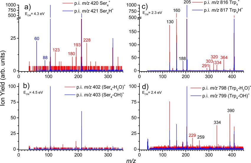
Page 2 of 7                                                                                      Eur. Phys. J. D (2020) 74: 85

                                Fig. 1. Sketch of the experimental apparatus and approach.

   It is, therefore, not completely clear how the cluster
structure and internal energy, activation methods or cata-
lysts (e.g. [18,25]) influence the cluster ion polymerization
of amino acids to form small peptides. Here we used a
novel He cluster ion assembly technique to prepare amino
acid cluster ions of yet another character. Using the CID
approach, we probe the stability of these precursor ions
(p.i.) as well as cations corresponding to neutral loss of
18 Da from nonprotonated or protonated parent cations.
   We selected two molecules for the study, L-serine
(Ser) containing an OH group on the side chain and
L-tryptophan (Trp) that does not contain an OH group on
the side chain. Another important difference of the stud-
ied amino acids is in their polarity. While Ser is polar, Trp
is nonpolar which may further influence the noncovalent
bonding character.
                                                                Fig. 2. Mass spectrum of positively charged ions formed upon
                                                                pickup of L-serine into charged He droplets. The dominant ions
                                                                can be assigned to protonated serine clusters (C3 H7 NO3 )n H+ .
2 Methods                                                       The numbers above selected peaks designate the cluster size n
                                                                of the corresponding ion.
The experiments were performed on a modified tandem
mass spectrometer Waters Q-TOF Ultima combined with
a novel cluster source developed in our laboratory and          He2 (IE = 22.22 eV) [27], Ser (IE ∼ 10 eV [28]) and Trp
recently described in [26]. A sketch of the setup is shown      (7.4 eV) [29], even excited charged states can be popu-
in Figure 1.                                                    lated, which enable dissociation. However, as known from
   The cluster ion assembly process was started by the for-     experiments ionizing neutral clusters inside He droplets
mation of a He droplet beam by expanding He gas at a            [30], these states can be quenched in the cold He envi-
stagnation pressure of 20 bar through a 5.7 µm diameter         ronment and for the most part molecular cluster ions are
nozzle cooled down to 8.9 K, into a first vacuum chamber        formed. In the case of amino acids the parent cations are,
of the setup (at ∼10−4 mbar under operation conditions).        however, metastable with respect to loss of a dehydro-
The droplets were multiply charged by electron ioniza-          genated amino acid radical and therefore the spectrum is
tion and a part of their m/z distribution was deflected by      dominated by protonated species [31].
a quadrupole bender towards the pickup cell containing             The amino acid cluster ions were released from the He
vapors of the studied amino acid. Cluster ions of amino         matrix by evaporation of the helium in the evaporation
acids were assembled inside the He droplets after pickup        cell of the instrument – a RF-hexapole filled with room
of their respective molecular precursors L-Serine (Sigma        temperature He gas. In simple MS mode, the cluster ions
Aldrich 99.5%) or L-Tryptophan (Sigma Aldrich 99.5%).           were transported to the reflectron time of flight mass spec-
   After pickup, charge transfer from He+   2 to the first      trometer and analyzed. This way the initial mass spectra
arriving dopant is highly exothermic. For the studied sys-      were obtained as depicted in Figures 2 and 3. Depending
tem with ionization energies for He(IE = 24.59 eV) and          on the pressure in the evaporation cell and the collision
Eur. Phys. J. D (2020) 74: 85                                                                                         Page 3 of 7

Fig. 3. Mass spectrum of positively charged ions formed upon
pickup of L-tryptophan into charged He droplets. The domi-
nant ions can be assigned to protonated tryptophan clusters
(C1 1H1 2N2 O2 )n H+ . The numbers above selected peaks desig-
nate the cluster size n of the corresponding ion.

energy, some He atoms may remain attached to the amino
acid clusters which is an indication of cold molecular clus-
ter ions [26]. The resulting cluster size distribution under
these gentle evaporation conditions is free of intensity         Fig. 4. Ion yield as a function of CID energy in the center of
anomalies or magic numbers, even for sodium cluster ions         mass frame for (a) M4 H+ and (b) M+  4 precursor ions. Depicted
that exhibit prominent intensity anomalies due to elec-          is always the precursor ion yield M4 H+ or M+
                                                                                                             4 , the yield of ions
tronic shell closures and spin pairing [32,33]. Pressures        formed by evaporation of monomer units M+   n
Page 4 of 7 Eur. Phys. J. D (2020) 74: 85
Trp+4 , while the order reverses in the case of protonated
cluster cations.
3.2 Water loss channel
The “water loss” channel was proposed to be the doorway
for peptide bond formation in small amino acid clusters.
However, it is worth mentioning that the m/z of ions form-
ing after 18 Da loss from protonated clusters is the same
as the m/z of ions forming after 17 Da loss from nonprot-
nated clusters and the initial formation step of these ions
cannot be unambiguously identified.
Figure 5 displays the CID energy dependence of frag-
mentation of the cluster cations. We can compare decom-
position (fragmentation+evaporation) for M+ 4 and M4 H
+
with the ions formed after neutral loss of 18 Da. The
slope of the M+ 4 and M4 H
+
ions represents the slope of
van der Waals cluster evaporation as we demonstrated
in Figure 4. We can see that (M4 –OH)+ decomposes the
same way while (M4 –H2 O)+ requires more energy to frag-
ment, which may indicate the presence of a covalently
bound complex. While this is consistent with previous
studies proposing cluster ion polymerization in amino acid
clusters, it is worth mentioning two facts: first, in our case
of reactions inside He droplets, the (Mn –H2 O)+ fragmen-
tation channel has very low intensity. Second, most of the
studies of amide bond formation inside amino acid clus-
ter ion cations were performed for protonated structures,
which are the van der Waals bound (M4 –OH)+ according
Fig. 5. Cluster ion stability, with respect to fragmentation to our results.
and evaporation, demonstrated by plotting the ratio of p.i. ion
yield to the total CID ion yield detected for a particular p.i.
as a function of the collision energy. Panel (a) shows Trp ions, 3.3 CID of amino acid clusters
panel (b) shows Ser ions.
To further explore the character of bonding and relevance
for peptide formation, we explore CID fragmentation at
fragmentation of M+ + higher collision energies. The low signal levels of the small
4 cluster cations and M4 H cluster
cations by comparing ion yields of the ions formed after clusters do not allow to measure CID at acceleration volt-
evaporation of monomer units with fragment ions, which ages above 50 V, corresponding to a c.m. energy for the Ser
are formed by the dissociation of monomer units. and Trp tetramer of 4.3 eV and 2.3 eV, respectively. The
At 0 eV collisional energy, the decomposition of Ser clus- respective fragmentation patterns are shown in Figure 6.
ters by evaporation of monomer units is more efficient At this energy the Ser tetramer still fragments primarily
than that of Trp. This can be attributed to higher ini- by the loss of monomer units and therefore we additionally
tial internal energies of the clusters [35] gained during He measured CID fragmentation pattern for the Ser trimer at
evaporation in the case of Ser. c.m. energy of 5.6 eV, which is shown in Figure 7.
The energy dependence of the decomposition is much The section is divided according to the p.i.:
steeper for Trp. With increasing collision energy, the (i) Ser+ +
4,3 /Ser4,3 H
initial internal energy becomes less important and we
can observe that Trp clusters decompose with higher as already mentioned, the Ser fragmentation pattern
efficiency. This is consistent with the structure of the can be better resolved in Figure 7. The fragmentation
molecules, where two OH groups of Ser provide more pattern of protonated ions Mn H+ correlates with previous
hydrogen bonding motifs. MS and MS/MS measurements of the molecule [23,24] as
At the studied energies, evaporation of monomer units is well as Ser containing peptides [36–38]. Three main frag-
more probable than molecular fragmentation via covalent ments can be observed below the monomer mass, which
bond breakage. For M+ 4 the intensities of fragments due are m/z = 88, 70 and 60 with respective assignments
to evaporation of one or more monomers are 8% and 20% (Ser–OH)+ , (Ser–OH–H2 O)+ and (Ser–COOH)+ . The
for Trp and Ser, respectively. For M4 H+ the molecular fragmentation patterns of protonated and nonprotonated
fragmentation yields are even lower, 2% for Trp and 0.7% clusters below the m/z of the monomer are practi-
for Ser. Taken together, Ser+ 4 is fragmenting more than cally identical. The spectrum above the m/z of theEur. Phys. J. D (2020) 74: 85 Page 5 of 7
Fig. 6. CID fragmentation patterns of M+
4 and M4 H
+
cluster ions of Ser (a) and Trp (c). CID fragmentation patterns of their
+ +
dehydrated analogs (M4 –H2 O) and (M4 –OH) for Ser (b) and Trp (d).
monomer shows significant differences. While Mn H+ frag-
ments mainly by the evaporation of monomer units, M+ n
exhibit significant fragmentation of the monomer units.
The most pronounced ions are m/z = 193, 180 and 123.
The m/z = 193 cation may be assigned to the protonated
dipeptide. The later two ions m/z = 180 and 123, how-
ever, cannot be assigned to peptide fragments but are
rather fragment cluster cations (Ser2 -formaldehyde)+ and
(Ser+H2 O)+ .
(ii) (Ser4,3 –H2 O/OH)+
again below the m/z of the monomer, fragmentation
patterns of (Mn –OH)+ and (Mn –H2 O)+ ions are sim-
ilar to that of M+ n and Mn H
+
ions. However, we can
see that more energy is required to fragment these ions.
Above the m/z of the monomer, (Mn –OH)+ fragments to
m/z = 124 and 142, corresponding to (Ser+H2 O)H+ and
(Ser+(H2 O)2 )H+ . (Mn –H2 O)+ fragments to m/z = 192
corresponding to the dipeptide cation forming by evapo-
ration of two molecules from the tetramer (Fig. 6) or one
molecule from the trimer (Fig. 7), and m/z = 118 which
again cannot be assigned to a peptide fragment but has
rather the form of a fragment cluster cation (Ser+CH)+ . Fig. 7. CID fragmentation patterns of Ser+ 3 and Ser3 H
+
clus-
The strong signal of the m/z = 192 fragment after the ter cations (a) and their dehydrated analogs (Ser3 –H2 O)+ and
dissociation of the (Mn –H2 O)+ is in contrast to the frag- (Ser3 –OH)+ .
mentation of the (Mn –OH)+ cation, illustrating again that
the cations formed after neutral loss of 18 Da are more
strongly bound in the case where the additional proton is Trp Mn H+ fragments to m/z = 188, 160 and 130
not available. ions. In contrast to Ser, the (M+(H2 O)n )+ ions are not
observed. The ion with m/z = 130, corresponding to
(iii) Trp+
4 /Trp4 H
+
the side chain cation, is the most intense fragment afterPage 6 of 7 Eur. Phys. J. D (2020) 74: 85
electron ionization [39] or photoionization [40,41] of iso- 4 Conclusions
lated gas phase Trp. The ion with m/z = 188, formed after
NH3 loss from the protonated TrpH+ , is very stable and We prepared Ser and Trp cluster cations inside He droplets
was observed as the only product of TrpH+ metastable using a novel cluster ion assembly technique. We show that
ion decay [42] after fast atom bombardment of amino the cluster ions prepared this way can be thermalized with
acid solutions. After collisional activation of TrpH+ , the the He matrix or vibrationally activated, depending on
m/z = 130 ion was observed [42,43], which is again in the conditions set in the RF-hexapole which liberates the
agreement with the present spectra. In previous works dopant cluster ions from the host helium droplets.
on CID of amino acids and peptides also m/z = 159 Using CID at low c.m. energies, we demonstrated that
cations, corresponding to the important immonium cation noncovalent bonding, as well as molecular stability against
were reported, however, with low intensities ∼2%. In the dissociation, is higher for Ser+
n clusters.
present spectra, we detect m/z = 160 cations instead and The CID energy dependencies show that except for the
with high intensities, corresponding to neutral loss of the (Mn –H2 O)+ p.i. all the studied p.i. types M+ n , Mn H
+
carboxyl group. M+ n dissociates into the same fragments
and (Mn –OH)+ are only weakly, presumably van der
below the m/z of the monomer, but with higher inten- Waals, bound. This observation is confirmed also by CID
sity. This is the opposite to the behavior observed for Ser, mass spectra at 50 eV lab frame CID energy inducing
where the M+ n ions fragment less. Above the mass of the
molecular fragmentation. At this energy, M+ +
n , Mn H and
monomer there is a new region of fragment cluster ions (Mn H–H2 O)+ fragment nearly exclusively by evapora-
with m/z = 291, 303, 320, 334, 364. These ions corre- tion of monomer units, while a significant amount of
sponding to masses m/z = 364 (Trp2 –CO2 )+ , m/z = (Mn –H2 O)+ p.i. dissociate to fragment ions with m/z val-
334 (Trp+R)+ , m/z = 320 (Trp+R–CH)+ , m/z = 291 ues typical of dipeptides.
(Trp2 –R+C)+ , where R=C9 NH8 , can be assigned directly However, except for the ions detected at the m/z of
to cationic complexes of Trp with ionic fragments of a dipeptides, there is no other evidence of peptide formation
second molecular unit of the cluster. For m/z = 303 in the fragmentation spectra. Therefore, other structures
(tentatively (Trp+C4 H5 NO2 )+ ) such assignment is not of the same m/z cannot be excluded in the present exper-
straightforward since it requires an opening of the ring iment. Optical spectroscopy techniques could give a clear
or participation of a third molecular unit of the cluster. answer on the formation of peptide bonds.
(iv) (Trp4,3 –H2 O/OH)+
Open access funding provided by University of Innsbruck and
fragmentation of the (Mn –OH)+ precursor ions is the Medical University of Innsbruck. This work was supported
same as for the Mn H+ ions. For (Mn –H2 O)+ , fragments by EFRE (K-Regio project FAENOMENAL, grant number
are the same below the m/z of the monomer. Above EFRE 2016-4) and the Austrian Science Fund FWF (project
the mass of the monomer, fragmentation of the M+ n and number P31149). J.K. acknowledges the support from Czech
(Mn –H2 O)+ ions is significantly different. New cluster Ministry of Education Youth and Sports via OP RDE. Grant
fragment ions are m/z = 229, m/z = 259 and m/z = 390. no. CZ.02.2.69/0.0/16 027/0008355. We thank Thomas F.M.
Mass 390 Da corresponds to the dipeptide and similar Luxford for proofreading the revised manuscript.
to Ser case, the intensity of the signal of this cation is
much stronger after CID of (Trp4 –H2 O)+ than of the
(Trp4 –OH)+ . The ion with m/z = 334 is (Trp+R)+ , Author contribution statement
which can be again directly assigned to complexes of Trp
with ionic fragments of a second molecular unit of the L.T. and J.K. performed the experiments and analyzed
cluster. The ion m/z = 229 stoichiometrically corresponds the data. J.K. prepared draft of the manuscript that has
to (Trp+C2 H)+ , however, may have a different structure. been improved by P.S. All authors read and corrected the
The ion with m/z = 259 cannot be assigned in the present final and revised versions of the manuscript.
experiment.
Generally, we can see that only ions that can be directly Open Access This is an open access article distributed
assigned to the formation of dipeptides in the clusters under the terms of the Creative Commons Attribution
are parent dipeptide ions that are formed with much License (https://creativecommons.org/licenses/by/4.0/),
higher intensities from M+ + which permits unrestricted use, distribution, and reproduction
n in comparison to Mn H pre-
cursors. Fragment ions below the m/z of the monomer in any medium, provided the original work is properly cited.
can be assigned to the fragmentation of peptides, but it is
caused only by the fact that known peptide residue ions Publisher’s Note The EPJ Publishers remain neutral
are mostly identical to monomer or protonated monomer with regard to jurisdictional claims in published maps and
dissociation products. The fragments observed at masses institutional affiliations.
above the m/z of the monomer can be assigned to cluster
ion complexes of molecules and fragments and practically References
none of them will be easily attributable to the dissociation
of dipeptide ions [44,45]. The question therefore remains, 1. A.E. Counterman, D.E. Clemmer, J. Phys. Chem. B 105,
if the detected ions corresponding to the dipeptide m/z 8092 (2001)
do really have dipeptide structure, which unfortunately 2. R.G. Cooks, D. Zhang, K.J. Koch, F.C. Gozzo, M.N.
cannot be answered in the present experiments. Eberlin, Anal. Chem. 73, 3646 (2001)Eur. Phys. J. D (2020) 74: 85 Page 7 of 7
3. R. Hodyss, R.R. Julian, J.L. Beauchamp, Chirality 13, 703 27. A. Kramida, Y. Ralchenko, J. Reader, NIST ASD
(2001) Team, NIST Atomic Spectra Database (ver. 5.7.1)
4. X. Kong, I.A. Tsai, S. Sabu, C.C. Han, Y.T. Lee, H.C. [Online], https://physics.nist.gov/asd [2019, Novem-
Chang, S.Y. Tu, A.H. Kung, C.C. Wu, Angew. Chem. Int. ber 5] (National Institute of Standards and Technology,
Ed. 45, 4130 (2006) Gaithersburg, MD, 2019)
5. F. Ferreira da Silva, P. Bartl, S. Denifl, T.D. Märk, A.M. 28. D.M. Close, J. Phys. Chem. A 115, 2900 (2011)
Ellis, P. Scheier, ChemPhysChem 11, 90 (2010) 29. F. Gaie-Levrel, G.A. Garcia, M. Schwell, L. Nahon, Phys.
6. M. Nihamkin, A. Kaiser, I. Nemtsov, P. Martini, P. Scheier, Chem. Chem. Phys. 13, 7024 (2011)
Y. Mastai, Y. Toker, Int. J. Mass Spectrom. 446, 116215 30. A. Mauracher, O. Echt, A. Ellis, S. Yang, D. Bohme,
(2019) J. Postler, A. Kaiser, S. Denifl, P. Scheier, Phys. Rep. 751,
7. Z. Takats, S.C. Nanita, R.G. Cooks, Angew. Chem. Int. 1 (2018)
Ed. 42, 3521 (2003) 31. M.R. Lalanne, G. Achazi, S. Reichwald, A. Lindinger, Eur.
8. S. Gronert, R.A.J. O’Hair, A.E. Fagin, Chem. Commun. Phys. J. D 69, 280 (2015)
17, 1944 (2004) 32. W.D. Knight, K. Clemenger, W.A. de Heer, W.A.
9. K.J. Koch, F.C. Gozzo, S.C. Nanita, Z. Takats, M.N. Saunders, M.Y. Chou, M.L. Cohen, Phys. Rev. Lett. 52,
Eberlin, R.G. Cooks, Angew. Chem. Int. Ed. 41, 1721 (2002) 2141 (1984)
10. I. Weissbuch, M. Lahav, Chem. Rev. 111, 3236 (2011) 33. W.A. de Heer, Rev. Mod. Phys. 65, 611 (1993)
11. K. Ruiz-Mirazo, C. Briones, A. de la Escosura, Chem. Rev. 34. V. Scutelnic, M.A.S. Perez, M. Marianski, S. Warnke,
114, 285 (2014) A. Gregor, U. Rothlisberger, M.T. Bowers, C. Baldauf,
12. M. Speranza, Int. J. Mass Spectrom. 232, 277 (2004) G. von Helden, T.R. Rizzo, J. Seo, J. Am. Chem. Soc.
13. M. Klussmann, H. Iwamura, S.P. Mathew, D.H. Wells, 140, 7554 (2018)
U. Pandya, A. Armstrong, D.G. Blackmond, Nature 441, 35. D.A. Hales, P.B. Armentrout, J. Cluster Sci. 1, 127
621 (2006) (1990)
14. H. Wincel, R.H. Fokkens, N.M.M. Nibbering, Rapid 36. H.J. Svec, G.A. Junk, J. Am. Chem. Soc. 86, 2278
Commun. Mass Spectrom. 14, 135 (2000) (1964)
15. S. Lee, S.J. Valentine, J.P. Reilly, D.E. Clemmer, J. Am. 37. W. Heerma, W. Kulik, Biomed. Environ. Mass Spectrom.
Chem. Soc. 133, 15834 (2011) 16, 155 (1988)
16. S. Lee, R.R. Julian, S.J. Valentine, J.P. Reilly, D.E. 38. Y. Liang, P. Neta, X. Yang, S.E. Stein, J. Am. Soc. Mass
Clemmer, Int. J. Mass Spectrom. 316–318, 6 (2012) Spectrom. 29, 463 (2018)
17. W.M. McGee, S.A. McLuckey, Proc. Natl. Acad. Sci. 111, 39. S.E. Stein, director, in NIST Mass Spec Data Cen-
1288 (2014) ter “Mass Spectra” in NIST Chemistry WebBook, NIST
18. E.C. Griffith, V. Vaida, Proc. Natl. Acad. Sci. 109, 15697 Standard Reference Database Number 69, edited by P.J.
(2012) Linstrom, W.G. Mallard (National Institute of standards
19. M. Pech, K.H. Nierhaus, ChemBioChem 13, 189 (2012) and Technology, Gaithersburg MD 20899, June 2014),
20. P.A. Leclercq, D.M. Desiderio, Org. Mass Spectrom. 7, 515 p. 127959, http://webbook.nist.gov
(1973) 40. K.R. Wilson, M. Jimenez-Cruz, C. Nicolas, L. Belau, S.R.
21. C.W. Tsang, A.G. Harrison, J. Am. Chem. Soc. 98, 1301 Leone, M. Ahmed, J. Phys. Chem. A 110, 2106 (2006)
(1976) 41. T.R. Rizzo, Y.D. Park, D.H. Levy, J. Am. Chem. Soc. 107,
22. A. Singh, S. Kaur, J. Kaur, P. Singh, Rapid Commun. 277 (1985)
Mass Spectrom. 28, 2019 (2014) 42. W. Kulik, W. Heerma, Biomed. Environ. Mass Spectrom.
23. G. Junk, H. Svec, J. Am. Chem. Soc. 85, 839 (1963) 15, 419 (1988)
24. G.E. Reid, R.J. Simpson, R.A. OHair, J. Am. Soc. Mass 43. P. Zhang, W. Chan, I.L. Ang, R. Wei, M.M.T. Lam,
Spectrom. 11, 1047 (2000) K.M.K. Lei, T.C.W. Poon, Sci. Rep. 9, 6453 (2019)
25. R. Frański, Amino Acids 51, 1241 (2019) 44. I.A. Papayannopoulos, Mass Spectrom. Rev. 14, 49
26. L. Tiefenthaler, J. Ameixa, P. Martini, S. Albertini, (1995)
L. Balluf, M. Zankl, M. Goulart, F. Laimer, F. Zappa, 45. B. Paizs, S. Suhai, Mass Spectrom. Rev. 24, 508
P. Scheier, RSI 91, 033315 (2020) (2005)You can also read