Consensus on Wound Antisepsis: Update 2018 - Semantic Scholar
←
→
Page content transcription
If your browser does not render page correctly, please read the page content below
Consensus Guidelines

                                                        Skin Pharmacol Physiol 2018;31:28–58                          Received: June 29, 2017
                                                                                                                      Accepted after revision: September 15, 2017
                                                        DOI: 10.1159/000481545
                                                                                                                      Published online: December 21, 2017

Consensus on Wound Antisepsis:
Update 2018
Axel Kramer a Joachim Dissemond c Simon Kim b Christian Willy d
Dieter Mayer e Roald Papke a Felix Tuchmann f Ojan Assadian g
a
  Institute of Hygiene and Environmental Medicine and b Department of Trauma, Reconstructive Surgery and
Rehabilitation Medicine, University Medicine Greifswald, Greifswald, c Department of Dermatology, Venerology and
Allergology, University Hospital Essen, Essen, and d Department of Trauma Surgery, Orthopedics, Reconstructive,
Plastic and Hand Surgery, Bundeswehr Hospital, Berlin, Germany; e Department of Surgery, Kantonsspital
Freiburg, Freiburg, Switzerland; f Department of Dermatology and g Department of Infection Control and Hospital
Epidemiology, Medical University of Vienna, Vienna General Hospital, Vienna, Austria

Keywords                                                                      taurolidine, and silver ions have been updated. For critically
Wound antisepsis · Wounds-at-Risk Score · Antiseptics ·                       colonized and infected chronic wounds as well as for burns,
Drug · Medical device · Octenidine · Polihexanide ·                           polihexanide is classified as the active agent of choice. The
Hypochlorite · Iodophors · Taurolidine · Silver ions ·                        combination 0.1% OCT/phenoxyethanol (PE) solution is suit-
Acetic acid · Negative pressure wound therapy with the                        able for acute, contaminated, and traumatic wounds, includ-
instillation of antiseptics · Physical body warm atmospheric                  ing MRSA-colonized wounds due to its deep action. For
plasma · Silver sulfadiazine · Dyes · Mercury compounds ·                     chronic wounds, preparations with 0.05% OCT are prefera-
Hydrogen peroxide                                                             ble. For bite, stab/puncture, and gunshot wounds, polyvinyl-
                                                                              pyrrolidone (PVP)-iodine is the first choice, while poli-
                                                                              hexanide and hypochlorite are superior to PVP-iodine for the
Abstract                                                                      treatment of contaminated acute and chronic wounds. For
Wound antisepsis has undergone a renaissance due to the                       the decolonization of wounds colonized or infected with
introduction of highly effective wound-compatible antimi-                     MDROs, the combination of OCT/PE is preferred. For perito-
crobial agents and the spread of multidrug-resistant organ-                   neal rinsing or rinsing of other cavities with a lack of drainage
isms (MDROs). However, a strict indication must be set for                    potential as well as the risk of central nervous system expo-
the application of these agents. An infected or critically colo-              sure, hypochlorite is the superior active agent. Silver-sulfadi-
nized wound must be treated antiseptically. In addition, sys-                 azine is classified as dispensable, while dyes, organic mer-
temic antibiotic therapy is required in case the infection                    cury compounds, and hydrogen peroxide alone are classi-
spreads. If applied preventively, the Wounds-at-Risk Score                    fied as obsolete. As promising prospects, acetic acid, the
allows an assessment of the risk for infection and thus ap-                   combination of negative pressure wound therapy with the
propriateness of the indication. The content of this updated                  instillation of antiseptics (NPWTi), and cold atmospheric
consensus recommendation still largely consists of discuss-                   plasma are also subjects of this assessment.
ing properties of octenidine dihydrochloride (OCT), poli-                                                                              © 2017 S. Karger AG, Basel
hexanide, and iodophores. The evaluations of hypochlorite,
                                                                                                                                                                    165.225.72.124 - 12/21/2017 10:11:22 AM

                           © 2017 S. Karger AG, Basel                         Prof. Dr. med. Joachim Dissemond
                                                                              Department of Dermatology, Venerology and Allergology, University Hospital Essen
                                                                              Hufelandstrasse 55
E-Mail karger@karger.com
                                                                              DE–45147 Essen (Germany)
www.karger.com/spp
                                                                                                                                                                    Downloaded by:

                                                                              E-Mail joachim.dissemond @ uk-essen.de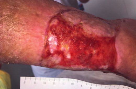
Renaissance of Xenobiotic Wound Antiseptics                        Table 1. MIC (μg/mL) at 24 h exposure of selected antibiotics and
                                                                      antiseptics against microbial test strains
    Wound antiseptics lost some of their importance for
                                                                      Microbial strain      Cefuroxime        OCT [50]      PHMB [50]
more than a century due to the toxicity of Lister’s carbolic                                [269, 270]
wound spray, the toxic side effects of the next generation of
antiseptics such as mercury- or arsenic-based compounds,              S. aureus             0.5 to 64         2             0.5
and the initial euphoria after the introduction of the antibi-        MRSA                  16                1             0.5
otic penicillin G. Reasons for the renaissance of antiseptics         E. faecalis           2 to 128          1             2
                                                                      VRE                   –                 4             4
are the development of effective and well-tolerated antisep-          E. coli               8 to >400         8             0.5
tic substances, the pandemic spread of multidrug resistant            P. aeruginosa         >400              8             2
organisms (MDROs), a comparatively high rate of sensiti-              C. albicans           –                 1             1
zation to locally applied antibiotics, the microbicidal in-
stead of microbiostatic effect of antiseptics, the locally de-
limited effect with no or – in the case of polyvinylpyrrol-
idone-iodine (PVP-I) – few systemic consequences when
correctly applied considering contraindications, and last                Since the microbicidal effect of antibiotics can only be
but not least, the absence of resistance development for              examined in a suspension test and has thus seldom been the
those antiseptic agents which damage pathogens irrevers-              focus of studies, only a few published reports on minimal
ibly. For example, so far, no resistance has been observed            inhibitory concentration (MIC) exist which could serve as
against antiseptics with unspecific effects, such as the de-          a basis for comparison. Nevertheless, this literature shows
struction of the bacterial cell as a whole, or the inhibition of      that many antiseptics are vastly more effective compared to
its function with destruction of the cell membrane or block-          antibiotics (Table 1). For gentamicin, which is also ap-
age of negative surface charges. This is the case for octeni-         proved for topical use as a cream, the MIC for sensitive S.
dine dihydrochloride (OCT), polihexanide (PHMB), PVP-                 aureus was 0.5–1 μg/mL, for Pseudomonas aeruginosa 2 μg/
I, and oxidizing agents, such as hypochlorous acid, or active         mL [12], and for Enterobacteriaceae 25–75 μg/mL [13]. In
substances from the class of peroxides/peroxy acids, such as          contrast, for fluoroquinolones for example, the MIC against
hydrogen peroxide (H2O2). Microbiostatic antiseptics, how-            sensitive Escherichia coli is 0.008–0.02 μg/mL, which is
ever, show transferable resistances, and can be partially             much lower than for OCT or PHMB, where the MIC may
cross-resistant with certain antibiotics. Examples are the ac-        be up to 1,000 μg/mL for resistant strains [14]. The use of
tivation of efflux pumps [1, 2] for chlorhexidine digluconate         this antibiotic would not be recommendable.
(CHD) and quaternary ammonium compounds, and a ge-                       The German Society for Wound Healing and Wound
netically coded periplasmatic Ag(I)-binding protein and 2             Treatment (Deutsche Gesellschaft für Wundheilung und
efflux pumps for silver ions [3]. This is also true for topi-         Wundbehandlung) only recommends microbiological
cally applied antibiotics such as mupirocin [4], silver sulfa-        diagnostics for chronic wounds if there are signs of a sys-
diazine [5–7], neomycin, and bacitracin [8], which have all           temic infectious event originating from the wound area
lost their significance as wound antiseptics, except for mu-          [15]. For this reason, the local application of antibiotics
pirocin [9], which is still used for the decolonization of            used for the treatment of systemic infections should be
MRSA (methicillin-resistant Staphylococcus aureus). Espe-             avoided in order to circumvent the development of resis-
cially the over-the-counter sale (without prescription) of            tance and sensitization [16]. The WHO also does not rec-
mupirocin as a wound antiseptic is considered to be a major           ommend topical use of or rinsing with antibiotics in this
cause for the increase in resistance development [5, 10],             case [17] (Table 1).
which can locally exceed more than 20% of examined hos-
pital-associated MRSA strains [11].
                                                                         Evidence Regarding Wound Antiseptics
    The local application of antibiotics for locally confined wound
infections and colonization is to be avoided, not only because of         An infected or critically colonized wound must be microbio-
the promotion of resistance development, but also because of          logically remediated in order to heal properly [18–20]. It must be
their microbiostatic mode of action and concentrations that are       determined whether the topical use of antiseptics is sufficient or
hard to adjust. Any systemic escalation of the infection, such as     if a systemic antibiosis is necessary due to septic spreading. If a
positive blood cultures, must be treated with systemic antibiotics    wound is at risk of becoming infected, antiseptics can prevent the
in combination with topic antiseptics, if necessary.                  emergence of infection [21].
                                                                                                                                            165.225.72.124 - 12/21/2017 10:11:22 AM

Consensus on Wound Antisepsis:                                        Skin Pharmacol Physiol 2018;31:28–58                             29
Update 2018                                                           DOI: 10.1159/000481545
                                                                                                                                            Downloaded by: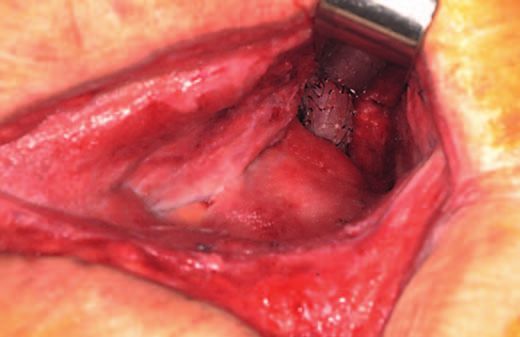
Although the proper treatment of wounds has been a                It is always considered to be a pharmacological mode
challenge since the beginning of mankind, sufficient evi-        of action if wound healing is supported by the antiseptic
dence is lacking for the choice of antiseptics to prevent        effects on cell-adherent pathogens, possibly with associ-
wound infections as well as treat wounds, especially             ated biochemical or immunological consecutive reac-
chronic ones. Many dermatological patients suffer from           tions. This is also true if the active substance binds to the
different types of erosive skin lesions, e.g., follicular bac-   wound tissue and offers a so-called remanent effect by
terial infections [22, 23] or eczema [24]. Often, local an-      gradual release [33, 34]. If the main mode of action of
tibiotics are used instead of considering antiseptic op-         wound rinsing solutions or wound dressings is based on
tions for treatment, although according to expert opin-          physical means, e.g., rinsing, absorption, moisture regu-
ion, the latter are more effective and do not pose a risk of     lation, or irreversible physicochemical binding of micro-
resistance development [22]. This leads to the conclusion        organisms, they are classified as MDs. In practice, the
that further studies and observations must be undertaken         transition between MPs and MDs is fluid, since physical
to examine the potential of antiseptic treatment for these       and pharmacological modes of actions cannot be strictly
conditions. Since only a small number of clinical trials are     separated. Since the classification does bear consequenc-
available as the basis for decisions, all available results      es for pharmacological-toxicological and clinical testing
from studies ranging from in vitro experiments up to             as well as user protection, the correct classification is im-
clinical studies, including meta-analyses, must be collect-      portant in terms of ethics and reimbursement. This de-
ed to form a plausible synopsis [18]. For this reason, all       marcation is further complicated by the approval of some
clinical studies available in PubMed were taken into ac-         antiseptics such as PHMB as a preservative in antisepti-
count for Tables 6–9, regardless of the evidence level.          cally effective concentrations and their use in wound
                                                                 treatment preparations without further declaration [33,
                                                                 34]. Because of this, all comparisons made within this
     Classification of Compounds for Wound Antisepsis            consensus recommendation require further careful eval-
                                                                 uation and interpretation.
   Products declared as wound antiseptics are classified
as pharmacological drugs (PDs). If the mechanical effects
such as rinsing (solutions) or absorption (gauzes) are the          Indications
primary mode of action and the antiseptic effect is only
provided by the addition of preservatives, the product is           The use of antiseptics for prophylactic or therapeutic indica-
classified as a medical device (MD). The distinction from        tions in wound treatment is possible for the following objectives:
                                                                 – Prevention of infection of acute wounds, e.g., after trauma,
PDs is based on the primary mode of action and the in-              bite, or gunshot wounds
tention for use as described by the manufacturer. PDs act        – Prevention of postsurgical wound infections (surgical site in-
pharmacologically, metabolically, and/or immunologi-                fections; SSI)
cally, while MDs primarily act physically. The pharmaco-         – Decolonization of wounds colonized with MDRO
logical mode of action can take various manifestations:          – Treatment of clinically manifested wound infections, includ-
                                                                    ing so-called critical colonization
– The binding to adhesion proteins or their biochemical          – Preparation for debridement or wound cleaning of chronic
   or immunological destruction can inhibit or prevent              wounds in outpatient facilities
   the attachment of bacteria [25, 26]. As long as patho-
   gens residing and multiplying in the upper cell layers           The interaction between microorganisms and wounds
   are killed, the effect is considered pharmacological,         can take place on different levels (Table 2). The clinically
   since reproduction cannot take place without adhe-            characterized term “critical colonization” reflects the
   sion on receptors and interaction with the tissue.            hard to define condition of the transition between physi-
– Wound healing can be supported by biochemical                  ological wound colonization and the pathological condi-
   means, such as interaction with inflammation media-           tion of a manifest local infection [35].
   tors. This was observed for PVP-I [27], OCT [28], and            Although almost all wounds, especially chronic ones,
   hypochlorous acid [29].                                       are contaminated, not all patients develop an infection.
– Healing can also be supported for aseptic wounds.              Since physiological colonization is either irrelevant or –
   This was observed for liposomal PVP-I [30] and                due to colonization resistance – even beneficial for the
   PHMB [31, 32], although the exact mechanism has yet           process of wound healing [36], the Wounds-at-Risk
   to be explained.                                              (WAR) Score [37], which is the sum of different points,
                                                                                                                                      165.225.72.124 - 12/21/2017 10:11:22 AM

30                   Skin Pharmacol Physiol 2018;31:28–58                              Kramer/Dissemond/Kim/Willy/Mayer/
                     DOI: 10.1159/000481545                                            Papke/Tuchmann/Assadian
                                                                                                                                      Downloaded by: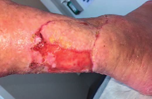
Table 2. Classification of the microbial status of wounds

                        Term                        Characteristics

                        Contamination               Microorganisms are present and have attached to the tissue (microbial attachment)
                                                    without (initial) proliferation
                        Colonization                Microorganisms are present and are proliferating; a clinically significant
                                                    immunological host reaction is (initially) absent
                        Critical colonization       Microbial proliferation without the formation of classical signs of infection but
                                                    delayed wound healing due to toxin production/or the wound is colonized with
                                                    antibiotic resistant strains without signs and symptoms of infection
                        Local infection             Clinically observable, immunological host reaction with the typical signs of infection
                                                    including redness (erythema 1 – 2 cm measured from the wound margin) with
                                                    tendencies of increase could be equivalent to spreading infection with the risk of
                                                    generalization, swelling, increased local skin/tissue temperature, pain, functional
                                                    impairment, and increase in exudate quantity and viscosity, for example, perceptible
                                                    odor and stagnation in wound healing
                        Systemic infection          In addition to the local inflammatory reactions, signs of a systemic host reaction
                                                    such as leukocytosis, increase in C-reactive protein and fever

was introduced in order to evaluate the infection risk                       Tolerability
(Table 3).
                                                                              The tolerability of antiseptics in wounds is supposed to be
    If the WAR Score reaches or exceeds 3 points, an antiseptic           equal to Ringer solution, physiological saline, or an inert hydro-
treatment is justified.                                                   gel. Ideally, wound healing is promoted.

                                                                              A good point of orientation would be to follow the prac-
   Criteria for Choosing Antiseptic Agents                                tical approach of not applying anything to chronic wounds
                                                                          which should not be applied to the eyes. This is true for
   Efficacy                                                               PVP-I up to 5% and for PHMB up to 0.02% [40–42], but
   When treating acute wounds, a microbicidal effect and                  not for silver sulfadiazine, CHD, or OCT (0.1%). If adja-
broad spectrum of activity are desirable. Only in certain                 cent tissues can be exposed in the wound treatment, such
cases does the substance have to be virucidal and addi-                   as cartilage, central nervous system (CNS), or peritoneum,
tionally effective against bacterial spores. For chronic                  the compatibility must be clarified. Furthermore, sensiti-
wounds, the spectrum of activity must only encompass                      zation potential including anaphylaxis risk should be low
Gram-positive and Gram-negative bacteria if no special                    or absent; there should also be no risk of long-term adverse
circumstances have been diagnosed. There should be no                     effects such as mutagenicity, carcinogenicity, or teratoge-
risk for the development of resistance, especially cross-                 nicity. If the quotient of bactericidal efficacy and tolerabil-
resistance towards antibiotics.                                           ity against mouse fibroblasts in vitro, both tested under the
   The efficacy of antiseptics is expected to result in killing of test   same conditions, is >1, the tolerance for the antiseptic of
organisms ≥3 log10 [38, 39] for a typical type of organic load            eukaryotic cells is better than that of bacteria. This is true
within the declared exposure time.                                        for OCT, PHMB, and almost for PVP-I (Table 4). A de-
                                                                          tailed observation about the selective antiseptic effect can
   In some cases, the efficacy is additionally tested without             be made when, in cocultures of human cells and bacteria,
an organic load typical for wounds, although this does not                the prokaryotic cells are destroyed, while the eukaryotic
correspond to the application situation, unless the load is               cells survive, or bacteria in a comparable solution are killed
significantly reduced, for example by repeated rinsing. With-             without damage to human cells. This is demonstrated for
out an organic load, the efficacy is expected to be ≥5 log10              sodium hypochlorite (NaOCl) [43], PHMB [44], and PVP-
versus bacteria and ≥4 log10 versus Candida albicans [38].                I [45]. Analogously, the treatment of epidermis equivalents
                                                                                                                                               165.225.72.124 - 12/21/2017 10:11:22 AM

Consensus on Wound Antisepsis:                                            Skin Pharmacol Physiol 2018;31:28–58                            31
Update 2018                                                               DOI: 10.1159/000481545
                                                                                                                                               Downloaded by:
Table 3. Assessment of risk for wound infection [37]
Risk class Risk condition (based on risk status and different indications) Point score
1 Acquired immunosuppressive disease (e.g., diabetes mellitus) Per risk
Acquired immune defect due to medical therapy such as cyclosporine, 1 point
methotrexate, glucocorticoids, or antibodies
Solid tumor disease
Systemic hematological disease
Postsurgical wound healing disorder, which results in (unplanned) secondary
healing
Potentially heavily contaminated wounds (e.g., perineum, genitals)
Problematic hygienic conditions related to social or occupational environment
(e.g., agriculture, lorry driver)
Patient age >80 years
Young age of patient (premature infants, babies, infants)
Wounds persisting for >1 year
Wound dimensions of >10 cm2
Chronic wounds of any etiology having a depth of >1.5 cm
Extended inpatient status >3 weeks
2 Severe acquired immune defects (e.g., HIV infection) Per risk
Heavily contaminated acute wounds 2 points
Bite, stab, and gunshot wounds penetrating 1.5 – 3.5 cm
3 Severe innate immunodeficiency such as Wiskott-Aldrich, Di-George syndrome, Per risk
immunodeficiency after stem cell transplantation, AIDS, immunosuppressive 3 points
therapy [271]
Burn wounds with involvement of >15% BSA
Traumatically contaminated wound after debridement
Wounds that have a direct connection to organs or functional structures
(e.g., including joints) or which contain foreign material
Bite, stab, and gunshot wounds penetrating >3.5 cm
derived from human keratinocytes with OCT alone or in Table 4. Biocompatibility index as a quotient of IC50 for L929 cells
combination with test organisms demonstrated no cyto- and the required MIC for a reduction factor of ≥3 log10 [173]
toxic effect in viable keratinocytes [51]. In contrast, H2O2
Compound E. coli S. aureus
inhibits mammalian cells beginning with a concentration
of 8.5 mg/L [46], thus inhibiting fibroblasts, whereas bac- OCT 1.7 2.1
teria still survive [47]. However, this is not transferable to PHMB 1.5 1.4
the endogenous formation of H2O2 occurring in the con- PVP-I (aqueous solution, referring on I2) 0.9 1.0
text of the nonspecific immune response, for example by CHD 0.7 0.7
Triclosan 0.2 0.5
granulocytes. Thus, 0.003% H2O2 already inhibits the cy- Ag-protein (referring Ag+) 0.2 0.1
tolytic activity of natural killer cells, but the killer cells re- Ag(I)-sulfadiazine and silver nitrate not measurable
main vital [48]. Even if, for example, H2O2 is formed in
noncytotoxic concentrations in medical honey by glucose
oxidase, this is not comparable with the external antiseptic
application of pure H2O2 alone [49].
In acute wounds the fast-acting effect of the antiseptic is at the Taking into account the different properties of anti-
forefront, under certain circumstances with a necessary depth ef- septic active ingredients (Table 5), the following develop-
fect, for example in patients with bite, puncture, or gunshot injuries. ment trends are apparent. PVP-I has partly lost impor-
For chronic wounds, a longer exposure time is acceptable for reach- tance due to the introduction of more recent and ad-
ing the antiseptic effect due to repeated application and/or remain- vanced substances. By solving the stability problem, the
ing on the wound. Wound healing should also be promoted here.
combination of sodium hypochlorite/hypochlorous acid
165.225.72.124 - 12/21/2017 10:11:22 AM
32 Skin Pharmacol Physiol 2018;31:28–58 Kramer/Dissemond/Kim/Willy/Mayer/
DOI: 10.1159/000481545 Papke/Tuchmann/Assadian
Downloaded by:Table 5. Properties of wound antiseptics relevant for antimicrobial agents used on wounds
Compound Antimicrobial Deep Development Wound healing Cartilage Sensibilization Systemic
onset time effectb of resistance tolerability risk
Ag+ ≥24 ha [272] 3 Yes Inhibition [191, 192] ? No Yes [273]
CHD 3 – 10 ha [43] 1 No inhibition [274] No Yes (rare), ?e
anaphylaxis
(n >200)
AA 15 – 30 s [49] 2 No At 0.15% supportive ? No
[229, 275, 276]
No
OCl– 30 s to 5 minc 2 Supportive [278] ?
[277]
OCT 3 – 10 ha [39] 1d No inhibition [30, 36] No [279] No
PHMB 3 – 10 ha [39] 2 Supportive [94] ≤0.005% [279] Yes (rare),
anaphylaxis
(n = 3)
PVP-I 30 mina [39] 3 Partial inhibition [172] Yes [279] Yes Yes
1, superficial effect due to high protein binding; 2, shallow penetration depth; 3, larger than 2.
a
Test-carrier (Tc) with organic load [39, 272]. b Due to a lack of experimental data, theoretical extrapolation based on physicochem-
ical properties or demonstrated absorption. c Without load. d In combination with phenoxyethanol 2 or 3. e Possibility of separation of
4-chloraniline from the chlorhexidine molecule [76].
(HOCl/OCl–) or sodium hypochlorite (NaOCl) is avail- I, PHMB, and CHD [51]. In relation to cytotoxicity, OCT
able as an additional option. Acetic acid (AA) or combi- is superior to PVP-I [52, 53]. When tested on metal carri-
nations with fruit acids such as lactic, malic, citric, fu- ers under load (artificial wound fluid), PVP-I solution was
maric, or oxalic acid are gaining increasing interest, in effective within in a time frame of 5 min, whereas gels
particular due to their efficacy against P. aeruginosa and based on OCT (0.05%) or PHMB (0.04 or 0.02%) needed
the promotion of wound healing, but also due to their 30 min or 3 h (PHMB 0.02%) to take effect [39]. PVP-I was
availability in countries with limited resources. also most effective in an in vitro wound model with S. au-
reus, followed by OCT and PHMB [54]. However, OCT
showed superior efficacy in a biofilm model with P. aeru-
Properties of Selected Antiseptic Active Agents ginosa PVP-I [55]. The S. aureus biofilm was almost com-
pletely eliminated within 5 min [56]. Even in experimental
Iodophores and modern compounds such as OCT, PHMB, burns in rats, OCT significantly exceeded both PHMB and
and stabilized hypochlorite meet the requirements for antiseptic PVP-I tested against P. aeruginosa [57]. CHD interacts
activity in vitro. Remanent effects are displayed only by OCT,
PHMB, and CHD. Wound healing is enhanced by PHMB, hypo- antagonistically with gentamicin and synergistically with
chlorite, and AA depending on the concentration. For PVP-I OCT [58]. In vitro, phagocytosis and growth factors, such
there is an increased risk of sensitization as well as absorptive side as the platelet-derived growth factor, are stimulated by
effects, particularly in thyroid disorders. OCT [59], which can be beneficial for wound healing.
Side Effects
Octenidine When OCT/phenoxyethanol (PE) was used in the epi-
cutaneous patch test, a negative response was found for
In vitro and Animal Experiments OCT, while a positive response to PE and cocamidopro-
OCT shows superior efficacy [50] in the quantitative pyl betaine was detectable. However, the distinction be-
suspension test without protein load compared with PVP- tween allergic and irritating reactions was inconclusive
165.225.72.124 - 12/21/2017 10:11:22 AM
Consensus on Wound Antisepsis: Skin Pharmacol Physiol 2018;31:28–58 33
Update 2018 DOI: 10.1159/000481545
Downloaded by:Table 6. Summary of clinical study findings for OCT
Type of wound Comparison Result Study design Sample Year
size, n
VLU W vs. W with OCT vs. OCT Wound healing and time to heal Prospective 17/17/15 2016 [280]
wound gel, no difference in was significantly better in both open-label cohort
infection rate at beginning OCT study arms, lowest costs in study
wound-gel study section
VLU W with OCT vs. W with OCT showed significantly rapid RCT 40/40 2015 [281]
silver healing and reduction of pain,
microbial eradication after 28 and
50 days
Neoplastic ulcer OCT-soaked W; Significantly faster wound Prospective 30 2013 [282]
comparison baseline vs. healing, significant eradication observational study
after 3 weeks of treatment of potentially pathogenic
Gram-positive and
Gram-negative bacteria, no
adverse events
Split-skin harvest OCT-hydrogel vs. hydrogel Significant reduction of wound Double-blind RCT 31/30 2012 [36]
sites colonization, no difference in
time to heal
VLU OCT/PE vs. Ringer lactate No difference in time to heal; for Double-blind RCT 60/66 2012 [283]
solution OCT fewer adverse events
Second-degree Irrigation of wound with Significant reduction of pain in Prospective RCT, 30/30 2011 [284]
burn OCT/PE, thereafter OCT gel OCT study arm, tendentially contralateral site
vs. silver sulfadiazine improved wound healing; served as control
possibly no difference observable
due to initial OCT treatment in
both study sections
Musculoskeletal OCT irrigation and drainage After 5 – 24 days eradication of all Prospective 8 2010 [285]
infection + OCT-soaked gauze pathogens, no adverse events observational study
Neoplastic ulcer OCT/PE-soaked gauze; Eradication of S. epidermidis and Prospective 16 2008 [286]
comparison of baseline vs. P. aeruginosa, reduction of observational study
after 3 weeks of treatment necrosis, exudate, erythema, and
edema; 1× persistence of
P. aeruginosa, 1× persistence of
E. coli, and 2× persistence of
E. faecalis
VLU, venous leg ulcer; W, wound dressing.
[60]. On the basis of the low absorption determined only is particularly suitable for antisepsis in patients with burn
after removal of the upper skin barrier, no systemic ab- injuries. OCT is superior to silver and PVP-I in the latter
sorption is to be expected when applied to wounds [61]. case (Table 6). In the surgical treatment of traumatic am-
putation and splinter injuries which were colonized with
Clinical Studies MDRO, an antibiotic treatment was not necessary after se-
OCT is available as a solution and a gel. The antiseptic rological and microbiological exclusion of a florid system-
(OCT/PE) itself as well as the rinse and the gel are well tol- ic infection by antiseptic wound care with OCT/PE in con-
erated, as shown by studies (Table 6) and case reports [62– junction with negative pressure wound therapy (NPWT)
64]. As an MD (rinse), it is suitable for wound cleansing [65, 66]. With the introduction of a new treatment algo-
[62] and supports biofilm removal [59]. Especially the gel rithm for chronic lower leg and foot ulcers in a surgical
165.225.72.124 - 12/21/2017 10:11:22 AM
34 Skin Pharmacol Physiol 2018;31:28–58 Kramer/Dissemond/Kim/Willy/Mayer/
DOI: 10.1159/000481545 Papke/Tuchmann/Assadian
Downloaded by:outpatient clinic, OCT-based antiseptics were implement- PHMB, wound gel 0.1%, and wound dressings 0.1%. Re-
ed instead of obsolete agents such as CHD, ethacridine, duction by ≥3 log10 of typical wound contamination on
H2O2, silver sulfadiazine, or local antibiotics, with an al- test specimens was achieved by wound gel with a concen-
most 3-fold reduction in the total cost [67]. OCT was also tration of 0.1% in 30 min, 0.04% in 3 h, and 0.02% in
effective in patients with inflammatory acne vulgaris [68]. 10 h. Enterococcus faecium was not sufficiently eliminat-
ed in 24 h [39]. As different concentrations of PHMB
Caveats solutions were not examined on test specimens, it is not
During the past few years, several misapplications of possible to deduce the optimal concentration for wound
OCT/PE have been recorded. In these cases, the compound treatment. A single irrigation of contaminated traumatic
was applied in puncture wounds, bite wounds, or abscess wounds for 3 min was shown to be effective in a clinical
cavities by syringe with pressure into the wound channel trial on the prevention of SSI [21], and treatment dura-
and deep tissue, instead of only superficial application. The tion should not fall below this as long as there are no
subsequent edematous swellings with tissue damage re- other results. Using wound gel, exposure for at least 3 h
quired partial surgical revision [69]. Only superficial ap- is needed [39].
plication by means of swabs or spray is recommended [70].
Any unwanted tissue reactions are improbable in this case, Results from in vitro and Animal Tests
because no local pressure necrosis was observed upon The efficacy of PHMB does not substantially differ
flushing locally limited skin soft-tissue infections in the from that of OCT. PHMB is equally effective against
hand area (n = 10) with applied drainage [71]. Since OCT methicillin-sensitive S. aureus and MRSA [77]. Higher
is practically not reabsorbed, any insertion into the skin or pH levels, which typically develop in wounds (6.5–8.5)
insertion canals is to be avoided. According to the manu- [78], decrease the efficacy of PVP-I but significantly im-
facturer, the use of OCT/PE for wound treatment without prove that of PHMB. This suggests that PHMB might be
medical supervision should not be extended for more than advantageous for the management of wound infections,
2 weeks, as the only data available are from a continuous as both S. aureus and P. aeruginosa exhibited increased
application period of up to approximately 14 days. susceptibility to the antiseptic with rising pH levels. The
inhibitory activity of chlorhexidine and OCT was only
Contraindications marginally affected by the pH in vitro, although a statisti-
Peritoneal lavage, retroperitoneal and intravenous cally significant improvement was observed against S. au-
application, allergy, application to hyaline cartilage, and reus at pH 9 for OCT [79].
CNS structures are contraindications. Interaction with In combination with undecylenamidopropyl betaine
CNS structures is recorded for CHD, and is considered (Betaine), the antimicrobial effect is enhanced because
valid for OCT as well until further data are available [59]. of altered physical properties [80, 81], while in vitro cy-
totoxicity is reduced [80] and cleaning performance is
improved [82]; the latter one could not be confirmed in
Polihexanide a newer study because of the interference of the surfac-
tant with the protein measurement [322]. Intracellular
In 1979, Good [72] combined PHMB, which until elimination is remarkable, as shown for E. coli [83],
then had only been used as disinfectant, with polyethyl- MRSA [84], and Acanthamoeba species [85]. Thus,
ene glycol 4000 to achieve improved wetting for use on PHMB (0.02%) is the preferred agent for the treatment
wounds [72, 73]. In the 1980s, PHMB was introduced by of Acanthamoeba keratitis [85]. Efficacy against P. ae-
Willenegger in Switzerland [74]. PHMB can be seen as a ruginosa can still be observed in the presence of 4% al-
virtually detoxified CHD, as the molecular structure of bumin [86], 4.5% blood + 4.5% albumin [87], and
PHMB monomers closely resembles the structure of wound exudates, while the expression of elastase is in-
CHD molecules, except for the terminal NH-group of hibited at the same time [88]. In a wound model on pigs,
CHD consisting of 4-chloroaniline, which is a potential MRSA was significantly reduced after 72 h by PHMB in
human carcinogen [75]. This similarity explains both the a wound treatment matrix based on collagen, while sil-
comparable antiseptic efficacy and the worse tolerability ver dressings were ineffective [89]. PHMB was effective
of CHD compared to PHMB, due to the release of 4-chlo- against biofilm in vitro [90] and in animal models [91].
roaniline in vivo [76]. Depending on the manufacturer, Loaded onto nanocellulose, PHMB was antimicrobially
wound irrigation solutions release 0.02, 0.04, or 0.1% more effective than PVP-I [92]. No antagonism could
165.225.72.124 - 12/21/2017 10:11:22 AM
Consensus on Wound Antisepsis: Skin Pharmacol Physiol 2018;31:28–58 35
Update 2018 DOI: 10.1159/000481545
Downloaded by:be shown against oxacillin, penicillin G, ampicillin, ce- Undesirable Effects
fazolin, cefuroxime, imipenem, gentamicin, erythro- Two cases of a possible anaphylactic reaction triggered
mycin, doxycycline, levofloxacin, linezolid, or vanco- by PHMB could not be verified in the skin-prick test
mycin [93]. Both in cell culture and animal wound [103]. One patient with a grade III anaphylactic reaction
models (rat, pig), wound healing was improved [31, 94– had IgE against both PHMB and CHD. Due to the similar
96]. Results of in vitro and animal tests (rat) are prom- structures, it is discussed that sensitization was caused by
ising for the combination of PHMB and sericine, an a prior treatment with CHD, so a known allergy against
ameliorator of wound healing, in a dressing [97]. Capil- CHD might be linked to a risk for PHMB anaphylaxis
lary density was significantly increased in the cremaster [104]. In the second case, only IgE against PHMB was
muscle (rat) by exposure to PHMB and OCT, while the proven [105]. A further suspected case of anaphylaxis was
diameter of arterioles was significantly increased only reported after wound application [106]. Contact allergies
by PHMB [98]. The irritation potency of 0.02% PHMB are rare, with a frequency of ≤0.08% in regard to the fre-
is lower than that of antibiotic eye drops [99]. quent use of PHMB, especially as a preservative [107].
In 2011, the Committee for Risk Assessment of the This suggests that antiseptic substances should be limited
European Chemicals Agency (ECHA) raised the suspi- to medical applications.
cion that PHMB was a category-2 carcinogen (Carc. 2).
Consequently, all products containing PHMB in a con- Clinical Trials
centration of at least 1% had to be labeled accordingly PHMB is available as a solution, hydrogel, and in
from January 1, 2015. Compositions containing 0.1% re- wound dressings [108]. It is well tolerated [109], anti-
quired an annotation on the safety data sheet. This clas- septically effective against MRSA and VRE (vancomy-
sification by the ECHA as category 2 “suspected of caus- cin-resistant Enterococcus) [110–112], can be used for
ing cancer” lacks scientific proof. The 2 studies used for wound irrigation, is suitable as an antiseptic for criti-
the evaluation were feeding studies using extremely high cally colonized and infected chronic wounds, including
PHMB concentrations far in excess of the no-observed- burns [37, 94, 113–124], and, in combination with
(adverse-)effect level. Only in the highest tested concen- NPWT, can be used for instillation (NPWTi). It is supe-
tration of 4,000 ppm did the frequency of cases with hem- rior to Ag+ and PVP-I regarding wound healing [123]
angiosarcoma significantly increase, but at ≤1,200 ppm (Table 7). Upon application of wound dressings impreg-
this was not the case. Neither genotoxicity nor epigenetic nated with 0.2% PHMB, epidermally applied Staphylo-
changes [100] could be shown; therefore, it is very likely coccus epidermidis were completely eliminated in 24 h
that hemangiosarcoma was triggered by enforced prolif- [125]. The same was shown for P. aeruginosa in an ani-
eration of the endothelium, as was proven for PHMB in mal model [126]. Application for pre- and postoperative
wound healing. For risk assessment, it is critical that there wound treatment significantly reduced the rate of SSI
be no systemic absorption up to the detection threshold (Table 7). Cytotoxically, wound dressings do not differ
of 10 μg for PHMB, so a health hazard can be excluded from PHMB-free dressings [127]. After a 4-week unsuc-
for antiseptic use according to regulations [detailed state- cessful treatment of a diabetic foot ulcer with PHMB/
ment with references in 101, 102]. betaine gel, the healing process commenced after a
It is important to note that, in this context, the ECHA 4-week treatment with OCT gel [128]. In cases with hu-
specifically excluded PHMB from the labeling require- man papillomavirus infection, the viral elimination was
ments when used invasively or on the skin surface for significantly improved by local treatment with PHMB,
wound irrigation or with dressings. The reason for this examined after 3 and 6 months, which could open a new
exemption is the regulatory classification of hazardous area of application [129].
substances and their preparation to ensure occupation-
al and environmental safety. Particularly exposure to Caveats
larger amounts, which is possible during the produc- Due to the relatively strong binding onto tissue struc-
tion of these substances, has to be considered. Person- tures, the same restrictions as those for OCT should ap-
nel should be appropriately protected against critical ply, although no clinical reports are available yet. This is
exposure. supported by the appearance of grayish, inert tissue after
retroperitoneal, mediastinal, and partially inguinal appli-
cation for more than 5–10 days. This tissue had to be re-
moved in order to permit the formation of granulation
165.225.72.124 - 12/21/2017 10:11:22 AM
36 Skin Pharmacol Physiol 2018;31:28–58 Kramer/Dissemond/Kim/Willy/Mayer/
DOI: 10.1159/000481545 Papke/Tuchmann/Assadian
Downloaded by:Table 7. Summary of clinical study findings for PHMB
Type of wound Comparison Result Study design Sample Year
size, n
Traumatic soft tissue 0.04% PHMB, 1% SSI rate: 1.9/4.8/11.7/5.9; PHMB was Longitudinal 3,264/ 2017 [21]
injuries PVP-I, 4% WPO, more effective in preventing infection cohort study 2,552/
Ringer solution in deep incisional wounds (A1 and 643/645
A2 SSI), in contusion wounds only in
A2 SSI
Pressure and VLU PHMB/betaine Significant improvement of Single-blinded 143/146 2016 [287]
solution vs. NaCl inflammation and wound healing, RCT
solution no difference in pain scores
Wounds in elderly PHMB/betaine Significant decolonization with 32% Prospective 200/99 2016 [112]
patients solution success (p < 0.05) controlled
nonrandomized
open-label study
Nonhealing wounds PHMB 0.5% vs. moist Superficial infection 38 vs. 47%, (ns), Prospective open 15/16 2015 [288]
after cardiothoracic gauze soaked with deep infection 44 vs. 40% (ns), randomized
surgery Ringer solution wound healing after 15 ± 5 vs. 16 ± 3 cohort study
days (ns); wound healing in 67 vs.
44% (ns); PHMB: patients without
complete wound healing showed
better epithelialization, after 12 h
CRP was significantly lower than
controls
Chronic wounds PHMB gel vs. betaine Significant reduction in wound size, Multicenter 120 2014 [289]
gel pain, fibrin slough, and necrosis; observational
reduction of exudate study
Grade II burns PHMB gel vs. betaine Less pain medication, good progress Observational 20 2014 [290]
gel in wound healing with formation of study
granulation tissue and
epithelialization; reduction of
erythema after 2 days; no infection
during mean treatment of 11.2 days
Postsurgical NPWTi with 0.04% Reduced duration of treatment Prospective 16 2014 [291]
subcutaneous PHMB-soaked gauze case-control
abdominal infections vs. NPWT study
Wounds after W vs. W+PHBM PHMB: significant decrease of SSI Cohort study 692/707 2013 [292]
cardiothoracic
surgery
Critically colonized PHMB vs. Ag-W PHMB: significantly faster pain RCT 21/18 2012 [293]
and infected chronic reduction and elimination of
wounds microorganisms
Entry point of W vs. W+PHBM PHMB: significant decrease of SSI RCT 18/22 2012 [294]
external fixator
Lower-limb and foot W vs. W+PHBM PHMB: significantly faster pain Double-blinded 22/23 2011 [295]
ulcers reduction and elimination of RCT
microorganisms, tendentially
faster wound healing
Burns W vs. W+PHBM PHMB: significant pain reduction RCT 30/30 2011 [296]
and fewer dressing changes
165.225.72.124 - 12/21/2017 10:11:22 AM
Consensus on Wound Antisepsis: Skin Pharmacol Physiol 2018;31:28–58 37
Update 2018 DOI: 10.1159/000481545
Downloaded by:Table 7 (continued)
Type of wound Comparison Result Study design Sample Year
size, n
Split-skin harvest CHD W vs. PHMB W PHMB: significantly faster RCT 21/21 2011 [297]
sites reepithelialization and lower pain
score
Infected orthopedic NPWTi with PHMB 86% of patients with acute and 80% Prospective 32 2011 [298]
implants 0.04% of patients with late-onset infections multicenter
kept their implant during a follow-up observational
time of 4 – 6 months study
VLU NaCl vs. PHMB PHMB: significantly faster bacterial Prospective 20/20 2010 [299]
solution elimination cohort study
Postsurgical wounds W vs. W+PHMB PHMB: significant reduction in SSI, Historic 9,372/ 2008 [300]
0.2% particularly of MRSA infection comparison 10,202
(first and possibly
second dressing after
surgical procedure)
Chronic wounds NaCl vs. 0.1% PHMB PHMB: after 2 weeks significantly Randomized 64/78 2008 [301]
gel faster bacterial elimination, faster nonblinded
wound healing, less pain, less cohort study
exudate, improved granulation
Burns PVP-I, 1% silver PHMB: improved epithelialization Parallel 4 2007 [113]
nitrate, 0.04% PHMB and elimination of malodor; deep intervention on
necrosis and slough observed in contralateral
PVP-I and silver nitrate study section symmetric wounds
Acute contaminated PHMB-wetted gauze Significant faster bacterial Double-blinded 28/22 2006 [302]
wounds (0.04%) vs. Ringer elimination, reduction of RCT
solution inflammation
Infected wounds W vs. W+PHBM PHMB: improved control of wound RCT 21/21 2004 [303]
colonization
NPWTi, negative pressure wound therapy with instillation of antiseptics; W, wound dressing.
tissue, even after infection control independent of PHMB electrochemically converted for its production. The acti-
use as a single substance or in combination with betaine vated solution is also called electrolyzed water [130]. The
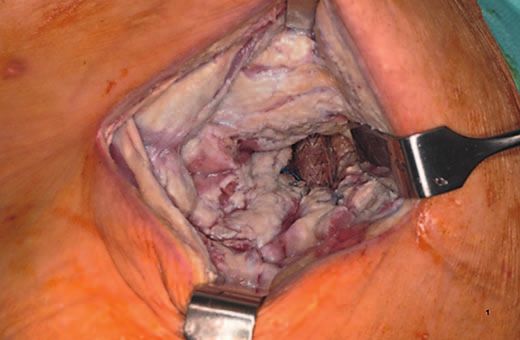
(Fig. 1a, b). currently used concentration amounts are 0.004% each
for NaOCl and HOCl, anda b
Fig. 1. Retroperitoneal instillation with PHMB on an aortal endoprosthesis infection. a Situs after 14 days and
multiple excisions of the yellowish-brownish slough within 10 days. b Situs after a 16-day instillation with phys-
iological NaCl solution. Source: D. Mayer.
a b
Fig. 2. A 90-year-old patient after excision of an ulcus hypertonicum Martorell and coverage with a split-skin
mesh. a In due course, the formation of slightly bleeding, instable hypergranulations, which are possibly a sign
of incipient infection. b After 2.5 weeks of daily dressing change and coverage with a Betadine gauze, the hyper-
granulations regressed and stable granulation tissue developed with a marked swelling of the wound vicinity.
Source: D. Mayer.
The combination of PHMB/betaine was slightly less ef- inhibited without intracellular impairment, possibly con-
fective than NaOCl/HOCl against biofilm [132]. The tributing to an anti-inflammatory effect [29]. NaOCl/
speed of effect was superior to PVP-I, OCT, and PHMB HOCl was not or barely irritating on chick chorioallan-
[130, 133–137]. It can be assumed that the efficacy is re- toic membranes [323]. Furthermore, no evidence for cy-
duced by protein or blood contamination, which can be totoxicity could be found in a 3D model of the skin [132].
reversed by repetitive extensive wound irrigation. The There is no evidence for toxic risks [139]. The feeding of
survival rate of rats with experimental peritonitis was sig- laboratory animals with 5 ppm is a safe alternative instead
nificantly increased compared to a treatment with NaCl of sterile water [140]. There is no evidence that NaOCl
without undesirable effects [138]. By stabilization of the poses a carcinogenic hazard [141, 142].
cell membrane, the release of cytokines from mast cells is
165.225.72.124 - 12/21/2017 10:11:22 AM
Consensus on Wound Antisepsis: Skin Pharmacol Physiol 2018;31:28–58 39
Update 2018 DOI: 10.1159/000481545
Downloaded by:Undesirable Effects In contrast to PVP-I, cadexomer-iodine (C-I) uses a
Rinsing of the mediastinum in heart surgery with hydrophilic, modified starch polymer to embed iodide
NaOCl/HOCl prior to wound closure was significantly ions. The advantages of C-I are similar to those of PVP-I;
associated with perioperative alterations of the ECG, in- however, PVP-I and C-I show different properties re-
cluding ST elevation, but without hemodynamic distur- garding the reactivity of iodine and water absorption
bances [143]. [155]. C-I did not become as widespread in German-
speaking regions as PVP-I did.
Clinical Trials
Case studies on NaOCl/HOCl report decolonization In vitro and Animal Experiments
of MRSA infections on skin and the base of the skull [144, The microbicidal effect is observed for all vegetative
145], decolonization of MRSA, P. aeruginosa, and E. coli pathogens, including mycobacteria, yeasts, and dermato-
in chronic diabetic ulcers [146], and successful adjuvant phytes, enveloped and nonenveloped viruses (including
application in the treatment of necrotizing soft tissue in- rabies especially in combination with alcohols), as well as
fection [147], osteitis [145], and osteomyelitis [148]. In protozoa, and, with a longer exposure time (2–24 h), also
cases of peritonitis, partially with peritoneal abscess (n = bacterial spores [156]. Depending on the test model, the
7), no bacterial growth was detectable 3–7 days after ir- efficacy of PVP-I in vitro can be higher than, comparable
rigating twice daily for 9–12 days [149]. Postoperative to, or less than OCT and PHMB; 10% sheep blood does
complications including SSI were significantly reduced in not affect the efficacy. In 10% serum albumin as well as
patients with peritonitis without symptoms of intoler- 4.5% sheep blood + 4.5% serum albumin + 1% mucin, the
ance [150]. The irrigation of infected chronic wounds was exposure time doubles, similar to OCT [38, 39]. In con-
well tolerated [151], also in combination with NPWT trast to OCT and PHMB, PVP-I has no remanent effect.
[152]. The combination NaOCl/HOCl with a hydropho- Extended antiseptic effects, shown in vitro, are not due to
bically coated wound dressing, to which microorganisms a true remanent effect in PVP-I, in contrast to OCT or
adhere and bind irreversibly, appears promising and to CHD, but are an artifact of the modified release kinetics
lack the subsequent physiological immune response im- of the iodine from the PVP molecule, which follows the
pairment that is triggered by OCl– (Table 8). second order of kinetics.
In vitro, PVP-I inhibits the formation and release of in-
flammatory mediators due to the reducted expression of
Iodophore bacterial exotoxins, the inhibition of excessive mediator
molecule release, and the activity of human immune effec-
The introduction of iodophores, complexes of iodine tor cells, as well as the inactivation of tissue-destroying en-
and macromolecules, in 1956 sparked a renaissance of an- zymes [157, 158]. Through chemical reactions with the
tisepsis. However, demands for stricter indications were physiological H2O2 peroxidase systems, oxidation products
already made in 1984, with a call for each specialty to with a higher efficacy than that of molecular iodine can be
more rigorously watch for undesired effects to prefer an formed in wounds [156]. C-I ex vivo and in animal models
antiseptic agent with similar antimicrobial spectrum but has a strong effect against biofilm-forming S. aureus and P.
fewer undesirable effects [153]. Especially the risk of thy- aeruginosa [159]. The contrasting effects against biofilms
roid gland dysfunction, but also the relatively high po- are attributed to the different availability of active iodine in
tency for allergic sensitization, has led to a restricted ap- the various forms administrated [160]. In animal experi-
plication of PVP-I during recent years. ments, the healing of skin wounds was significantly delayed
The macromolecular carrier system of PVP and the by 2% PVP-I [161]. For PVP-I-L, proliferation and im-
release of iodine after degradation by reacting agents re- provement of microcirculation have been demonstrated in
sult in lower iodine absorption, cytotoxicity, and sensiti- in vitro and animal experiments [30, 162, 163]. In animal
zation, and thus in better tolerability than aqueous or al- models, the application of C-I promotes epithelial cell re-
coholic iodine solutions. In aqueous solutions, only a generation and thus wound healing [164, 165]. In PAOD
thousandth of the total iodine is free and microbicidally (peripheral arterial occlusive disease)-associated ulcers, C-I
active. The development of liposomal PVP-I composi- was tolerated without irritation [166]. In accordance with
tions (PVP-I-L) on the basis of hydrogel improved the this, in histological tests, no tissue damage was observed in
wound tolerability [30, 154]. the treatment of chronic exudative wounds [167]. There is
no evidence of neurotoxicity, mutagenicity, carcinogenici-
165.225.72.124 - 12/21/2017 10:11:22 AM
40 Skin Pharmacol Physiol 2018;31:28–58 Kramer/Dissemond/Kim/Willy/Mayer/
DOI: 10.1159/000481545 Papke/Tuchmann/Assadian
Downloaded by:Table 8. Summary of clinical study findings for the combination NaOCl/HOCl
Type of wound Comparison Result Study design Sample size, Year
n
Explorative laparotomy/ NaOCl/HOCl vs. OCl: significant reduction of fever and RCT 50/50 2013 [150]
peritonitis NaCl prevention of SSI
Diabetic foot ulcers, NaOCl/HOCl vs. OCl: faster granulation and RCT 100/100 2011 [304]
VLU, burns PVP-I epithelialization, faster reduction of wound
size, reduction of surrounding edema and
erythema, better cosmetic results in burn
wounds; PVP-I: minute skin irritation and
pain in burns
Chronic wounds, SSI NaOCl/HOCl vs. OCl: significant wound size reduction, RCT 50/50 2011 [305]
PVP-I fewer persisting infections due to
P. aeruginosa, S. aureus, and Klebsiella spp.
Diabetic foot ulcers NaOCl/HOCl vs. OCl: significantly better wound healing, RCT 20/20 2010 [306]
PVP-I control of infection, significantly more
interventions in the PVP-I study section
Chronic wounds NaOCl/HOCl vs. OCl: significant wound size reduction, RCT 15/15 2009 [307]
PVP-I better control of microbial colonization; was
well tolerated
Diabetic wounds NaOCl/HOCl vs. OCl: significantly reduced hospitalization Blinded 50/50 2007 [278]
NaCl (soaked and wound size, improvement in wound RCT
gauze) score
Diabetic foot ulcers NaOCl/HOCl vs. OCl: significant reduction of malodor, Blinded 21/16 2007 [308]
PVP-I significant reduction of soft tissue infection, RCT
improved granulation, lower occurrence of
erythema
Diabetic foot ulcers NaOCl/HOCl vs. OCl: reduced treatment time RCT 110/108 2005 [309]
PVP-I
Burns NaOCl/HOCl vs. OCl: 11% reduction in use of antibiotics, Retrospective 64/64 2005 [310]
Ag 50% reduction in hospitalization cohort study
SSIs NaOCl/HOCl vs. OCl: significantly reduced hospitalization Retrospective 46/42 2001 [311]
PVP-I and reduction of pain cohort study
ty, or teratogenicity [156, 168]. In the cell culture (fibro- swelling, and renal impairment [170]. In the case of C-I
blasts), 0.45% C-I was found to be noncytotoxic [167]. application, temporary pain may also occur [171].
Side Effects Clinical Studies
Iodophores display a high sensitization potential [169]. Clinically, wound healing was generally not impaired
In adults with no known thyroid disease, and in contrast by PVP-I. However, in some cases the control group had
to premature infants and newborns as well as small chil- a better outcome [172], probably promoted by C-I [165],
dren, irreversible damage to the thyroid gland is not to be although PVP-I showed worse results than OCT and
expected after a single antiseptic application of PVP-I. PHMB in terms of biocompatibility [173]. On the one
However, even in patients who do not have a thyroid con- hand, PVP-I has been shown to be less comfortable than
dition, PVP-I should not be used for more than 7 days due medical honey and less effective in reducing the wound
to a risk of thyroid dysfunction [156]. Rare extrathyroidal size than silver dressings [174], but on the other hand, it
side effects have been described, such as iodine acne, run- was superior to silver and C-I dressings regarding the
ny nose, conjunctivitis, gastroenteritis, bronchitis, parotid amount of pain during medical dressing changes [175].
165.225.72.124 - 12/21/2017 10:11:22 AM
Consensus on Wound Antisepsis: Skin Pharmacol Physiol 2018;31:28–58 41
Update 2018 DOI: 10.1159/000481545
Downloaded by:In a prospective randomized controlled trial (RCT), cross-linking of muramyl peptides in the bacterial cell
PVP-I-L was significantly more effective and tolerable wall by transferring methylol groups from the taurolidine
than a CHD-impregnated layer on a mesh graft [30]. Over- molecule. This is intended to reduce the release of inflam-
all, iodophores offer no significant advantages over PHMB, matory mediators. In peritonitis, the inflammatory-in-
active ingredients and wound dressings containing silver, duced serum levels of TNF-α and interleukin-1 decreased,
medical honey, and nonantiseptic treatments. Exploitation and the survival rate increased after the application of
of the antiseptic and cytotoxic properties of iodine in the taurolidine [178, 179]. Furthermore, the activity of fibro-
treatment of pathological granulation tissue or hypergran- blasts, the hydroxyproline tissue levels, and the mechani-
ulation tissue is of particular interest in wound healing. cal stability in anastomoses increased [180]. In a mono-
This is due to the mode of action, i.e., the pathophysiolog- layer cell culture of human amniotic cells, no cytotoxicity
ical approach of preventing tissue destruction by combat- was detected even with complete replacement of the cell
ing the “low-grade” infection (Fig. 2a, b), unlike in conven- culture medium by taurolidine 2% [181]. On peritoneal
tional methods, such as silver nitrate etching or surgical explants, taurolidine Ringer 0.5% was completely toler-
resection. Within 2–3 weeks of treatment with iodine ated (with a slight increase of growth promotion). Re-
gauze, fragile, bleeding hypergranulation tissue transforms garding the tolerability of peritoneal explants, taurolidine
into stable, healthy, vital granulation tissue. 2% was comparable to 0.04% PHMB [182]. Despite the
good in vitro tolerability of taurolidine, epithelialization
Caveats was significantly delayed in secondary wound healing in
Considering the broad availability of new antiseptics, the animal model (rat) [183].
the application of iodophores must be evaluated carefully
[176]. If PVP-I is continuously used, thyroid function Clinical Studies
must be checked in patients with euthyroid goiter, or in Despite the the expectactions due to the mechanism of
patients with any known thyroid disease, during preg- action and the proven partial reduction of the bacterial
nancy and lactation, and before extensive use in prema- count in the peritoneum, the outcome did not verifiably
ture and newborn infants, as well as in infants up to 6 improve after a prophylactic peritoneal lavage [184], nor
months old. Because of its cytotoxicity, repeated use is not did it improve the outcome when treating sepsis and vari-
recommended in chronic wounds, especially on trans- ous forms of peritonitis [185–188], compared to rinsing
planted mesh grafts (this does not apply to PVP-I-L). with physiological NaCl solution. After a first unsuccessful
treatment of septic ulcers with 0.04% PHMB or 8-quinoli-
Contraindications nol, bacteria were eliminated after changing to taurolidine
PVP-I allergy, hyperthyroid goiter, dermatitis herpeti- 2% (soaked dressings) after 2, 6, and 7 days. Although a
formis Duhring, use before and after radioiodine treat- patient showed a slower elimination of bacteria, his status
ments, as well as peritoneal lavage [156] contradict the continuously improved and the wound showed good epi-
use of C-I. Hashimoto’s thyroiditis, pregnancy, lactation, thelization after 28 days, such that the patient could be
and an age below 12 years are additional contraindica- transferred to outpatient treatment [189]. Because of as-
tions for C-I [171]. sociated pain, taurolidine had to be combined with a local
anesthetic. Due to limited data, currently taurolidine can-
not be recommended for wound antisepsis.
Taurolidine
Taurolidine was introduced in 1981, and although ini- Silver Ions
tial results seemed promising, scientific studies still show
unsatisfactory results. Silver-releasing compounds have been used since an-
cient times for wound treatment. However, silver in its
In vitro and Animal Experiments elemental form has no antimicrobial effect. Antimicro-
Due to the slow elimination of the formaldehyde mol- bial activity develops only after the silver atoms lose an
ecule, the antiseptic efficacy of taurolidine only begins af- electron and become positively charged silver ions.
ter 6–24 h in vitro [177]. Therefore, antiseptic efficacy can
only be expected in long-term applications. Another
mode of action is based on the antiendotoxin effect of the
165.225.72.124 - 12/21/2017 10:11:22 AM
42 Skin Pharmacol Physiol 2018;31:28–58 Kramer/Dissemond/Kim/Willy/Mayer/
DOI: 10.1159/000481545 Papke/Tuchmann/Assadian
Downloaded by:You can also read