Cryptochrome expression in avian UV cones: revisiting the role of CRY1 as magnetoreceptor - Nature
←
→
Page content transcription
If your browser does not render page correctly, please read the page content below
www.nature.com/scientificreports
OPEN Cryptochrome expression in avian
UV cones: revisiting the role
of CRY1 as magnetoreceptor
Atticus Pinzon‑Rodriguez* & Rachel Muheim
Cryptochromes (CRY) have been proposed as putative magnetoreceptors in vertebrates.
Localisation of CRY1 in the UV cones in the retinas of birds suggested that it could be the candidate
magnetoreceptor. However, recent findings argue against this possibility. CRY1 is a type II
cryptochrome, a subtype of cryptochromes that may not be inherently photosensitive, and it
exhibits a clear circadian expression in the retinas of birds. Here, we reassessed the localisation and
distribution of CRY1 in the retina of the zebra finch. Zebra finches have a light-dependent magnetic
compass based on a radical-pair mechanism, similar to migratory birds. We found that CRY1
colocalised with the UV/V opsin (SWS1) in the outer segments of UV cones, but restricted to the tip of
the segments. CRY1 was found in all UV cones across the entire retina, with the highest densities near
the fovea. Pre-exposure of birds to different wavelengths of light did not result in any difference in
CRY1 detection, suggesting that CRY1 did not undergo any detectable functional changes as result of
light activation. Considering that CRY1 is likely not involved in magnetoreception, our findings open
the possibility for an involvement in different, yet undetermined functions in the avian UV/V cones.
Cryptochromes (CRY) are flavoproteins well known for their role in the regulation of circadian activity in diverse
animals (reviewed b y1–3). They have also been proposed as the candidate magnetoreceptor proteins underlying
the light-dependent magnetic compass of animals4. Magnetoreception of the light-dependent magnetic com-
pass is suggested to be based on a radical-pair process which involves a light-induced electron transfer between
two reaction partners, and thereby the creation of an excited-state radical pair which the magnetic field can act
upon4–8. Assuming that such radical-pair-based magnetoreceptors are arranged in an ordered, spherical array,
with different receptors aligned at different angles to the magnetic field, the animal would be able to perceive a
sensory pattern centrally symmetric to the magnetic field lines4,9–11. Such a magnetic modulation pattern would
provide information about the spatial alignment, but not the polarity, of the magnetic field lines, which is used
by animals with an inclination compass, such as birds, amphibians and insects10,12–14. In birds, the eye with its
semi-spherical shape and the highly ordered array of cells in the retina has been considered as the most likely
location where magnetoreception may take p lace4,8,11.
Cryptochromes are the only known vertebrate photopigments that are able to form spin-correlated radical
pairs upon light excitation that last long enough for a magnetic field of the strength of the Earth’s magnetic
field to affect the interconversion between the singlet and triplet excited states of the radical p airs4,15,16. Several
members of the cryptochrome gene family have been found to be expressed in the retinas of both migratory
and non-migratory b irds17–24: the (vertebrate) type II cryptochromes Cry 1, with its two isoforms CRY1a and
CRY1b, and CRY2, and the type IV cryptochromes CRY4a and CRY4b. Until recently, CRY1a was considered
the most promising candidate magnetoreceptor in b irds25. Evidence of CRY1a presence in the outer segments
of SWS1-opsin (OPNSW1) expressing ultraviolet/violet (UV/V) cones across the retinas of domestic chick-
ens (Gallus domesticus) and European robins (Erithacus rubecula) agreed with the involvement of CRY1a in
radical-pair-based magnetoreception26–28. In birds exposed to full-spectrum or monochromatic light with peak
wavelengths between 373 nm UV and 590 nm yellow light prior to dissection CRY1a and SWS1 colocalised in
the retina, whereas in birds exposed to 645 nm red light or in darkness only SWS1 opsin, but no CRY1a, could
be detected27,28. These findings at least partially agree with behavioural experiments showing that the light-
dependent magnetic compass in birds is only operational under wavelengths between about 370 nm UV and
565 nm green light, but not under lights of longer wavelengths29–32. Apart from the presence of CRY1a under
590 nm yellow light, the wavelength-dependent presence of CRY1a is also compatible with the photocycle and
formation of different redox states of the cryptochrome cofactor FAD (Flavin Adenine Dinucleotide). Thereby,
Department of Biology, Lund University, Biology Building B, 223 62 Lund, Sweden. *
email: atticus.pinzon_
rodriguez@biol.lu.se
Scientific Reports | (2021) 11:12683 | https://doi.org/10.1038/s41598-021-92056-8 1
Vol.:(0123456789)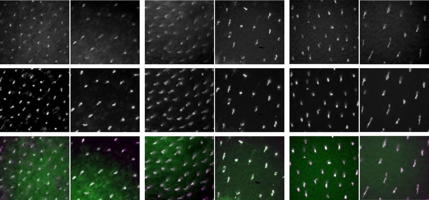
www.nature.com/scientificreports/
Figure 1. Alignment of the C-terminal amino acids of mouse (Mus musculus) CRY1 with chicken (Gallus
gallus) CRY1 and several zebra finch (Taeniopygia guttata) cryptochromes. The green box highlights the target
sequence detected by the ABCAM CRY1 antibody used in this paper, as well as the target sequence used by
Niessner26,27,56, and Bolte44. Accession numbers: mouse CRY1 (NP_031797.1), chicken CRY1 (NP_989576.1),
zebra finch CRY1a (XP_030118992.2), zebra finch CRY1b (XP_030118993.2), zebra finch CRY2a
(XP_030130159.1), zebra finch CRY2b (XP_012429630.1), zebra finch CRY4 (XP_002198533.1).
the fully oxidized state F ADOX is reduced to a semi-reduced state, the neutral semiquinone radical FADH●, by
the absorption of light in the UV and blue spectrum (peaks at about 360 nm and 470 nm). The neutral semi-
quinone radical FADH● in turn is reduced to the fully reduced state FADH- by the absorption of light from
the UV to red (peaks at about 495 nm and 580 nm), which is then re-oxidized in darkness to the fully oxidized
state FADOX33–37. The radical pairs can only be affected by the magnetic field in the semiquinone form, formed
either during photoreduction of F ADOX to FADH● or during re-oxidation of FADH- to F ADOX35,38. In both
Arabidopsis and Drosophila cryptochromes, light activation of FAD has been shown to result in a conformational
change of the C-terminal end of the cryptochrome p rotein39–43. Since Niessner and colleagues observed CRY1a
labelling only after exposures to UV to yellow light, but not after exposure in darkness or under red light, and
their antibody was directed against the C-terminal segment of the cryptochrome, they proposed that their anti-
body only detected the protein after a light-triggered conformational c hange25–28. It is worth mentioning that
during the revision of this manuscript, Bolte el at.44 confirmed that CRY1a is present in the outer segments of
UV cones in European robins, Eurasian blackcaps (Sylvia atricapilla) and domestic chickens, using an antibody
directed against the same target as Niessner et al.26. However, contrary to Niessner et al.’s results, the detection
of the CRY1a signal did not differ between light- and dark-adapted retinas, arguing against the involvement of
a conformational change of CRY1a44.
Despite the presented evidence for a role of CRY1 in avian light-dependent magnetoreception, it has been
questioned more recently whether vertebrate type II cryptochromes, which include avian CRY1, have the func-
tional properties to act as the primary magnetoreceptors. Type II cryptochromes are widely considered to be
integral parts of the negative feedback loop of the circadian clock in vertebrates by inhibiting CLOCK/BMAL1-
driven2,3,45,46. Furthermore, they are believed to have a very low binding affinity to FAD and to not be intrinsically
photosensitive47,48. Last, but not least, CRY1a, CRY1b and CRY2 have been shown to exhibit circadian expression
patterns in the retinas of several bird species, which does not exclude, but makes a role in avian magnetorecep-
tion less likely20,22,23,49–52.
In view of these findings, we reassessed the suitability of CRY1 as magnetoreceptor by localising CRY1
proteins in the retinas of zebra finches (Taeniopygia guttata, Reichenbach 1862). Zebra finches have a light-
dependent magnetic compass based on a radical-pair mechanism, similar to migratory b irds32,53–55. We recently
showed that in the zebra finch retina expression of Cry1 and Cry2 genes, unlike Cry4 genes, exhibits a clear
circadian expression profile, suggesting a role in the circadian regulation of physiological processes rather than
in magnetoreception23. Here, we examined the cellular localisation and distribution of CRY1 protein across the
zebra finch retina and tested whether the detection of CRY1 protein was wavelength dependent by examining
the abundance of CRY1 after exposure to monochromatic lights.
Results
CRY1 antibody. To detect the presence of CRY1 in the zebra finch retina we used a commercial polyclonal
antibody designed to target a peptide unique to CRY1 (Fig. 1). The target sequence is almost identical, albeit
shorter, to the sequence used by Niessner26,27,56, and more recently by Bolte44, to detect CRY1 in retinas of other
bird and mammal species. Even though the western blot analysis on total protein extracted from the retinas of
the zebra finch with our antibody revealed a single band at a lower molecular weight than the expected size (see
supplemental information), the immunofluorescent signal location and pattern coincides with that indepen-
dently reported by Niessner26,27,56, and Bolte44 and colleagues, strongly supporting that the antibody used in this
paper is likely detecting CRY1.
It is important to note that the alignment shown in Fig. 1 includes different isoforms for CRY1 and CRY2.
When we started the current study, no isoforms of CRY1 were known for the zebra finch, which is why we
make no differentiation between CRY1 and CRY1a throughout the text. Nevertheless, the alignment clearly
Scientific Reports | (2021) 11:12683 | https://doi.org/10.1038/s41598-021-92056-8 2
Vol:.(1234567890)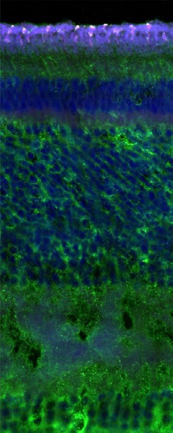
www.nature.com/scientificreports/
Figure 2. Cross section of the zebra finch retina. (A) Overview of the cross section observed with bright
field microscopy (first panel) and confocal fluorescence microscopy (second to fifth panel). The fluorescence
panels are merged into an artificially coloured merged image (fifth panel) in which blue corresponds to DAPI
(including autofluorescence due to collapsed retinal pigment epithelium), green to CRY1 and magenta to SWS1,
with colocalisation of CRY1 and SWS1 resulting in white. (B) Detail of a cross section imaged as in (A). PL
(photoreceptor layer), OS (outer segments), IS (inner segments), OLM (outer limiting membrane), ONL (outer
nuclear layer), OPL (outer plexiform layer), INL (inner nuclear layer), IPL (inner plexiform layer) and GCL
(ganglion cell layer). Bar is 20 µm in (A) and 10 µm in (B).
shows that the detected epitope corresponds to the CRY1a isoform, as is also the case for the protein detected
by Niessner26,27,56, and Bolte44.
CRY1 expression in UV cones. Evaluation of cross sections of the zebra finch retina revealed CRY1
immunolabelled cells exclusively in the photoreceptor layer (Fig. 2A, B, third panel). Some non-specific signal
in the inner nuclear layer and inner plexiform layer did not seem to be associated with any other retinal cells.
We believe it to be background noise present in that channel, or faint autofluorescence since it is also noticeable
in the negative control without primary antibody (Fig. S2C). The strong signal visible in the photoreceptor layer
in the DAPI channel is most likely due to the collapse of the pigment epithelium layer during dissection and
sectioning (also noticeable from the flattened appearance of the outer segments). The retinal pigment epithelium
contains lipofuscin, a known source of autofluorescence in the vertebrate retina57,58, and perhaps broken oil
droplets (Fig. S2C).
The CRY1 signal colocalised with the SWS1 immunolabelled cells (Fig. 2A, B, fourth and fifth panel), con-
firming that CRY1 is present exclusively in UV cones of zebra finches, and in no other photoreceptors. Like
SWS1, CRY1 was located in the outer segments of the UV cones, but it was less densely packed than SWS1 and
Scientific Reports | (2021) 11:12683 | https://doi.org/10.1038/s41598-021-92056-8 3
Vol.:(0123456789)
www.nature.com/scientificreports/

                           A                                        CRY1                                    SWS1                                       Merge

                            Peripheral retina

                           B                                        CRY1                                    SWS1                                       Merge
                            Central retina

                                                           Figure 3.  Outer segments of the zebra finch UV cones in a whole mount retina. (A) Outer segments of
                                                           photoreceptors in the peripheral retina. They appear bent and elongated because of the low density of
                                                           photoreceptors and the absence of pigment epithelium, which, when present, helps to keep them straight. The
                                                           signal from CRY1 appears only at the tip of the outer segment (first panel), while the UV opsin is visible across
                                                           the full length of the outer segment (second panel). (A) Outer segments of the photoreceptors in the central
                                                           retina. Here, the photoreceptors are not bent, because their high density keeps them packed and straight despite
                                                           the absence of pigment epithelium. The merged images are coloured as in Fig. 1. Bar 10 µm.

                                                           accumulated towards the tip of the outer segment (Fig. 3). This difference in localisation of CRY1 and SWS1
                                                           signals in the outer segment is clearly visible in the bent photoreceptors of the flat-mounted, peripheral retina
                                                           (Fig. 3A). It is not visible in flat mounts of the central retina due to the photoreceptors being straight (Fig. 3B).
                                                              The distribution of CRY1-positive cells was identical to the distribution of UV cones across all retinas exam-
                                                           ined (Fig. 4A). The evaluation of transects to quantify the colocalisation of CRY1 and SWS1 (Fig. 4B) did not
                                                           reveal any differences in distribution between left or right eyes, or sex of the birds. In all evaluated retinas we
                                                           found the same pattern of a peak density of the CRY1/SWS1 positive cells in the vicinity of the fovea and a
                                                           concentric decline towards the periphery of the retina (Fig. 5A). The density of the co-labelled cells was slightly
                                                           higher in the central, dorsal-temporal area compared to the other areas of the retina at equal distances from the
                                                           fovea (Fig. 5B).

                                                           Effect of pre‑exposure to monochromatic light on CRY1 localisation. We found no observable
                                                           differences in the colocalisation of CRY1 and SWS1 opsin, neither in the central fovea nor the periphery in
                                                           any of the retinas of birds exposed to monochromatic light of 461 nm (blue), 521 nm (green) or 633 nm (red)
                                                           prior to and during dissection (Fig. 6). CRY1 was detected in all cases irrespective of the wavelength during
                                                           pre-exposure. There were no differences in CRY1 localisation between left and right eyes, or between male and
                                                           female individuals.

                                                           Discussion
                                                           The aim of this study was to reassess the suitability of CRY1 as the primary magnetoreceptor in the retina of
                                                           birds. Our objectives were to identify the distribution and the cellular localisation of the CRY1 proteins in the
                                                           retinas of zebra finches and to test whether the localisation of CRY1 was wavelength dependent. Our findings
                                                           confirm earlier reports from migratory songbirds and chickens that CRY1 is located in the outer segments of UV

          Scientific Reports |                  (2021) 11:12683 |               https://doi.org/10.1038/s41598-021-92056-8                                                    4

Vol:.(1234567890)
www.nature.com/scientificreports/
A B Dorsal
CRY1 T2
10
19
11 25 34
8 T1
35
18
40
44
Temporal
27
Nasal
SWS1 39
Fovea
39 77
27
40
19 38
10 Pecten 27
21 17
10
T3
17
11
Ventral
Figure 4. Example of a whole-mounted retina of a zebra finch. (A) Whole-mounted retina showing a positive
signal for CRY1 (upper image in green) and for SWS1 opsin (lower image in purple). The image is from
a left eye retina, mounted with the photoreceptors side up. The banded pattern is an artefact of the digital
reconstruction of the entire image from smaller images at higher magnification. The darker regions towards
the centre of the retina correspond to thicker remains of pigment epithelium. (B) Schematic of the entire
retina shown in (A). The grey lines and dots show the sampling transects (T1, T2 and T3) and locations where
the immunopositive signals were counted. The numbers (× 100) give the counts of positive CRY1/SWS1 cells
calculated for a 1 mm2 area at the respective locations of the retina. Bar: 2 mm.
cones throughout the avian retina. However, we could not substantiate earlier reports of wavelength-dependent
effects on CRY1 localisation.
CRY1 localisation in outer segments of UV cones. Our observation that CRY1 was present in all
SWS1-labelled UV cones in the retinas of all zebra finches examined confirms earlier reports of CRY1a in UV
cones of European robins, Eurasian blackcaps and V cones of chickens26–28,44. The occurrence of CRY1 proteins
in only the UV cones, and no other retinal tissue, in zebra finches strongly suggests that our CRY1 antibody
exclusively labelled the C-terminal of the CRY1a isoform of the protein (cf.25–28). CRY1b, in contrast, has been
found in the retinal ganglion cells, displaced ganglion cells, and inner segments of some unspecified photorecep-
tors in several species of migratory songbirds and pigeons59,60 (see also61).
The localisation of CRY1 near the tip of the outer segments of the UV cones is supported by the close proxim-
ity to the UV opsins labelled with SWS1. The SWS1 antibody, like opsin antibodies in general, is known to only
label the opsins once they are fixed in the membrane of the discs of the outer segment of the cones, but not while
being transported from the nucleus in the inner segment to the base of the outer segment, where they are inte-
grated in the membrane of the discs of the outer segment (reviewed b y62). Our findings only partly agree with the
26
study by Niessner and c olleagues , which reported CRY1a detection along the full length of the outer segments
of V cones in chickens, and not only at the tip of the outer segments. The same happens with Bolte’s results in
blackcaps and robins, where the CRY1 signal also appears in the full length of the outer s egment44, and not just
at the tip of the UV cones as in our results. This difference may originate either from species-specific results, or
because the antibodies, despite being designed against a similar region, are not identical. The antiserum used by
Niessner26 and B olte44 was made against a 20 aa peptide in the C-terminal region of the CRY1a sequence, while
the antibody we used in this study was a commercial polyclonal anti-CRY1 designed against 12 aa peptide, in
the same region of the mouse CRY1 sequence and fully homologous to their 20 aa peptide (Fig. 1). This leaves
an 8 aa peptide that is recognized by Niessner’s and Bolte’s antibodies but not ours. Regardless of such differ-
ences, the localisation of the CRY1 protein far from the nucleus in the inner segment argues for a function not
directly involved in the negative feedback loop of the circadian clock, one of which could be magnetoreception.
According to the original radical-pair model, the putative magnetoreceptors should ideally be fixed along
at least one molecular axis and be evenly distributed across a hemisphere, like the avian retina, to allow for the
comparison of reaction yields of radical pairs with different alignments relative to the magnetic fi eld4,8,11 (note,
55,63–65
however, that some of these requirements are not necessarily needed ). These requirements are met by
Scientific Reports | (2021) 11:12683 | https://doi.org/10.1038/s41598-021-92056-8 5
Vol.:(0123456789)
www.nature.com/scientificreports/
Figure 5. Quantification of the co-localisation of CRY1 and SWS1 in the zebra finch retina. (A) Example of
sampled areas across Transect 1 of a whole-mounted retina (see Fig. 3B for position of sampling transects and
counting locations). The upper row shows CRY1 positive cells, the middle row SWS1 positive cells, and the
bottom row the merged images (CRY1 in green, SWS1 in purple and colocalisation is seen as white). (B) Mean
number (± standard deviations) of immunopositive cells per mm2 for each sampling location along the three
transects. The counts are based on six retinas from four individual birds (see Table S1). Bar: 30 µm.
Scientific Reports | (2021) 11:12683 | https://doi.org/10.1038/s41598-021-92056-8 6
Vol:.(1234567890)www.nature.com/scientificreports/
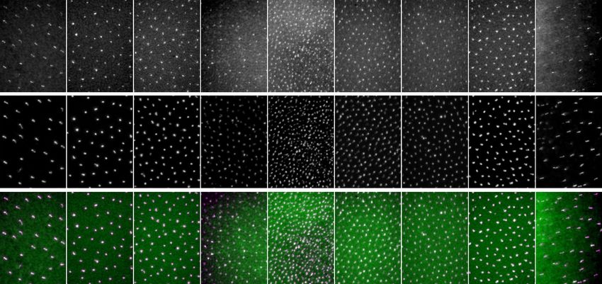
Figure 6. Colocalisation of CRY1 and SWS1 in retinas of zebra finches exposed to monochromatic
illumination. The upper row shows CRY1-positive cones, the middle row SWS1-positive cells, and the bottom
row shows the merged images (CRY1 in green, SWS1 in purple and colocalisation is seen as white). The panels
illustrate examples from birds exposed to 461 nm blue light (left), 521 nm green light (centre) and 638 nm red
light (right). For each wavelength spectrum, an example from the central retina and one from the peripheral
retina is shown. Bar: 30 µm.
the localisation of CRY1 in the UV cones of the zebra finches, assuming that the CRY1 proteins are aligned
roughly along the same axis in the cone outer segments. UV/V cones are in theory ideal candidate locations for
cryptochrome-based magnetoreceptors, as pointed out e arlier26,66. Their transparent oil droplets do not filter
out UV l ight67,68, thus light in the UV and blue spectrum can reach the FAD in the cryptochromes, which show
a high absorption of light in this wavelength range (reviewed b y2,37,38). Also, UV/V cones are the least abundant
photoreceptors in the avian retina (max. 10%, depending on species; e.g.,68–70), which would minimize interfer-
ence of light-dependent magnetoreception with vision (but see below).
Distribution of CRY1 across retina. We found an up to nine times higher density of CRY1/SWS1 cones
in the fovea of the zebra finch retina compared to the periphery, which agrees well with the general density dis-
tributions of cones in retinas of passerine birds, which usually peak in one or two foveas and decrease towards
the periphery of the retina71–74. The fovea is an important area in the visual field with a high density of cones and
none or few rods75. This area of improved visual acuity is used for various visual tasks that require high-reso-
lution vision, like food detection or obstacle avoidance76. The higher density of CRY1-positive UV cones in the
fovea of the zebra finch retina would suggest that, if they were magnetoreceptors, magnetic compass information
to be perceived at a higher spatial resolution when viewed through the fovea. This may improve the detection of
the magnetic field but could also pose a possible caveat in that the magnetic modulation pattern could interfere
with important visual tasks. This will largely depend on how the signals from the CRY1 and UV receptors are
processed [see66, and discussion on the localisation of CRY1 below].
Effect of monochromatic light on CRY1 localisation. One of the key indications for an involvement
of CRY1 in avian magnetoreception was based on the observation that CRY1 could only be detected in chicken
retinas after exposure to UV to yellow light, but not after exposure to red light or darkness27,28. Exposure of zebra
finches to 461 nm (blue), 521 nm (green) or 633 nm (red) light for one hour prior to dissection did not result
in any differences in immunodetection. We detected the CRY1 protein in the retinas of zebra finches exposed
to any of the light conditions, irrespective of the spectrum and the location in the retina (centre or periphery;
Fig. 5). This came as a surprise, since our primary antibody was designed to detect the same region (C-terminal
of CRY1), as the one used by Niessner and colleagues27,28. In both cases, the same antibody against SWS1 was
used, in similar concentrations. Niessner and colleagues argued that their CRY1a antibody detected the CRY1a
protein only after it had undergone a conformational change in the C-terminal upon activation by UV to yel-
low light, since they detected CRY1a only after exposing the birds to UV to yellow light prior to dissection, but
not after exposure to darkness or red light27,28. Nevertheless, Bolte and colleagues44 found the same discrepancy
when testing their antibody on light and dark adapted retinas. No difference was noted after the different light
treatments, which made them conclude that there was no evidence of the antibody detecting only the light-
activated form of CRY1a.
The assumption that CRY1 undergoes a conformational change was based on evidence from plant (Arabi-
dopsis) and type I (Drosophila) cryptochromes, which are both known to be directly light sensitive41–43,77. We
Scientific Reports | (2021) 11:12683 | https://doi.org/10.1038/s41598-021-92056-8 7
Vol.:(0123456789)www.nature.com/scientificreports/
are not aware of any studies showing that vertebrate type II cryptochromes undergo a conformational change
in the C-terminal as a result of light activation. On the contrary, they are suggested to be vestigial flavoproteins
which do not stably bind FAD and use the C-terminal for interactions with other clock proteins instead45–47,78.
Assuming that the CRY1 in the outer segments of the UV/V cones are located too far away from the nucleus to
be directly involved in the circadian clock, the C-terminal should be detectable to antibodies under any light
condition. We observed that the signal from the immunolabelled cells in some regions of our whole mounts was
masked by the presence of remnants of the pigment epithelium. Despite being removed to the most extent and
bleached to avoid darkening of the preparation, the pigment epithelium interfered with the fluorescent signal
of the marked cells, making it almost not differentiable from the background, and therefore easy to be misin-
terpreted as non-labelled tissue. Our own immunostainings of retinas of robins and chickens will have to show
whether this may be the explanation for the discrepancies between studies, or whether differences between the
antibodies or bird species are responsible.
Localisation of CRY1 in the avian UV/V cones suggests unique function. Based on the cellular
localisation of the CRY1 proteins at the tip of the outer segments of the UV cones, CRY1 could well be the
thought-after candidate magnetoreceptor of the light-dependent magnetic compass25. However, the high density
of the CRY1-containing UV cones in the central retina is not necessarily in support of a role in magnetorecep-
tion, even though it does not exclude this possibility. The molecular and functional properties of CRY1 also
argue against its involvement in light-dependent, radical-pair-based magnetoreception. Nevertheless, the possi-
bility remains that CRY1 might be involved in signal transduction further downstream in the signalling cascade.
If CRY1 is not involved in light-dependent magnetoreception in birds, why is it present in the outer segment
of all UV cones across the entire retina of the zebra finch, but in none of the other photoreceptors or retinal cells?
Together with the reports of CRY1a existance in UV cones of European robins and V cones of chickens26–28 our
findings suggest that CRY1 likely has a very specific function which is unique to cones expressing the SWS1
pigment and which is not required in any of the other cone or rod photoreceptors, unless other cryptochromes
are expressed in those photoreceptors instead. However, to date there is no convincing evidence that either
CRY1b or CRY2 are expressed in avian photoreceptors. CRY1b has been reported in ganglion cells and a few dis-
placed ganglion cells in retinas of pigeons (Columba livia), European robins and Northern wheatears (Oenanthe
oenanthe)59,60, and possibly in the inner segment of photoreceptors, but this latter observation was only made
by one of the groups59 and could not be substantiated by the other group60. Thus, all evidence points towards a
very specific function of CRY1 in only UV/V cones. Interestingly, CRY1 has also been found in short-wavelength
sensitive SWS1 (S1) cones in representatives of some groups of mammals (Canidae, Mustelidae and Ursidae
within Carnivora, and Hominidae, some Cercopithecidae, and possibly Lemuridae and Callitrichidae within
Primata)56. It might suggest that the expression of CRY1 is a more widespread feature of SWS1-expressing cones,
common to birds and mammals, and possibly vertebrates in general.
SWS1-expressing photoreceptors are unique in that they absorb light in the UV to V s pectrum79–81, which
is the visible light spectrum with the highest energy and has been shown to damage the retina of v ertebrates82.
The vertebrate ancestral state of SWS1 opsins is suggested to be UV sensitive, but was independently replaced
by V sensitivity in various lineages (reviewed b y79,83). These include birds which likely possessed an ancestral
V-sensitive pigment with certain lineages secondarily regaining UV sensitivity83. Since both the UV and V
cones of birds contain transparent oil droplets which contain no carotenoids and do not filter short-wavelength
light72, CRY1 located in these cones could possibly be involved in UV/V-light protection. However, in mam-
mals UV-light below 400 nm is often absorbed by the cornea and lens84, and there does not appear to be any
relationship between CRY1 expression and the degree of UV light below 400 nm reaching the SWS1 cones in
different groups of m ammal60.
Other possible functions of the CRY1 proteins in the outer segments of UV/V cones in birds could be linked
to the growing body of evidence that cryptochrome proteins, independent of their role as circadian clock regula-
tors, are directly involved in various metabolic processes, like e.g., glucocorticoid signalling85, modulation of the
cAMP pathway86, and DNA damage response (reviewed by87).
Conclusions
Based on the overall detection of CRY1 in the UV cones of the entire retina of zebra finches, CRY1 fulfils the
requirements of the radical-pair model for a spatial distribution in a hemisphere, thus could be a candidate
magnetoreceptor of the light-dependent magnetic compass in birds based on this criterion alone. However,
considering that CRY1 is expressed in a clear circadian rhythm and might not be able to harvest light on its own,
it is unlikely the primary magnetoreceptor initiating the radical-pair mechanism, even though the possibility
remains that CRY1 might be involved in signal transduction further downstream in the signalling cascade.
Nevertheless, the unique localisation of CRY1 at the tip of the UV cones in zebra finches suggests an exclusive
function, which however remains to be determined.
Materials and methods
The experimental procedures carried out in this work were planned and performed in accordance with the
ARRIVE guidelines, where applicable.
The birds were handled and terminated for tissue extraction following Swedish ethical guidelines approved
by the Malmö-Lund Animal Ethics Committee (permits M 24–16, and M 108–16).
Experimental animals and light exposure conditions. The zebra finches belonged to a permanent
captive breeding colony at Stensoffa Field Station, near Lund (Sweden). The birds were kept indoors under full-
Scientific Reports | (2021) 11:12683 | https://doi.org/10.1038/s41598-021-92056-8 8
Vol:.(1234567890)www.nature.com/scientificreports/
spectrum, indoor light conditions under a constant 12 h:12 h light–dark cycle for at least 7 days prior to dissec-
tion. The birds belonged to a heterogeneous group of sexually mature individuals (> 6 months old, 12 males and
seven females) (Table S1. All retina samples were collected during the summer and autumn of 2017 and 2018
11:00 and 12:00 CET. 4 extra males were dissected for antibody validation controls and Western Blots.
Control birds (seven males and three females) were taken directly from the holding cages for tissue collec-
tion. Birds selected for the monochromatic light experiments (five males and four females distributed among
the different monochromatic light conditions—Table S1) were exposed in individual cages to one of the three
wavelength spectra for 1 h prior to tissue collection. The dissections were done under the same light spectra as
they were treated (full spectrum white light or monochromatic light). We used the same monochromatic light
conditions as in our previous study on light-dependent magnetic compass orientation in zebra fi nches32: mono-
chromatic light with peak wavelengths at 463 nm blue, 521 nm green or 638 nm red produced by a LED array
(OF-BLR5060RGB300, OPTOFLASH, Łódź, Poland) with a total light irradiance of 15–18 × 1016 quanta s −2 m−2.
Tissue collection & whole mount preparation. Birds were sacrificed by cervical dislocation followed
by decapitation and enucleation of both eyes. Retina dissection, fixation and whole mount preparation followed
the method described in88,89. Briefly, the eyes were enucleated and immediately submerged in phosphate-buff-
ered saline (PBS) where they remained for the duration of the dissection until fixation. To access the eyecup,
a circular cut was made around the ora serrata to remove the cornea and lens. The exposed eyecups were then
fixed by immersion in 4% paraformaldehyde in 0.1 M phosphate buffer (PB), pH 7.4, for 2 h at room tempera-
ture. The fixed retinas were then placed in PBS, and the sclera was carefully detached and removed from the
retina. To flatten the retina, six cuts were made from the periphery of the eyecup towards the centre, to alleviate
the curvature. Once flat, the pecten was cut away and the pigment epithelium was mechanically removed as
much as possible. Since in most samples a portion of the pigment epithelium was strongly attached to the retina,
we bleached the dissected retinas in a solution of 3% H2O2 in 0.1 M PB overnight in the dark and at room tem-
perature. The bleached retinas were then rinsed and stored in 0.1 M PB until use. A shorter fixation (20 min)
was used in a few samples (see Table S1), where the bleaching step was replaced by a dissection in PBS at 37 °C.
The aim was to detach the pigment epithelium without the need for the bleaching step, exposing the tissue to a
less harsh protocol.
Cryosectioning. The samples were cryosectioned following standard methods90: fixed eyecups were cryo-
protected by immersion in a 25% sucrose solution overnight at 4 °C. After this, the eyecups were changed from
the sucrose solution and immersed in freezing medium (Neg-50, Richard-Allan Scientific, Thermo Fisher, Hvi-
dovre, Denmark) at room temperature for 10 min to fully coat the tissue. The tissue was further embedded
in freezing medium and frozen at − 60 °C. Semi-thin Sects. (10 µm) were cut sequentially in a Microm HM
560 cryostat (Microm, Walldorf, Germany) along the dorsal–ventral axis. The resulting sections were thawed/
mounted in chrome alum gelatine-coated microscope slides, dried overnight at room temperature, and stored
at − 20 °C until further use.
Antibodies and immunostaining. To identify which cells expressed CRY1, we used a rabbit polyclonal
antibody against the C-terminal end of the mouse CRY1 sequence (ab3518. ABCAM, Cambridge, UK; concen-
tration 1:100). Since the corresponding blocking CRY1 peptide from ABCAM was no longer available, we syn-
thesized a custom peptide spanning amino acids 594–606 of the Mouse Cryptochrome I (QSVGPKVQRQSSN;
Capra Science, Ängelholm, Sweden) for blocking controls and antibody validation. Specificity of the CRY1 anti-
body was verified by Western Blot analysis (see supplementary information). To detect UV-cones in the zebra
finch retina, we used a goat polyclonal antibody against the N-terminus of the blue opsin in humans (OPN1SW;
sc-14363. Santa Cruz, Dallas, TX, USA; concentration 1:250), which labels the SWS1 opsin in the UV and V
cones in birds. Zebra finches have UV cones, not V cones91, so we are certain that the signal of the SWS1 anti-
body labelled only UV cones. Primary antibodies were diluted in 1% Triton X-100, 0.1 M PB solution.
Cryosections were incubated with the primary antibodies overnight at room temperature in a humid chamber.
After rinsing the slides in PBS + TritonX100, the sections were incubated with the corresponding secondary fluo-
rescent antibody [anti-rabbit Alexa 647 (1:1000) to label CRY1; anti-goat Alexa 555 (1:1000) to label SWS1 opsin;
ThermoFisher] for 1 h in darkness at room temperature. The sections were rinsed again in PBS + TritonX100,
followed by PBS and then mounted with Fluoromount-G containing DAPI (SouthernBiotech, Birmingham,
USA). The secondary antibody spectrum was selected to avoid autofluorescence artefacts raising from the pig-
ment epithelium (see “Results”).
The whole mounted retinas were incubated with the primary antibodies overnight at room temperature under
constant gentle rocking. After rinsing the retinas in 0.1 M PB, the tissue was incubated with the corresponding
secondary fluorescent antibody [anti-rabbit Alexa 488 (1:1000) to label CRY1; anti-goat Alexa 555 (1:1000) to
label SWS1 opsin; ThermoFisher] for 2 h in darkness at room temperature and with constant gentle rocking. The
tissue was then rinsed again in 0.1 M PB and placed on gelatinized glass slides, with the photoreceptor side up,
and mounted with Fluoromount-G (SouthernBiotech). Control retinas were incubated in the absence of primary
antibody or with CRY1 pre-mixed with its corresponding blocking peptide to assess specificity (Figure S2).
Image acquisition and analysis. To evaluate the presence and intracellular localisation of the CRY1 pro-
teins, we analysed both cross sections and whole mounted retinas with a Leica SP8 DLS confocal microscope
(63 × /1.4 objective) with LAS-X software (Leica, Wetzlar, Germany). In addition to the immunolabelled retinal
cross-sections, we acquired images of the periphery and the centre of the whole mounted retina from z-stacks
Scientific Reports | (2021) 11:12683 | https://doi.org/10.1038/s41598-021-92056-8 9
Vol.:(0123456789)www.nature.com/scientificreports/
spanning 9 µm, starting from the base towards the tip of the outer segments of the UV cones, up to the point
where the signal disappeared.
To assess the distribution of CRY1-positive cells and cones expressing SWS1 opsin throughout the retina,
mounted slides were inspected on an AXIOPHOT Fluorescence Microscope (25 × /0.8 and 40 × /1.3 Plan-Neofluar
objectives; Zeiss, Oberkochen, Germany). The images were captured with a NIKON DS-fi1c CCD camera with
NIS-elements software (Nikon. Tokyo, Japan). Composed images of entire retinas were reconstructed using
Adobe Photoshop CS6 (San Jose, CA, USA). We established three transects with seven sampling points each to
sample the photoreceptor density across the control retinas and to evaluate the degree of co-expression of CRY1
and SWS1. Each transect covered the retina from one periphery, passing through the central fovea all the way
to the opposite periphery, so that they all intersected each other at the fovea. At each sampling point, images
of each secondary antibody fluorescence were taken, and the number of positive signals was quantified using
a custom particle counting software written in Matlab R2016b (The MathWorks Inc., Natick, MA, USA). The
program counted positive cells on a selected region of interest of 0.1 mm2. Counts from equivalent transect points
from all retinas examined were averaged and presented as a histogram, with standard deviation as error bars.
Data availability
The data generated and analysed in the present study are included in the manuscript and its supplementary
information files, or available on request from the corresponding author.
Received: 16 November 2020; Accepted: 3 June 2021
References
1. Cashmore, A. R. Cryptochromes: Blue light receptors for plants and animals. Science 284, 760–765 (1999).
2. Chaves, I. et al. The cryptochromes: Blue light photoreceptors in plants and animals. Annu. Rev. Plant Biol. 62, 335–364 (2011).
3. Haug, M. F., Gesemann, M., Lazović, V. & Neuhauss, S. C. F. Eumetazoan cryptochrome phylogeny and evolution. Genome Biol.
Evol. 7, 601–619 (2015).
4. Ritz, T., Adem, S. & Schulten, K. A Model for photoreceptor-based magnetoreception in birds. Biophys. J . 78, 707–718 (2000).
5. Schulten, K., Swenberg, C. E. & Weller, A. A biomagnetic sensory mechanism based on magnetic field modulated coherent electron
spin motion. Z. Phys. Chem. 111, 1–5 (1978).
6. Rodgers, C. T. & Hore, P. J. Chemical magnetoreception in birds: The radical pair mechanism. Proc. Natl. Acad. Sci. 106, 353–360
(2009).
7. Dodson, C. A., Hore, P. J. & Wallace, M. I. A radical sense of direction: Signalling and mechanism in cryptochrome magnetorecep-
tion. Trends Biochem. Sci. 38, 435–446 (2013).
8. Hore, P. J. & Mouritsen, H. The radical-pair mechanism of magnetoreception. Annu. Rev. Biophys. 45, 299–344 (2016).
9. Phillips, J. B., Muheim, R. & Jorge, P. E. A behavioral perspective on the biophysics of the light-dependent magnetic compass: A
link between directional and spatial perception?. J. Exp. Biol. 213, 3247–3255 (2010).
10. Phillips, J. B., Jorge, P. E. & Muheim, R. Light-dependent magnetic compass orientation in amphibians and insects: Candidate
receptors and candidate molecular mechanisms. J. R. Soc. Interface 7, S241–S256 (2010).
11. Solov’yov, I. A., Mouritsen, H. & Schulten, K. Acuity of a cryptochrome and vision-based magnetoreception system in birds.
Biophys. J. 99, 40–49 (2010).
12. Deutschlander, M. E., Phillips, J. B. & Borland, S. C. The case for light-dependent magnetic orientation in animals. J. Exp. Biol.
202, 891–908 (1999).
13. Wiltschko, W. & Wiltschko, R. Magnetic orientation and magnetoreception in birds and other animals. J Comp Physiol A 191,
675–693 (2005).
14. Muheim, R. & Liedvogel, M. The light-dependent magnetic compass. in Photobiology (ed. Björn, L. O.) 323–334 (Springer, New
York, 2015).
15. Henbest, K. B. et al. Magnetic-field effect on the photoactivation reaction of Escherichia coli DNA photolyase. Proc. Natl. Acad.
Sci. 105, 14395–14399 (2008).
16. Maeda, K. et al. Chemical compass model of avian magnetoreception. Nature 453, 387–390 (2008).
17. Mouritsen, H. et al. Cryptochromes and neuronal-activity markers colocalize in the retina of migratory birds during magnetic
orientation. Proc. Natl. Acad. Sci. 101, 14294–14299 (2004).
18. Möller, A., Sagasser, S., Wiltschko, W. & Schierwater, B. Retinal cryptochrome in a migratory passerine bird: a possible transducer
for the avian magnetic compass. Naturwissenschaften 91, 585–588 (2004).
19. Liedvogel, M. & Mouritsen, H. Cryptochromes—A potential magnetoreceptor: What do we know and what do we want to know?.
J. R. Soc. Interface 7, S147–S162 (2010).
20. Singh, D., Rani, S. & Kumar, V. Daily expression of six clock genes in central and peripheral tissues of a night-migratory SongBird:
Evidence for tissue-specific circadian timing. Chronobiol. Int. 30, 1208–1217 (2013).
21. Fusani, L., Bertolucci, C., Frigato, E. & Foa, A. Cryptochrome expression in the eye of migratory birds depends on their migratory
status. J. Exp. Biol. 217, 918–923 (2014).
22. Günther, A. et al. Double-cone localization and seasonal expression pattern suggest a role in magnetoreception for European Robin
cryptochrome 4. Curr. Biol. 28, 211-223.e4 (2018).
23. Pinzon-Rodriguez, A., Bensch, S. & Muheim, R. Expression patterns of cryptochrome genes in avian retina suggest involvement
of Cry4 in light-dependent magnetoreception. J. R. Soc. Interface. 15, 20180058 (2018).
24. Einwich, A., Dedek, K., Seth, P. K., Laubinger, S. & Mouritsen, H. A novel isoform of cryptochrome 4 (Cry4b) is expressed in the
retina of a night-migratory songbird. Sci. Rep. 10, 15794 (2020).
25. Wiltschko, R., Nießner, C. & Wiltschko, W. The magnetic compass of birds: The role of cryptochrome. Front. Physiol. 12, 584
(2021).
26. Niessner, C. et al. Avian ultraviolet/violet cones identified as probable magnetoreceptors. PLoS ONE 6, e20091 (2011).
27. Niessner, C. et al. Magnetoreception: Activated cryptochrome 1a concurs with magnetic orientation in birds. J. R. Soc. Interface
10, 20130638 (2013).
28. Niessner, C., Denzau, S., Peichl, L., Wiltschko, W. & Wiltschko, R. Magnetoreception in birds: I. Immunohistochemical studies
concerning the cryptochrome cycle. J. Exp. Biol. 217, 4221–4224 (2014).
29. Muheim, R., Bäckman, J. & Åkesson, S. Magnetic compass orientation in European robins is dependent on both wavelength and
intensity of light. J. Exp. Biol. 205, 3845–3856 (2002).
Scientific Reports | (2021) 11:12683 | https://doi.org/10.1038/s41598-021-92056-8 10
Vol:.(1234567890)www.nature.com/scientificreports/
30. Wiltschko, R., Stapput, K., Thalau, P. & Wiltschko, W. Directional orientation of birds by the magnetic field under different light
conditions. J. R. Soc. Interface. 7, S163–S177 (2010).
31. Wiltschko, R. et al. Orientation of migratory birds under ultraviolet light. J Comp Physiol A 200, 399–407 (2014).
32. Pinzon-Rodriguez, A. & Muheim, R. Zebra finches have a light-dependent magnetic compass similar to migratory birds. J Exp
Biol 220, 1202–1209 (2017).
33. Bouly, J.-P. et al. Cryptochrome blue light photoreceptors are activated through interconversion of Flavin Redox States. J. Biol.
Chem. 282, 9383–9391 (2007).
34. Liedvogel, M. et al. Chemical magnetoreception: Bird cryptochrome 1a is excited by blue light and forms long-lived radical-pairs.
PLoS ONE 2, e1106 (2007).
35. Kao, Y.-T. et al. Ultrafast dynamics of flavins in five Redox States. J. Am. Chem. Soc. 130, 13132–13139 (2008).
36. Ozturk, N. et al. Comparative photochemistry of animal type 1 and type 4 cryptochromes. Biochemistry 48, 8585–8593 (2009).
37. Liu, B., Liu, H., Zhong, D. & Lin, C. Searching for a photocycle of the cryptochrome photoreceptors. Curr. Opin. Plant Biol. 13,
578–586 (2010).
38. Wang, J., Du, X., Pan, W., Wang, X. & Wu, W. Photoactivation of the cryptochrome/photolyase superfamily. J. Photochem. Photobiol.,
C 22, 84–102 (2015).
39. Kondoh, M. & Terazima, M. Conformational and intermolecular interaction dynamics of photolyase/cryptochrome proteins
monitored by the time-resolved diffusion technique. Photochem. Photobiol. 93, 15–25 (2017).
40. Lin, C., Top, D., Manahan, C. C., Young, M. W. & Crane, B. R. Circadian clock activity of cryptochrome relies on tryptophan-
mediated photoreduction. Proc. Acad. Sci. https://doi.org/10.1073/pnas.1719376115 (2018).
41. Ozturk, N., Selby, C. P., Annayev, Y., Zhong, D. & Sancar, A. Reaction mechanism of Drosophila cryptochrome. Proc. Natl. Acad.
Sci. 108, 516–521 (2011).
42. Ganguly, A. et al. Changes in active site histidine hydrogen bonding trigger cryptochrome activation. PNAS 113, 10073–10078
(2016).
43. Zoltowski, B. D. et al. Structure of full-length Drosophila cryptochrome. Nature 480, 396–399 (2011).
44. Bolte, P. et al. Cryptochrome 1a localisation in light- and dark-adapted retinae of several migratory and non-migratory bird spe-
cies: no signs of light-dependent activation. Ethol. Ecol. Evolution 0, 1–25 (2021).
45. Czarna, A. et al. Structures of Drosophila cryptochrome and mouse cryptochrome1 provide insight into circadian function. Cell
153, 1394–1405 (2013).
46. Ozturk, N. Phylogenetic and functional classification of the photolyase/cryptochrome family. Photochem. Photobiol. 93, 104–111
(2017).
47. Kutta, R. J., Archipowa, N., Johannissen, L. O., Jones, A. R. & Scrutton, N. S. Vertebrate cryptochromes are vestigial flavoproteins.
Sci Rep 7, 44906 (2017).
48. Wang, X. et al. Comparative properties and functions of type 2 and type 4 pigeon cryptochromes. Cell. Mol. Life Sci. 75, 4629–4641
(2018).
49. Fu, Z., Inaba, M., Noguchi, T. & Kato, H. Molecular cloning and circadian regulation of cryptochrome genes in Japanese quail
(Coturnix coturnix japonica). J Biol Rhythms 17, 14–27 (2002).
50. Haque, R., Chaurasia, S. S., Wessel, J. H. 3rd. & Iuvone, P. M. Dual regulation of cryptochrome 1 mRNA expression in chicken
retina by light and circadian oscillators. NeuroReport 13, 2247–2251 (2002).
51. Kubo, Y., Akiyama, M., Fukada, Y. & Okano, T. Molecular cloning, mRNA expression, and immunocytochemical localization of
a putative blue-light photoreceptor CRY4 in the chicken pineal gland. J Neurochem 97, 1155–1165 (2006).
52. Trivedi, A. K., Malik, S., Rani, S. & Kumar, V. Pinealectomy abolishes circadian behavior and interferes with circadian clock gene
oscillations in brain and liver but not retina in a migratory songbird. Physiol. Behav. 156, 156–163 (2016).
53. Voss, J., Keary, N. & Bischof, H.-J. The use of the geomagnetic field for short distance orientation in zebra finches. NeuroReport
18, 1053–1057 (2007).
54. Keary, N. et al. Oscillating magnetic field disrupts magnetic orientation in Zebra finches, Taeniopygia guttata. Front Zool 6, 25
(2009).
55. Muheim, R., Sjöberg, S. & Pinzon-Rodriguez, A. Polarized light modulates light-dependent magnetic compass orientation in birds.
Proc Natl Acad Sci USA 113, 1654–1659 (2016).
56. Niessner, C. et al. Cryptochrome 1 in retinal cone photoreceptors suggests a novel functional role in mammals. Sci Rep 6, 21848
(2016).
57. Marmorstein, A. D., Marmorstein, L. Y., Sakaguchi, H. & Hollyfield, J. G. Spectral profiling of autofluorescence associated with
lipofuscin, Bruch’s membrane, and sub-RPE deposits in normal and AMD Eyes. Invest. Ophthalmol. Vis. Sci. 43, 2435–2441 (2002).
58. Double, K. L. et al. The comparative biology of neuromelanin and lipofuscin in the human brain. Cell. Mol. Life Sci. 65, 1669–1682
(2008).
59. Bolte, P. et al. Localisation of the putative magnetoreceptive protein cryptochrome 1b in the retinae of migratory birds and homing
pigeons. PLoS ONE 11, e0147819 (2016).
60. Niessner, C. et al. Seasonally changing cryptochrome 1b expression in the retinal ganglion cells of a migrating passerine bird. PLoS
ONE 11, e0150377 (2016).
61. Niessner, C. & Winklhofer, M. Radical-pair-based magnetoreception in birds: radio-frequency experiments and the role of cryp-
tochrome. J. Comp. Physiol. A 203, 499–507 (2017).
62. Pearring, J. N., Salinas, R. Y., Baker, S. A. & Arshavsky, V. Y. Protein sorting, targeting and trafficking in photoreceptor cells. Prog.
Retin. Eye Res. 36, 24–51 (2013).
63. Hill, E. & Ritz, T. Can disordered radical pair systems provide a basis for a magnetic compass in animals?. J. R. Soc. Interface 7,
S265–S271 (2010).
64. Lau, J. C. S., Wagner-Rundell, N., Rodgers, C. T., Green, N. J. B. & Hore, P. J. Effects of disorder and motion in a radical pair mag-
netoreceptor. J. R. Soc. Interface 7, S257–S264 (2010).
65. Lau, J. C. S., Rodgers, C. T. & Hore, P. J. Compass magnetoreception in birds arising from photo-induced radical pairs in rotation-
ally disordered cryptochromes. J. R. Soc. Interface 9, 3329–3337 (2012).
66. Bischof, H.-J., Niessner, C., Peichl, L., Wiltschko, R. & Wiltschko, W. Avian ultraviolet/violet cones as magnetoreceptors: The
problem of separating visual and magnetic information. Communicative & Integrative Biology 4, 713–716 (2011).
67. Bowmaker, J. K., Heath, L. A., Wilkie, S. E. & Hunt, D. M. Visual pigments and oil droplets from six classes of photoreceptor in
the retinas of birds. Vision. Res. 37, 2183–2194 (1997).
68. Hart, N. S., Partridge, J. C., Bennett, A. T. D. & Cuthill, I. C. Visual pigments, cone oil droplets and ocular media in four species
of estrildid finch. J Comp Physiol A 186, 681–694 (2000).
69. Hart, N. S., Partridge, J. C. & Cuthill, I. C. Visual pigments, oil droplets and cone photoreceptors distribution in the European
Starling, Sturnus vulgaris. J. Exp. Biol. 201, 1433–1446 (1998).
70. Hart, N. S., Partridge, J. C., Cuthill, I. C. & Bennett, A. T. D. Visual pigments, oil droplets, ocular media and cone photoreceptor
distribution in two species of passerine bird: the blue tit (Parus caeruleus L.) and the blackbird (Turdus merula L). J. Comp. A:
Sens. Neural Behav. Physiol. 186, 375–387 (2000).
71. Meyer, D. B. & May, H. C. The topographical distribution of rods and cones in the adult chicken retina. Exp. Eye Res. 17, 347–355
(1973).
Scientific Reports | (2021) 11:12683 | https://doi.org/10.1038/s41598-021-92056-8 11
Vol.:(0123456789)www.nature.com/scientificreports/
72. Hart, N. S. The visual ecology of avian photoreceptors. Prog. Retin. Eye Res. 20, 675–703 (2001).
73. Coimbra, J. P., Collin, S. P. & Hart, N. S. Variations in retinal photoreceptor topography and the organization of the rod-free zone
reflect behavioral diversity in Australian passerines. Journal of Comparative Neurology 523, 1073–1094 (2015).
74. Moore, B. A., Pita, D., Tyrrell, L. P. & Fernandez-Juricic, E. Vision in avian emberizid foragers: maximizing both binocular vision
and fronto-lateral visual acuity. J. Exp. Biol. 218, 1347–1358 (2015).
75. Meyer, D. B. The avian eye and its adaptations. in The Visual System in Vertebrates. Handbook of Sensory Physiology (ed. Crescitelli,
F.) vol. 7/5, 549–611 (Springer, Berlin, Heidelberg, 1977).
76. Bischof, H.-J. The visual field and visually guided behavior in the zebra finch (Taeniopygia guttata). J. Comp. Physiol. 163, 329–337
(1988).
77. Kondoh, M. et al. Light-induced conformational changes in full-length Arabidopsis thaliana cryptochrome. J. Mol. Biol. 413,
128–137 (2011).
78. Merbitz-Zahradnik, T. & Wolf, E. How is the inner circadian clock controlled by interactive clock proteins?. FEBS Lett. 589,
1516–1529 (2015).
79. Hunt, D. M., Carvalho, L. S., Cowing, J. A. & Davies, W. L. Evolution and spectral tuning of visual pigments in birds and mammals.
Philosophical Transactions of the Royal Society B: Biological Sciences 364, 2941–2955 (2009).
80. Davies, W. I. L. et al. Vertebrate ancient opsin photopigment spectra and the avian photoperiodic response. Biol. Let. 8, 291–294
(2012).
81. Hunt, D. M. & Peichl, L. S cones: Evolution, retinal distribution, development, and spectral sensitivity. Visual Neurosci. 31, 115–138
(2014).
82. Carvalho, L. S., Knott, B., Berg, M. L., Bennett, A. T. D. & Hunt, D. M. Ultraviolet-sensitive vision in long-lived birds. Proc. R. Soc.
Lond.B: Biol. Sci. 278, 107–114 (2011).
83. Shi, Y. & Yokoyama, S. Molecular analysis of the evolutionary significance of ultraviolet vision in vertebrates. Proc. Natl. Acad. Sci.
100, 8308–8313 (2003).
84. Douglas, R. H. & Jeffery, G. The spectral transmission of ocular media suggests ultraviolet sensitivity is widespread among mam-
mals. Proc. R. Soc. Lond.B: Biolog. Sci. 281, 20132995 (2014).
85. Lamia, K. A. et al. Cryptochromes mediate rhythmic repression of the glucocorticoid receptor. Nature 480, 552–556 (2011).
86. Zhang, E. E. et al. Cryptochrome mediates circadian regulation of cAMP signaling and hepatic gluconeogenesis. Nat. Med. 16,
1152–1156 (2010).
87. Michael, A. K., Fribourgh, J. L., Gelder, R. N. V. & Partch, C. L. Animal Cryptochromes: Divergent roles in light perception, cir-
cadian timekeeping and beyond. Photochem. Photobiol. 93, 128–140 (2017).
88. Coimbra, J. P., Marceliano, M. L. V., da Andrade-da-Costa, B. L. S. & Yamada, E. S. The retina of tyrant flycatchers: Topographic
organization of neuronal density and size in the ganglion cell layer of the Great Kiskadee Pitangus sulphuratus and the rusty
margined flycatcher Myiozetetes cayanensis (Aves: Tyrannidae). Brain Behav Evol 68, 15–25 (2006).
89. Coimbra, J. P. et al. Number and distribution of neurons in the retinal ganglion cell layer in relation to foraging behaviors of tyrant
flycatchers. J. Comp. Neurol. 514, 66–73 (2009).
90. Ekström, P., Garm, A., Pålsson, J., Vihtelic, T. S. & Nilsson, D.-E. Immunohistochemical evidence for multiple photosystems in
box jellyfish. Cell Tissue Res 333, 115–124 (2008).
91. Hart, N. Variations in cone photoreceptor abundance and the visual ecology of birds. J. Comp. Physiol. A 187, 685–697 (2001).
Acknowledgements
We thank Joao-Paulo Coimbra for acting as a co-advisor and teaching A.P.-R. all the technical skills, like avian
retina dissections and immunohistochemistry methods. This study would not have been possible without his
invaluable expertise in fine tuning of the immunohistochemistry protocol and help with the interpretation of
the findings. We thank Carina Rasmussen and Ola Gustafsson from the Biology Department of Lund University
for invaluable technical assistance with immunohistochemical procedures and confocal imaging, respectively.
Author contributions
A.P.-R. and R.M. designed the experiments. A.P.-R. carried out the lab work and analysed the data. A.P.-R. wrote
a first draft of the manuscript. A.P.-R. and R.M. wrote the final version.
Funding
Open access funding provided by Lund University. This work was funded by Vetenskapsrådet (2011-4765 and
2015-04869 to R.M.), Crafoordska Stiftelsen (2010-1001, 2013-0737 and 2019-0839 to R.M.) and The Colombian
Ministry of Science – Minciencias (formerly Colciencias: Grant 568 from Departamento Administrativo de
Ciencia, Tecnología e Innovación to A.P-R.).
Competing interests
The authors declare no competing interests.
Additional information
Supplementary Information The online version contains supplementary material available at https://doi.org/
10.1038/s41598-021-92056-8.
Correspondence and requests for materials should be addressed to A.P.-R.
Reprints and permissions information is available at www.nature.com/reprints.
Publisher’s note Springer Nature remains neutral with regard to jurisdictional claims in published maps and
institutional affiliations.
Scientific Reports | (2021) 11:12683 | https://doi.org/10.1038/s41598-021-92056-8 12
Vol:.(1234567890)You can also read