Deep Learning based Dimple Segmentation for Quantitative Fractography
←
→
Page content transcription
If your browser does not render page correctly, please read the page content below
Deep Learning based Dimple Segmentation for Quantitative Fractography

                                                                                            Ashish Sinha
                                                                       Department of Metallurgical and Materials Engineering
                                                                              Indian Institute of Technology Roorkee
                                                                                      Roorkee-247 667, India
                                                                                            asinha@mt.iitr.ac.in
arXiv:2007.02267v3 [eess.IV] 1 Oct 2020

                                                                                             K.S Suresh
                                                                       Department of Metallurgical and Materials Engineering
                                                                              Indian Institute of Technology Roorkee
                                                                                      Roorkee-247 667, India
                                                                                          ks.suresh@mt.iitr.ac.in

                                                                   Abstract                                    nature during deformation and stress accumulation. Defor-
                                                                                                               mation of metals caused due to application of load or cor-
                                              In this work, we try to address the challenging problem          rosive actions of nature, causes an accumulation of pores
                                          of dimple segmentation in titanium alloys using machine              predominantly in the central part of the neck of the frac-
                                          learning methods, especially neural networks. The frac-              ture, which coalesce with grain (can be thought as domains
                                          tographic images for this task are obtained using a Scan-            in magnetic field) conglomerates leading to the growth of
                                          ning Election Microscope (SEM). To determine the cause               the crack in a continuous fashion in the direction of load-
                                          of fracture in metals we address the problem of segmen-              ing. Thus, the central crack which grows by thinning and
                                          tation of dimples in fractographs i.e. the fracture surface          breaking connections between the pores, together with the
                                          of metals using supervised machine learning methods. De-             newly formed crack leaves traces on the surface in the form
                                          termining the cause of fracture would help us in material            of dimples, which indicates the history of the material frac-
                                          property, mechanical property prediction and development             ture [4] [5] [6].
                                          of new fracture-resistant materials. This method would also
                                          help in correlating the topographical features of the frac-
                                          ture surface with the mechanical properties of the material.
                                          Our proposed novel model achieves the best performance as
                                          compared to other previous approaches. To the best of our
                                          knowledge, this is one of the first work in fractography using
                                          fully convolutional neural networks with self-attention for
                                          supervised learning of deep dimple fractography, though it
                                          can be easily extended to account for brittle characteristics
                                          as well.

                                          1. Introduction
                                              Titanium is an important metal for making the plates                              Figure 1. Stress-Strain curve
                                          of body armour of soldiers, body implants, surgical instru-
                                          ments. In addition, titanium alloys are also used for making            The process of damage to a material can be depicted on
                                          aircrafts and spacecrafts due to it’s high strength and wear         stress-strain curve, whereas fracture is the final stage of de-
                                          resistance [1] [2] [3].                                              formation 1. These links between the stages of deformation
                                              Fracture patterns of metals in general and high-strength         are important when analyzing the causes of fracture using
                                          titanium and iron alloys in particular happens in a stage-like       fractographic analysis. Fractographic analysis uses physics

                                                                                                           1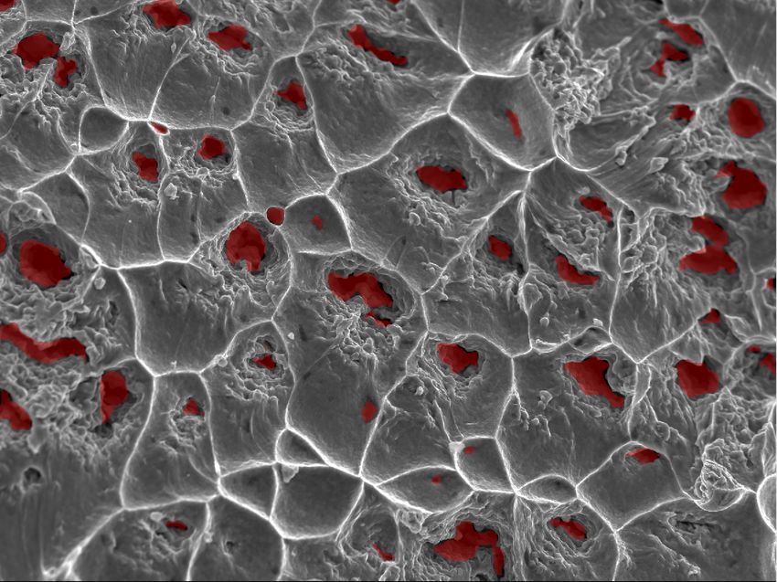
of solid body, material science, optic-digital methods to de-         structures in a progressive way. Most of the effort was put
termine the causes of fracture. Earlier, parameter measure-           in to learn a defined material system and different classes
ments of fracture surfaces were made manually or automat-             of micro structures.if the features of micro structures are
ically but the software was positioned by a operator. A large         known then digital analysis techniques on images would be
variety of materials made the generalization difficult [7].           helpful in characterising, segmenting and comparing differ-
                                                                      ent structures with high resolution. Presently the analytics
                                                                      of microstructures is mostly focused on finding the relation-
                                                                      ship between structure and properties by using the shape
                                                                      and size and appearance of the features. These approaches
                                                                      have moved forward to more of a machine learning ap-
                                                                      proach where the properties are found out by the choosing
                                                                      of correct algorithms. Some of the areas where machine
                                                                      learning techniques are used in metallurgy and materials
                                                                      science are: new material design, material property predic-
                                                                      tion, microstructure recognition, and analysis of failure in a
                                                                      material, etc.

         Figure 2. Steps of ductile fracture under stress             2.1. Computer Vision in Quantitative Fractography
                                                                          The main interest in fractography is due in finding the
    Administered by both the extrinsic (e.g. imposed
                                                                      correlation between the features of the surface that is frac-
loading, environmental conditions) and the intrinsic (mi-
                                                                      tured and the environment or conditions that lead to its fail-
crostructure) characteristics is the process of fracture of ma-
                                                                      ure. For centuries, this has remained qualitative in nature.
terials. The data regarding the affect of both intrinsic and
                                                                      Mostly, scientists examine the Scanning Electron Micro-
extrinsic characteristics of the fracture process is contained
                                                                      scope (SEM), Optical Microscope (OPM) images of sam-
in the surface of the fracture. Important means applied to
                                                                      ples for failure analysis. Therefore quantitative analysis
study the surface of fracture is fractography and obtain fac-
                                                                      brings the potential for improving and understanding the
tors approaching to failure and data relating properties of
                                                                      mechanisms that control the fracture process and also de-
material. To attain the topographic characterization of the
                                                                      termine the reliability of the models that in the current ma-
surface, fractography is used. Analyzing and classifying
                                                                      terial design system. With the latest advancement in the
the several mechanisms of fracture, and the interrelation-
                                                                      field of image analysis and moreover the availability of ma-
ship between them with the microstructure of material,the
                                                                      chine learning tools, it has become more easy to automate
situations approaching towards it’s failure, and its mechani-
                                                                      the event of finding important features and information from
cal nature are also some applications in which fractography
                                                                      the fractographs.
plays its part.
    Formation of dimples on the fracture surface is due to                The use of computer vision methodologies by [8] [9]
the ductile fracture of materials. Generally, the micro-pore          lead to identify the images which contained dendritic mor-
merger in the material during deformation in plastic region           phology to classify if the the direction was longitudinal or
leads to such dimples. After effect of the pore rupture and           transverse. Another use case has been automatic measuring
the destruction of the surrounding material are dimples as            the volume of ferritic(iron) volume fraction from the binary
shown in 2. Many models of nucleation, growth, and coa-               phase structures of ferritic and austenite (a phase of iron).
lescence of pores are known and their application under un-           In the field of fractography, many successful attempts have
certain condition leads to fracture of materials, the results         been made to build automated models for quantitative frac-
of which are usually complicated to compare.                          tography [7] used non-linear algorithms of machine learn-
                                                                      ing (ANN and SVM) and combined it with texture analysis
    The ponderous process which is quantitative and quali-
                                                                      to classify the images into there modes: ductile sudden, brit-
tative assessment of fracture surface is executed manually
                                                                      tle sudden and fatigue. A recent work [10] aims to quantify
by experienced and well-trained technicians, hence requir-
                                                                      fracture surface for materials with brittle fracture character-
ing significant labour. Quantitative estimations are likely
                                                                      istics. Our work focuses primarily on ductile materials.
to have more errors because there is a huge dependency on
human factor.                                                             In our project, we use different variants of deep neural
                                                                      networks to segment deep dimples and benchmark the re-
                                                                      sults, in ductile fracture materials on SEM images of tita-
2. Related Work
                                                                      nium alloys which can be further used to find the properties
   In the last century, metallurgists and material scientists         of fracture mechanism.
have acquired, analysed and compared images of micro-                     In previous works, the authors have proposed methods

                                                                  2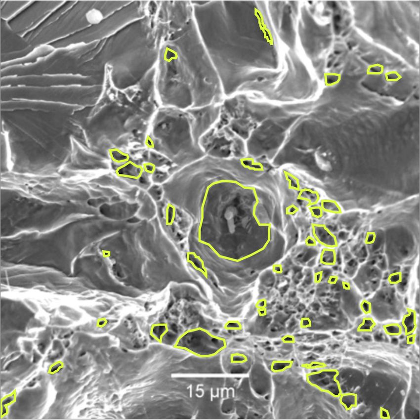
for fractographic recognition, control and calculation of pa-         in the structure. These defects can take many forms but the
rameters of the dimples of based on neural networks [11]              primary ones are the pores. To acquire micrographs, both
[12]. In [13], the authors trained 17 models of neural                optical as well as electron microscopy is used.
networks with various sets of hyper-parameters, then their
speed and accuracy were evaluated and the optimal neural              2.5. Dimple Fracture
network was selected. We propose a fully convolutional
U-Net [14] inspired deep neural network with position and                A dimple fracture is a type of material failure on a
channel based attention residual blocks with dense connec-            metal’s surface that is characterized by the formation and
tions [15] [16] [17] and squeeze and excitation [18] block in         collection of micro-voids along the granular boundary of
the bottleneck layer for the segmentation of deep dimples.            the metal i.e. the fracture path. The occurrence of dim-
                                                                      ple fractures is directly proportional to increased corrosion
   Our work explores the application of deep learning meth-
                                                                      rates. The material appears physically dimpled when exam-
ods in fractography, an active field of research in material
                                                                      ined under high magnification. There are three main types
science. Below, we briefly explain the terms necessary to
                                                                      of dimple fractures:
better understand our work.

2.2. Fractography                                                       • Shear fractures
   Fractography is a technique to understand the causes
of failures and also to verify theoretical failure predictions          • Tearing fractures
with real life failures. It can be used in forensics, for ana-
lyzing broken products which have been used as weapons,                 • Tensile fractures
such as broken bottles. Thus, a defendant might claim that
a bottle was faulty and broke accidentally when it impacted              All three of these fractures are characterized by tiny
a victim of an assault. Fractography could show the allega-           holes, known as micro-voids, which are microscopically lo-
tion to be false and that considerable force was needed to            cated in the interior of a piece of metal when under the force
smash the bottle before using the broken end as a weapon              of an external load. The greater the load, the greater the
to deliberately the victim. In these cases, the overall pat-          proximity and the total gap volume of these voids. The ap-
tern of cracking is important in reconstructing the sequence          pearance of such a fractured surface is referred to as a dim-
of events, rather than the specific characteristics of a single       ple rupture. A scanning electron microscope can be used to
crack, since crack grows by coalescing with other grains in           examine a dimple rupture at a magnification of about 2500x.
the microstructure of the metals.

2.3. Crack Growth                                                     3. Methodology
   The initiation and continuation of crack growth is de-                Here we discuss the fractographic analysis for detection
pendant on several factors such as bulk material properties,          of dimples in fracture metal surfaces using various deep
geometry of the body, geometry of the crack, loading rate,            learning models. We consider Titanium (Ti) alloys as our
loading distribution, load magnitude, environmental condi-            focus of discussion. In the next section we briefly discuss
tions, time and microstructure. Cracks are initiated, and as          the previous methods used for segmentation tasks but which
the cracks grow, energy is transmitted to the crack tip at an         are new in this domain, and then we explain our proposed
energy release rate G, which is a function of the applied             model.
load, crack length and the geometry of the body. All solid
materials, have an intrinsic energy release rate GC , where           3.1. Previous Approaches
GC is referred to as the fracture energy or fracture tough-
                                                                      3.1.1   U-Net
ness of the material. A crack will grow if G ≥ GC .
                                                                      The U-Net [14] is mainly employed for bio-medical image
2.4. Microstructure
                                                                      segmentation. It has two parts: a contracting encoder and
    Microstructure is a very small scale structure of a ma-           an expanding decoder. The encoder, a feature correction
terial, defined as the structure of a prepared surface of a           path, is a continuous stack of convolution and pooling lay-
material as revealed under an optical microscope above 25x            ers; used for image identification. The decoder, a feature
magnification. The microstructure of a material correlates            expanding path, is used for collecting exact localisation of
strongly with the strength, toughness, ductility, hardness,           fractures using transposed convolutions or deconvolutions.
wear resistance, etc of the material. A microstructure’s in-          The model is an end-to-end fully convolutional network.
fluence on the physical and mechanical properties of a ma-            The image of any size can be fed in the model as it lacks
terial is governed by the different defects present or absent         any densely connected layer.

                                                                  3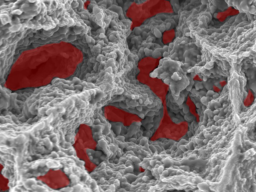
3.1.2 U-Net++ 4. Evaluation
This model uses dense block ideas of DenseNet [17] to im- 4.1. Dataset
prove upon U-Net [14].
This model architecture consists of an encoder sub-
network and a decoder sub-network after it. The skip con-
nections between each node comprises of three convolution
layers and a deep dense convolution block. A convolution
layer follows every concatenation layer. This convolution
layer considers output from preceding convolution layers of
the dense block and up-sampled output from lower dense
block, and fuses them.
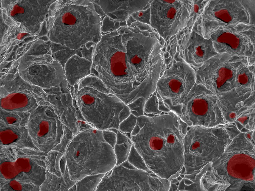
Figure 4. A sample fractograph and it’s GT showing dimples
3.1.3 Mask R-CNN
Mask R-CNN [19] is the skeletal that assists in object de- For conducting our experiments, we collected 216 high-
tection and localization. It completes the task in two steps: resolution SEM images of Ti alloys (tested under vari-
scanning the image and generating proposals to point the ous physical conditions) at 200x, 500x, 1000x and 2000x
probable locations of an object,bclassification on first step magnification. It is a very difficult process to obtain this
proposals, and generation of bounding boxes and masks. kind of data in such magnitude since it requires heavy pre-
processing with chemicals of the metal surface before it can
3.2. Proposed Model be viewed under a scanning electron microscope. Typically,
it required around a day for the polishing and obtaining a
Like the U-Net architecture, our proposed network is di- SEM image of the metal surface. We performed the exten-
vided into two parts, a contracting encoder and an expand- sive tedious task of annotating the SEM images with deep
ing decoder. For the encoder part, we employ 5 layers of dimples. During annotation, we classify the areas of the
residual convolutional blocks with channel and spatial at- SEM images that presented the most characteristic features
tention as proposed in [20] but instead of using attention of deep dimple (dark areas), while the areas with unclear
blocks in parallel we find that using them as proposed in classification or ambiguous features are labeled as back-
[15] gives better results in our case. We employ a dense ground. As visible from 4, the dark, small regions are cir-
connection [17] in each residual block used in the encoder cled and overlapped on the SEM image to give the reader a
of the model. Bottleneck blocks consists of 3 layers of better understanding of what deep dimples are. The larger
dense connection of residual convolution blocks followed surrounding region demarkated by the bright lines are the
by a squeeze and excitation block [18]. The encoder part shallow dimples, but in this work we are primarily inter-
starts with a convolution of filter size 5 and stride 1 followed ested in the deep dimples, hence shallow dimples are not
by a batch normalization layer [21] and a PRelu activation. discussed. The annotated SEM images were then cropped
The other convolutions have a filter size of 3. into slices of 128x128px size to generate around 17786
In the decoder part, we make use of the residual con- images. This methodology of data preparation is popular
volutional blocks similar to that of the encoder and use in medical imaging domain involving whole-scale images
parameter-free bilinear upsampling instead of transposed (WSI) and the domain of satellite images. For the purpose
convolutional operations to reduce the number of trainable of training our method, we used around 70% the total im-
parameters [22]. The overall model architecture is shown ages for training our model, 20% for validating our model
in 3. Each upsampling block is followed by a channel and and 10% of the remaining dataset was reserved for testing
spatial attention block. our model. Since there is no overlap in the training, vali-
The goal of this work is not to propose a novel archi- dation and testing datasets, we believe the model is able to
tecture but to establish a baseline for further development achieve generalization.
and also to show the application of deep learning models
4.2. Experiments
on an age old problem of dimple detection for quantitative
fractography so as to determine the cause of fracture in ma- In the experiments, we observed that training the model
terials which in turn will lead to the design of new and better for a total 150 epochs was enough to reach convergence.
materials. This work can be a reference for the material sci- The input dimensions of the image was a single channel
entists to further explore the domain of fractography with 128x128 SEM and the output dimension was also a single
machine learning. channel 128x128 segmented mask. After extensive hyper-
4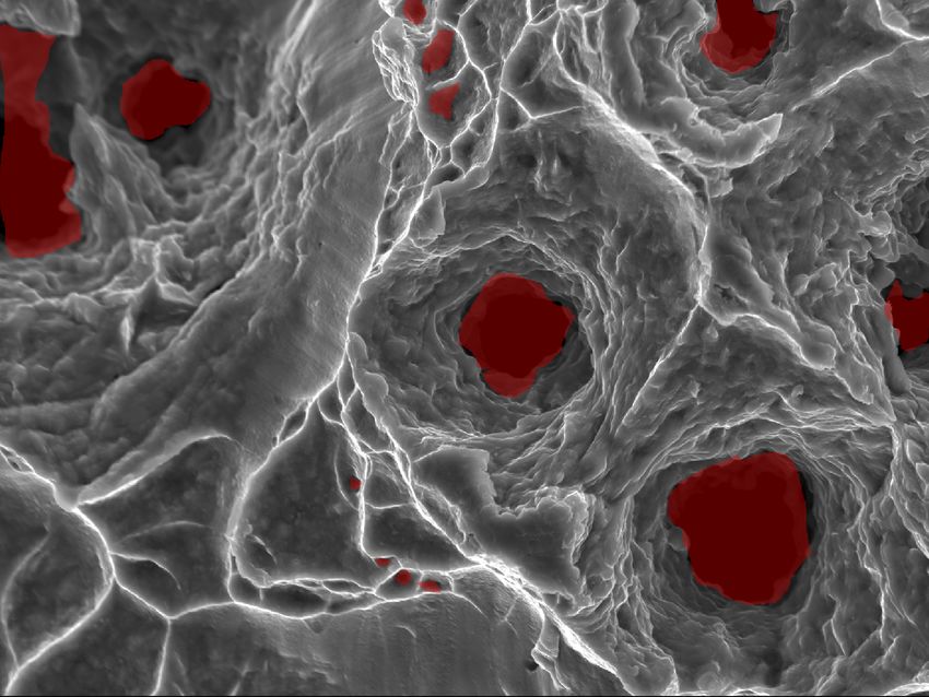
Figure 3. Our Proposed model

parameter tuning with optimizer, learning rates and weight            Method                                         Dice Score (DSC)
decay, we found out that Adam [23] optimizer as an opti-              U-Net                                          0.68509 (±7.88%)
mizer performs the best with a learning rate of 1e − 4, a             U-Net++                                        0.73423 (±6.51%)
weight decay of 1e − 6, β1 = 0.9 and β2 = 0.999. We                   Attentive U-Net                                0.81630 (±5.27%)
use an exponential decay for learning rate after every 10             ResU-Net with Dual Attention (ours)            0.86305 (±5.05%)
epochs. We used a weighted loss function consisting of
dice score and binary cross-entropy loss, the weights were           Table 1. Quantitative Results on Microstructures of Ti alloys
generated empirically with 1.25 for dice-score and 0.95 for
binary cross-entropy loss. The batch size for all the ex-
periments were kept to 1 due to memory constraints of our            4.3. Results
GPU. The hyper-parameters were consistent across all the                 We evaluate the performance of our proposed model
reported methods. We evaluate our model on the basis of              and the baseline model on the widely used metric of Dice-
dice score [24] and compare it to other state-of-the-art meth-       coefficient. The results of various methods are tabulated
ods on semantic segmentation tasks.                                  in table 1. It’s visible from the quantitative and qualitative
                                                                     results 5, 6 that our proposed model performs the best as
          Ltotal = λ1 ∗ (1 − Ldice ) + λ2 ∗ Lbce                     compared to other previous established methods. After get-
                                                                     ting the segmentation results, the results can be analyzed
   The code was written in PyTorch [25] and trained on a             to understand the materials inherent microstructure, grain
single Nvidia GeForce GTX 1080Ti. Each experiment took               boundary strength as well as the conditions leading to the
around 6-7 hours.                                                    failure of the material.
                                                                         Quantifying the probability of occurrence of the mecha-
                                    2 ∗ |A ∩ B|                      nisms of ductile fracture is cumbersome and highly biased
            DiceScore(DSC) =
                                     |A| + |B|                       on the user, this makes the task at hand more challenging.
                                                                     Moreover, the size of the features on the fracture surface
where A and B are predicted segmentation map and ground              changes drastically from one material to the other. Since,
                   truth, respectively                               simple hand-crafted features based model or deep learning

                                                                 5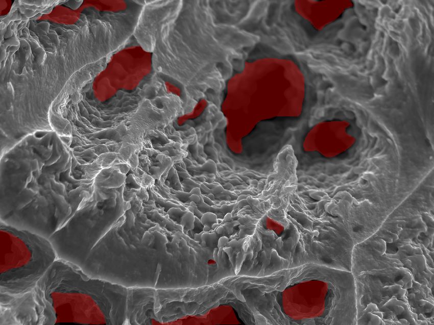
Figure 5. Qualitative Results. From Top to Bottom: GT, U-Net,
UNet++, Attention Unet, Res-Unet with Dual Attention (ours)

based classification or object detection algorithms are not
able to effectively tackle these challenges, we use semantic
segmentation algorithms to classify every pixel in the SEM
images which allows for the topographic characterization of
the fracture surface, making this approach best suited for a
fractographic analysis. This method tries to learn to classify
the background pixels even though they do not follow a cer-
tain pattern, which leads to certain misclassifications. The
results presented herein can be improved, given a larger,
annotated, training set. Depending on the area occupied by
the deep dimples or shallow dimples (beyond the scope of
this work) present in the SEM image, we can analyze if
                                                                     Figure 6. Predictions of our model on SEM images.
                                                                 6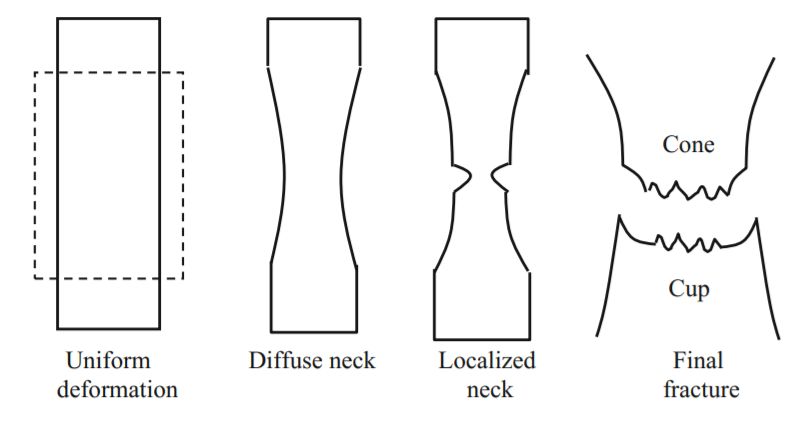
the material underwent brittle or ductile fracture and what                   computer vision. Engineering Failure Analysis, 59:237–252,
mechanism did the material follow. Thus, this work aims                       2016.
to reduce the time and effort of material science researchers             [8] Weiping Hu, Arnold Wiliem, Brian Lovell, Simon Barter,
for fractographic analysis.                                                   and Liangchen Liu. Automation of quantitative fractography
                                                                              for determination of fatigue crack growth rates with marker
5. Conclusion                                                                 loads. In 29th ICAF Symposium Nagoya, 2017.
                                                                          [9] Aritra Chowdhury, Elizabeth Kautz, Bülent Yener, and
    In this work, we have elucidated how fractures occur in                   Daniel Lewis. Image driven machine learning methods for
the material and how they can be fatal while under service.                   microstructure recognition. Computational Materials Sci-
We have presented an overview of the traditional as well as                   ence, 123:176–187, 2016.
modern approaches involved in detecting of these defects
                                                                         [10] Stylianos Tsopanidis, Raúl Herrero Moreno, and Shmuel
on the microscopic scale. We also present new methods for                     Osovski. Toward quantitative fractography using convolu-
the segmentation of deep dimples, which may serve as the                      tional neural networks. Engineering Fracture Mechanics,
first step towards categorizing the type of defect a material                 231:106992, 2020.
had by analyzing the segmented fractographic surface of the              [11] Igor Konovalenko, Pavlo Maruschak, Mykola Chausov, and
SEM image. This work presently focuses on dimple detec-                       Olegas Prentkovskis. Fuzzy logic analysis of parameters of
tion for ductile materials like Ti alloys, but can be easily                  dimples of ductile tearing on the digital image of fracture
extended to other kinds of defects and other materials too.                   surface. Proc. Engin, 187:229–234, 2017.
This is a robust method and the results can be improved pro-             [12] Pavlo Maruschak, Ihor Konovalenko, Mykola Chausov, An-
vided the availability of large data. This work is an aim to                  drii Pylypenko, Sergey Panin, Ilya Vlasov, and Olegas Pren-
foster machine learning and deep learning principles in au-                   tkovskis. Impact of dynamic non-equilibrium processes on
tomating the traditional methods applied in material science                  fracture mechanisms of high-strength titanium alloy vt23.
for fractography or material forensics. We hope to see bet-                   Metals, 8(12):983, 2018.
ter models in the future which can obtain the desired results            [13] Ihor Konovalenko, Pavlo Maruschak, Olegas Prentkovskis,
with limited data and can lead to reduced human labour and                    and Raimundas Junevičius. Investigation of the rupture sur-
material wastage, increased efficiency during production of                   face of the titanium alloy using convolutional neural net-
new materials.                                                                works. Materials, 11(12):2467, 2018.
                                                                         [14] Olaf Ronneberger, Philipp Fischer, and Thomas Brox. U-
References                                                                    net: Convolutional networks for biomedical image segmen-
                                                                              tation. In International Conference on Medical image com-
 [1] Hooyar Attar, Mariana Calin, LC Zhang, Sergio Scudino,
                                                                              puting and computer-assisted intervention, pages 234–241.
     and Jürgen Eckert. Manufacture by selective laser melt-
                                                                              Springer, 2015.
     ing and mechanical behavior of commercially pure titanium.
     Materials Science and Engineering: A, 593:170–177, 2014.            [15] Sanghyun Woo, Jongchan Park, Joon-Young Lee, and
                                                                              In So Kweon. Cbam: Convolutional block attention module.
 [2] Shima Ehtemam-Haghighi, KG Prashanth, Hooyar Attar,                      In Proceedings of the European Conference on Computer Vi-
     Anil K Chaubey, GH Cao, and LC Zhang. Evaluation of me-                  sion (ECCV), pages 3–19, 2018.
     chanical and wear properties of tixnb7fe alloys designed for
     biomedical applications. Materials & Design, 111:592–599,           [16] Kaiming He, Xiangyu Zhang, Shaoqing Ren, and Jian Sun.
     2016.                                                                    Deep residual learning for image recognition. In Proceed-
                                                                              ings of the IEEE conference on computer vision and pattern
 [3] Igor V Kabashkin and Irina V Yatskiv. Reliability and statis-            recognition, pages 770–778, 2016.
     tics in transportation and communication. 2010.
                                                                         [17] G Huang, Z Liu, KQ Weinberger, and L van der Maaten.
 [4] CD Beachem and GR Yoder. Elastic-plastic fracture by ho-                 Densely connected convolutional networks. arxiv 2017.
     mogeneous microvoid coalescence tearing along alternating                arXiv preprint arXiv:1608.06993.
     shear planes. Metallurgical Transactions, 4(4):1145–1153,
                                                                         [18] Jie Hu, Li Shen, and Gang Sun. Squeeze-and-excitation net-
     1973.
                                                                              works. In Proceedings of the IEEE conference on computer
 [5] GA Kardomateas. Fractographic observations in asymmetric                 vision and pattern recognition, pages 7132–7141, 2018.
     and symmetric fully plastic crack growth. Scripta Metallur-
                                                                         [19] Kaiming He, Georgia Gkioxari, Piotr Dollár, and Ross Gir-
     gica,, 20:609–614, 1986.
                                                                              shick. Mask r-cnn. In Proceedings of the IEEE international
 [6] E Merson, V Danilov, D Merson, and A Vinogradov. Con-                    conference on computer vision, pages 2961–2969, 2017.
     focal laser scanning microscopy: The technique for quanti-
                                                                         [20] Jun Fu, Jing Liu, Haijie Tian, Yong Li, Yongjun Bao, Zhi-
     tative fractographic analysis. Engineering Fracture Mechan-
                                                                              wei Fang, and Hanqing Lu. Dual attention network for
     ics, 183:147–158, 2017.
                                                                              scene segmentation. In Proceedings of the IEEE Conference
 [7] MX Bastidas-Rodriguez, FA Prieto-Ortiz, and Edgar Espejo.                on Computer Vision and Pattern Recognition, pages 3146–
     Fractographic classification in metallic materials by using              3154, 2019.

                                                                     7
[21] Sergey Ioffe and Christian Szegedy. Batch normalization:
Accelerating deep network training by reducing internal co-
variate shift. arXiv preprint arXiv:1502.03167, 2015.
[22] Jeffrey De Fauw, Joseph R Ledsam, Bernardino Romera-
Paredes, Stanislav Nikolov, Nenad Tomasev, Sam Blackwell,
Harry Askham, Xavier Glorot, Brendan ODonoghue, Daniel
Visentin, et al. Clinically applicable deep learning for di-
agnosis and referral in retinal disease. Nature medicine,
24(9):1342–1350, 2018.
[23] Diederik P. Kingma and Jimmy Ba. Adam: A method for
stochastic optimization, 2014.
[24] Lee R Dice. Measures of the amount of ecologic association
between species. Ecology, 26(3):297–302, 1945.
[25] Adam Paszke, Sam Gross, Francisco Massa, Adam Lerer,
James Bradbury, Gregory Chanan, Trevor Killeen, Zeming
Lin, Natalia Gimelshein, Luca Antiga, et al. Pytorch: An
imperative style, high-performance deep learning library. In
Advances in neural information processing systems, pages
8026–8037, 2019.
8You can also read