Density and types of probiotic bacterial filter media with different number of bioballs in silver pompano (Trachinotus blochii) culture with ...
←
→
Page content transcription
If your browser does not render page correctly, please read the page content below
Density and types of probiotic bacterial filter
media with different number of bioballs in silver
pompano (Trachinotus blochii) culture with
recirculation system
Mulyadi, Niken A. Pamukas, Usman M. Tang

      Department of Aquaculture, Faculty of Fisheries and Marine Science, Riau University,
     Pekanbaru, Indonesia. Corresponding author: Mulyadi, mulyadibrian26@yahoo.com

       Abstract. Cultivation intensification with a recirculation system tends to have a high stocking density
       and a large amount of feed, so that feed residue, metabolic waste, and oxygen consumption also
       increase. To speed up the decomposition of feed residue and metabolic waste, filtering is necessary,
       namely by means of a bioball. This study aimed to analyze the types and numbers of bacteria present in
       the bioball, to improve water quality and the optimal number of bioballs for bacterial living media. The
       study used a completely randomized design, with 1 factor, 5 levels of treatment and 3 replications. The
       treatment levels are: A=without bioballs (control), B=35 bioball filters per container, C=45 bioball filters
       per container, D=55 bioball filters per container and E=65 bioball filters per container. Silver pompano
       (Trachinotus blochii) fish specimens measuring 10-12.55 cm and a body weight of 24.3-28.9 g, were
       cultivated for 56 days, at a stocking density of 1 fish for a water volume of 4 L (a total of 20 fish in a
       volume of 80 L). During the rearing, fish were fed with Megami GR2 pellets with a composition of 46%
       protein, 10% fat, 2% crude fiber and 10% moisture content, 3 times a day adlibitum. The type and
       density of bacteria in the bioball was observed, together with the water quality parameters, such as:
       temperature, pH, dissolved oxygen, total ammonium nitrogen, nitrite and nitrate. The research data
       were analyzed statistically using ANOVA (P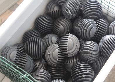
in water quality (Putra & Pamukas 2011; Prayogo et al 2012). Recirculation systems are
technologies used to maintain water quality in fisheries, in order to remain suitable for
aquatic organisms and to support the optimization of water utilization. The recirculation
system is able to reduce the level of ammonia concentration, restraining it within the
range of 31-43% (Djokosetiyanto et al 2006; Putra & Pamukas 2011).
        According to Lekang (2008) and Fadhil et al (2010), the use of a recirculation
system has several advantages including: more efficient water use, flexibility in culture
locations, more hygiene, relatively small space or land requirements, ease of controlling
and maintaining cultured organisms, ease of maintaining water quality, environmentally
friendly, pollution prevention and it can be functional permanently, without disturbing the
aquatic environment.
        Recirculating aquaculture system technology can also be adapted to the
aquaculture system and to the filter substrate in order to control the dissolved solids
(Fadhil et al 2010). The use of the right type of filter material will determine the success
of fish rearing in the recirculation system because it will determine the growth of non-
pathogenic bacteria on the material. Currently, biological filter substrates are widely
used, being more environmentally friendly.
        There are many biological filters that can be used, one of which is the bioball.
Bioball is a sphere with a diameter of 4 cm, a specific area of ±230 m² m-³ with a width
of each hole in a bioball of 0.92 mm, made of PVC material (Said 2002). Bioball functions
as a physiological filter which is a growing medium for bacteria that can remove ammonia
contained in water (Alfia et al 2013). Nelvia et al (2015) reported that the bioball filtered
Nitrosomonas sp. bacteria, which function to oxidize ammonia to nitrite, and Nitrobacter
sp., which function to oxidize nitrite to nitrate.
        Several studies related to improving the water quality in aquaculture with
recirculation and hydroponic systems have been carried out, such as: Mulyadi & Pamukas
(2011) used a recirculation system in nursery and rearing of fish (Mystus nemurus); Alfia
et al (2013) used a recirculation system with a bioball filter in tilapia (Oreochromis
niloticus) culture; Nelvia et al (2015) studied the addition of a bioball to the maintenance
media filter on the survival and growth of goldfish (Carassius auratus) fry. Based on the
description above, a series of studies on the number and types of bacteria cultured with
different numbers of bioballs and on the improvement of water quality in the cultiring and
recirculation systems are needed. This research was aimed at analyzing the types and
numbers of bacteria present in the bioball, to measure the water quality improvement
(temperature, pH, DO, TAN, nitrite and nitrate) and to determine the optimal number of
bioballs.

Material and Method

Location and time of research. This research was conducted between January and July
2019, in several laboratories, as follows: the fish rearing was carried out at the Batam
Marine Cultivation Fishery Center (BPBL), the water quality analysis was carried out at
the Laboratory of Fish and Environmental Health Testing of the Batam Marine
Aquaculture Fisheries Agency, the bacteria types identification and densities calculation
were performed at the Fish and Environmental Health Examiners Lab of the BPBL, the
polymerase chain reaction (PCR) test was performed at PT Genetics Science Indonesia,
West Jakarta.

Materials and tools. The main ingredients used in this research were 300 seeds of
Trachinotus blochii (Lacepede) with a size of 10-11 cm, originating from the BPBL, fed
with commercial pellets "GR-2" (46% protein, 10% fat, 13% ash content, 2% crude fiber
and 10% moisture content). The test feed was obtained from the BPBL. A round bucket
with a diameter of 60 cm, a height of 45 cm and a volume of 100 L was used as a
research vessel. Bioballs have a diameter of 4 cm, a specific area ±230 m² m-³ and a
witdth of each hole in a bioball of 0.92 mm, and are made of PVC, as a filter substrate
(Figure 1a).

AACL Bioflux, 2021, Volume 14, Issue 1.
http://www.bioflux.com.ro/aacl
                                            581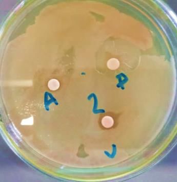
Experimental design. The research method used in this study was an experimental
method completely randomized design, with 1 factor, 5 levels of treatment and 3
replications (Steel & Torrie 1993). As a factor in the study was the difference in the
number of bioballs. The treatment level determination in this study is based on the study
of Nelvia et al (2015) on the goldfish fry (Carassius auratus) in closed recirculation
system, stating that the use of 50 bioballs gutter-1 can improve the water quality,
providing 100% survival rates and a growth performance up to a weight of 2.605 g and
to a length of 1.48 mm. Therefore, the treatment in the present study was established as
follows: A=0 (control), B=35, C=45, D=55 and E=65 bioball filters per container.

Research procedure. T. blochii measuring 10-12.55 cm in length and having a body
weight of 24.3-28.9 g were obtained from the BPBL and were adapted for 7 days before
being used as test specimens. Fish rearing containers were filled with sea water with a
salinity of 15‰, using a closed recirculation system. The rearing container was
connected to a 50 x 14 x 14 cm PVC gutter, as a filter container placed at the top of the
fish-raising containers. Then the water from the filter gutter flowed back through the PVC
pipe with a diameter of 2.5 cm to the rearing tub of T. blochii. Water from the fish
rearing containers flowed into the filter media (gutters), each gutter containing a number
of bioballs corresponding to the tested treatment (Figure 1b) and according to Nelvia et
al (2015), by using a water pump with a power of 18 watts. After passing through the
filter media (gutter), the water returned to the fish-raising tub through the drain pipe in
the filter container. In the rearing medium (KP-SUPER N, with Nitrosomonas sp. and
Nitrobacter sp.), trademark starter bacteria were added at a dose of 2.5 mL 200 L-1 week-1
(Hartini et al 2013), to accelerate bacterial growth in the bioball.

                        (a)                                   (b)
 Figure 1. (a) Bioball used as a filter substrate, (b) The design of the Trachinotus blochii
                        aquaculture recirculation system (original).

The test specimens that have been adapted to the culturing media were then randomly
placed into 15 rearing containers. The fish were given commercial feed at satiation,
containing: 46% protein content, 10% fat, 13% ash, 2% crude fiber, and 10% moisture,
at 07:00, 12:00 and 17:00 WIB.

Measured response. The response variables measured in this study were: density and
type of bacteria in the filter container were observed at the beginning, in the middle and
at the end of the study (SNI.01-2332.3-2006 and SNI.01-2332.3-2006); the
temperature, pH and salinity were observed every day; the dissolved oxygen (DO), total
ammonia nitrogen (TAN), nitrite (NO2) and nitrate (NO3) were observed every week.
Water quality measurement procedures refer to SNI in the Public Works Service (2013).

Data analysis. Bacterial density data were analyzed according to the RAL model (Steel &
Torrie 1993). To determine the effect of salinity on each measured variable, a diversity
analysis was performed using the SPSS 17.0 software. If P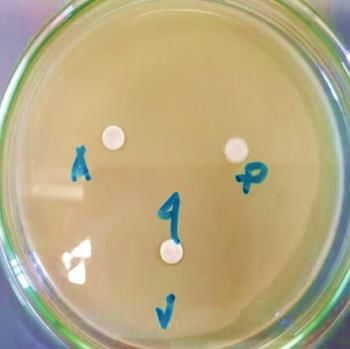
Results and Discussion

Density of bacteria in the filter container. The results of bacterial density
observations carried out on plate count agar (PCA) media using the total plate count
method can be seen in Figure 2.

                                                      800.00
                  Bacterial density (x 103 CFU mL-1

                                                      700.00
                                                      600.00
                                                      500.00
                                                      400.00
                                                      300.00
                                                      200.00
                                                      100.00
                                                                               Day
                                                        0.00
                                                                 1             28               56
                                                           0    89.33         108.33           208.33
                                                           35   90.00         156.67           337.67
                                                           45   89.67         176.33           388.33
                              Figure 2.55
                                        Bacterial density
                                                   88.67 in all treatments during the study.
                                                                        329.00                 667.67
Figure 2 shows that the65bacterial density
                                    89.33 from day 1 to137.33
                                                           day 28 in treatments307.67
                                                                                   with 0, 35,
45 and 65 bioball filters per container are relatively similar, in contrast to the treatment
with 55 bioball filters per container, which caused a significant increase of bacteria,
compared to the other treatments. This is due to the fact that the bacteria and T. blochii
fish are permanently adapting to the media in which they live and to the fact that the
nutrients resulting from the remaining feed and fish feces are still not sufficient to
support the maximum bacterial growth. After the 28 th day the number of bacteria in all
treatments continued to increase, reaching a maximum peak on the 56 th day. This is due
to the increase in organic matter from fecal matter and fish feed along with the growth of
the T. blochii specimens, which is a source of bacterial nutrition, thereby increasing the
rate of bacterial development. In accordance with the opinion of Sunarto (2003),
dissolved organic matter is needed by living bacteria, then Sidharta (2000) explains that
organic material contains carbon, nitrate, phosphate, sulfur, ammonia, and several
minerals which are nutrients for bacterial growth. Administering KP SUPER N probiotics
(routinely added in culture containers) causes continuous bacterial growth due to the
availability of organic materials that can be used optimally. The highest bacterial density
was found in the treatment of 55 bioball filters per container.
        The highest bacterial density was found in the treatment with 55 bioball filters per
container, at the end of the study, namely 667.7 x 10 5 CFU mL-1, demonstrating the
optimality of a configuration consisting of: 55 bioballs for the given size of the filter (50 x
14 x 14 cm) and for the given volume of the culture media (80 L). These parameters are
balanced in such a manner that bacteria receive a sufficient amount of oxygen, through
the bioball cavity. According to Nelvia et al (2015), the bioball structure (not too
compact) provides an opportunity for oxygen to enter the bioball cavity and to reach
bacteria. If the oxygen is sufficient, then the bacteria growth determines an optimal
functioning of the bioball.
        The lowest bacterial density, 208.3 x 10⁵ CFU mL-1, was found in treatment A
without using bioballs, starter bacteria (KP SUPER N) and additional nutrients, resulting
in a limited media for the growth and development of bacteria. The low density of

AACL Bioflux, 2021, Volume 14, Issue 1.
http://www.bioflux.com.ro/aacl
                                                                        583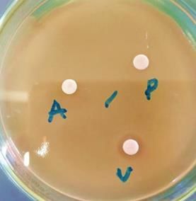
bacteria when using the highest number of bioball filters (65 container-1) is due a number
of bioballs almost exceeding the container capacity. According to Nelvia et al (2015)
bioball too densely disposed in the gutter and lacking of oxygen supply can cause
extinction of the bacteria in the bioball cavity. Consequently, the performance of the
bioball filter decreases, suspending the process of decomposing organic matter by
nitrifying bacteria.
         The biofilter system removes ammonia where it accumulates and reaches toxic
levels if it is not transferred by a nitrification process where the process, first oxidizing
ammonia to toxic nitrite, then to nitrate, which is relatively non-toxic. The above process
involves Nitrosomonas and Nitrobacter bacteria. Bacteria grow like a film on the surface
of the material (biofilter container) or adhere to organic particles. Biofilter consists of a
medium with a large surface for the growth of nitrite bacteria. In the recirculation
system, bacteria play a role in converting toxic substances (ammonia) to non-toxic
substances (nitrate). The performance of bacteria in the biofilter system is marked by an
increase in the average biochemical oxygen demand (BOD) value in the culture medium
(Nurhidayat et al 2012). The more bioballs, the more opportunities for bacteria to stick,
when a viable oxygen level is still preserved (Nelvia et al 2015).
         ANOVA results showed that the number of bioballs had an effect on the density of
bacteria on day 28 and 56. The Newman Keuls range study test identifies the bacterial
density differences between treatments. The results of the regression analysis between
the number of bioballs and bacterial density provide the following regression equation:

                                    Y = 227.9 + 3.851X
Where:
Y-bacterial density;
X-number of bioballs;
The calculated r was 0.557 and the R2 was 0.311.
        The results of the regression analysis showed that the number of bioballs had a
positive correlation with the bacterial density (r=0.557). The increase in the number of
bacteria in the filter container was influenced by the number of bioballs by 31.1 and
68.9%, which is thought to be influenced by the content of organic matter (Stepwise
regression analysis). Nelvia et al (2015) stated that the bioball is a breeding ground for
various bacteria needed in the breakdown of organic matter. According to Sunarto
(2003), dissolved organic matter is needed by bacteria to live. Furthermore, according to
Sidharta (2000) organic matter contains carbon, nitrate, phosphate, sulfur, ammonia and
several minerals which are nutrients for bacterial growth.

Identification of bacteria in filter containers. Based on the observations, there were
7 pure bacterial isolates. The identification of bacteria against the isolates found in the
filter media was carried out by physical, biochemical and sensitivity tests to see which
bacteria had the greatest inhibition zone against pathogenic bacteria.

Physics test. The results of observations of color, shape, edge, elevation and gram of
the seven isolates found are presented in Table 1.

                                                                                           Table 1
                             Observation results of bacterial characteristics

                                         Colony                           Cell
No     Isolate                                                                           Treatment
                    Color           Shape      Edge    Elevation   Gram type     Shape
 1        A1        Yellow         Circular   Entire   Umbonate    Negative      Stem      P2U1
 2        A2        Beige          Circular   Entire    Raised     Negative      Stem      P0U3
 3        A3        White          Circular   Entire    Raised     Negative      Stem      P3U2
 4        A4        White          Circular   Entire    Raised     Negative      Stem      P4U1
 5        A5        Beige       Filamentous Entire      Filiform   Negative      Stem      P1U1
 6        A6        White          Circular   Entire    Raised     Negative      Stem      P4U2
 7        A7        White          Circular   Entire    Raised     Negative      Stem      P3U3

AACL Bioflux, 2021, Volume 14, Issue 1.
http://www.bioflux.com.ro/aacl
                                                   584
Table 1 shows that all bacterial isolates have gram-negative, entire edges and are rod-
shaped. The dominant bacterial colony was white (4 isolates), 2 isolates were beige and
1 isolate was yellow. Colonies were circular or round (6 isolates) and filamentous (1
isolate). The elevation of the dominant bacterial colony was raised (5 isolates), 1
umbonate isolate and 1 filiform isolate.
Biochemical test. The results of observations of the Catalase, Oxidase, Motility,
oxidative/fermentation (O/F) glucose, Indole and triple sugar iron agar (TSIA) tests are
presented in Table 2.
Table 2
Bacterial biochemical test results
Biochemical characteristics
No. Isolate Treatment
Catalase Oxidase Motility O/F Indole TSIA
1 A1 + + + - - A/K P2U1
2 A2 + + - F - K/K P0U3
3 A3 + - + O - A/K P3U2
4 A4 + + + F - A/A P4U1
5 A5 + + + F + A/A P1U1
6 A6 + + + F - A/A P4U2
7 A7 + + - F - A/A P3U3
O-oxidative; F-fermentative; A/K-alkaline acid; K/K-alkaline; A/A-acid.
Table 2 shows a positive catalase enzyme test in all bacterial isolates, indicated by the
gas bubbles formation during their reaction with the H2O2 drops. According to Stoica
(2016), catalase is an enzyme contained in the majority of bacteria and it is involved in
the decomposition of hydrogen peroxide into water and oxygen. Hydrogen peroxide and
oxygen are used for electron transport during fermentation in both aerobic and
facultatively anaerobic bacteria.
Oxidase tests A1, A2, A3, A4, A5, A6 and A7 are positive, as shown by the color
change on the oxidase paper, which indicates activity, while A3 is negative. Five isolates
(A1, A3, A4, A5, A6) showed positive results in the motility test, due to the spread of
bacterial growth on the Sulfide Indole Motility (SIM) medium, and did not grow on the
part of the stick site bacteria. The O/F test results showed no color change in the
paraffin-covered media and a color change on the exposed media. Five bacterial isolates
(A2, A4, A5, A6, A7) were observed to be fermentative because the tube without paraffin
and the tube filled with paraffin changed their color to yellow. According to Fahri (2008),
in the O/F test, oxidative organisms occur at a color change in the open media, while
fermentative organisms can be indicated by no color change in the closed media.
The indole test shows 6 isolates (A1, A2, A3, A4, A6 and A7) giving negative results,
marked with a yellow color on the surface of the media, which means that the bacteria
are unable to break down the amino acid tryptophan, and no red ring after dropping the
Kovacs reagent in the sulfur, indole, motility (SIM) media. According to Acharya (2012),
the indole test was carried out to determine the ability of bacteria to break down amino
acid tryptophan and to form indole compounds. Tryptophan is hydrolyzed by
tryptophanase to produce three possible end products, one of which being the indole.
Indole production is detected by the Kovacs or Ehrlich's reagent, composed of 4-
(dimethylamino)benzaldehyde, which reacts with the indole to produce a red compound.
A positive reaction is indicated if there is a red color on the surface of the medium, which
indicates that the bacteria are able to break down the amino acid tryptophan. In the TSIA
test, 4 isolates (A4, A5, A6 and A7) showed acidic properties (A/A), 2 isolates (A1 and A3)
were acid alkaline and 1 isolate (A2) was alkaline.
Sensitivity test. The sensitivity test to pathogenic bacteria was carried out after
observing the physical and biochemical tests of the seven isolates. This test aimed to find
probiotic bacteria candidates for polymerase chain reaction (PCR) testing. The results of
the sensitivity test on the 7 isolates are presented in Figure 3. Inhibition zones produced
AACL Bioflux, 2021, Volume 14, Issue 1.
http://www.bioflux.com.ro/aacl
585by the seven bacterial isolates were found in different filter containers. Bacteria from
isolate A1 were able to inhibit the growth of Vibrio bacteria during the 24-hour incubation
period at a temperature of 37 0C, as indicated by the formation of a clear area around the
blank disk, but they were unable to inhibit the growth of Aeromonas and Pseudomonas
bacteria. After an incubation period of 48 hours, the inhibition zone remains clear, which
indicates that these bacteria can kill (bacteriocidal) Vibrio bacteria. Similarly, for the
bacteria from isolate A2, a clear zone was formed around the blank disk, indicating an
inhibition of Pseudomonas and Vibrio bacteria during the 48-hour incubation period,
meaning that A2 bacteria are bacteriocidal against Pseudomonas and Vibrio, but they
cannot inhibit the growth of Aeromonas bacteria.
Bacteria in A3 and A4 isolates were unable to inhibit the growth of Aeromonas,
Pseudomonas and Vibrio bacteria, as shown by a no inhibition zone formed on the blank
disk. In A5 and A7 isolates, an inhibition zone occurred against Aeromonas, Pseudomonas
and Vibrio bacteria during the 24-hour incubation period, but during the 48-hour
incubation period, the clear area became cloudy, indicating that these bacteria were only
able to inhibit (bacteriostatic) the three pathogenic bacteria . In A6 isolate, a clear zone
was also formed during the 48-hour incubation period against Aeromonas and Vibrio
bacteria, but no inhibition zone was formed against Pseudomonas bacteria. This indicates
that the A6 bacteria are able to kill (bacteriocidal) Aeromonas and Vibrio bacteria.This is
in line with Azaldin et al (2020), who states that sensitivity of a material to bacteria is
characterized by the presence of an inhibition zone around the blank disk. Furthermore,
according to Dwyana & Murniati (2020), this inhibition area is formed due to bioactive
compounds contained in microbes that produce exoenzymes which break down organic
matter and secrete hydrolytic enzymes, proteases, lipases, amylases, and cellulose so
that they are able to hydrolyze polysaccharides as a carbon source and electron donors.
A1 - Inhibition A2 - Inhibition A3 - Inhibition A4 - Inhibition
A = 0, P = 0 and A = 0, P = 8,88 mm and A = 0, P = 0 and A = 0, P = 0
V = 3.74 mm V = 3.49 mm V=0 and V = 0
(bacteriocidal) (bacteriocidal)
A5 - Inhibition A6 - Inhibition A7 - Inhibition
A = 4.32 mm, A = 1.62 mm, P = 0 A = 3.15 mm,
P = 3.87 mm and and V = 3.8 mm P = 2.58 mm and
V = 5.24 mm (bacteriocidal) V = 2.20 mm
Figure 3. Sensitivity test of bacterial isolates against pathogenic bacteria (A: Aeromonas,
P: Pseudomonas, V: Vibrio).
Madigan et al (2012) grouped bacteria based on their selective toxicity. According to
them, antimicrobial compounds had 3 kinds of effects on the microbial growth: 1).
Bacteriostatic inhibit growth but they do not kill. Bacteriostatic compounds inhibit protein
synthesis or bind to ribosomes; 2). Bacteriocides kill cells but not cause cell lysis or cell
AACL Bioflux, 2021, Volume 14, Issue 1.
http://www.bioflux.com.ro/aacl
586breakdown. 3). Bacteriolytic cause cells lysis or cell breakdown. Furthermore, Dwyana &
Murniati (2020) stated that the sensitivity test on bacteria indicated that these were
bacteriostatic after 24 hours of incubation period (clear zone formed on disc paper), while
after 48 hours they were classified as bacteriocidal.
According to Jannata (2014), the responses related to the bacterial growth
inhibition can be classified based on the inhibition zone diameter, as follows: weak (≤5
mm), medium (5-10 mm), strong (10-20 mm) and very strong (≥20 mm). Based on this
scale, isolates A5, A6 and A7 were classified as having a weak response to Aeromonas,
isolate A2 had a moderate response and isolates A5 and A7 had a weak response to
Pseudomonas bacteria. Furthermore, A5 isolate had a moderate response to Vibrio
bacteria, while isolates A1, A2, A6 and A7 were classified as having a weak response.
According to Sugita et al (1996), these bacteria are able to produce antimicrobial
compounds, such as: siderophores, bacteriocins, lysozymes, proteases, hydrogen
peroxidase, and organic acids, as a mechanism of competition for nutrients and energy,
inhibiting the growth of pathogens in the filter container. According to Rengpipat et al
(2000), the siderophore is a compound with a low molecular weight (The base pairs obtained were combined and trimmed using the Bioedit 7.0 software. DNA
sequencing analysis gives the following results for the nucleotide and protein FASTA
sequences (Figure 5).
> MUL_1
CTTGCGGTTAGCGCACTGCCTTCGGGTAACCCAACTCCCATGGTGTGACGGGCGGTGTGTACAAGGCCCG
GGAACGTATTCACCGCGGCATGCTGATCCGCGATTACTAGCGATTCCAACTTCATGCACCCGAGTTGCAGA
GTGCAATCCGAACTGAGATGGTTTTTGGAGATTAGCTCGACCTCGCGGTCTCGCTGCCCACTGTCACCACC
ATTGTAGCACGTGTGTAGCCCAGCCCGTAAGGGCCATGAGGACTTGACGTCATCCCCACCTTCCTCTCGGC
TTATCACCGGCAGTCCCCTTAGAGTGCCCAACTTAAGGCTGGCAACTAAGGGCGAGGGTTGCGCTCGTTG
CGGGACTTAACCCAACATCTCACGACACGAGCTGACGACAGCCATGCAGCACCTGTCTTGGGTCCAGCCTA
ACTGAAGGATACCGTCTCCGGTATCCGCGACCCAGATGTCAAGGGCTGGTAAGGTTCTGCGCGTTGCTTC
GAATTAAACCACATGCTCCACCGCTTGTGCGGGCCCCCGTCAATTCCTTTGAGTTTTAATCTTGCGACCGTA
CTCCCCAGGCGGGAAGCTTAATGCGTTAACTGCGCCACCGACAGGTAAACCTGCCGACGGCTAGCTTCCA
TCGTTTACGGCGTGGACTACCAGGGTATCTAATCCTGTTTGCTCCCCACGCTTTCGCACCTCAGCGTCAGT
ATCGAGCCAGTGAGCCGCCTTCGCCACTGGTGTTCCTCCGAATATCTACGAATTTCACCTCTACACTCGGA
ATTCCACTCACCTCTCTCGAACTCTAGATCGGCAGTATTAGAGGCAGTTCCGGGGTTGAGCCCCGGGATTT
CACCCCTAACTGACCGATCCGCCTACGCGCGCTTTACGCCCAGTAATTCCGAACAACGCTAGCCCCCTTCG
TATTACCGCGGCTGCTGGCACGAAGTTAGCCGGGGCTTCTTCTCCGGTTACCGTCATTATCTTCACCGGTG
AAAGAGCTTTACAACCCTAGGGCCTTCATCACTCACGCGGCATGGCTGGATCAGGCTTGCGCCCATTGTCC
AATATTCCCCACTGCTGCCTCCCGTAGGAGTCTGGGCCGTGTCTCAGTCCCAGTGTGGCTGATCATCCTCT
CAGACCAGCTACTGATCGTCGCCTTGGTGAGCCTTTACCTCACCAACTAGCTAATCAGACATGGGCTCATC
TAACTCCGATAAATCTTTCTCCCGAAGGACGTATACGGTATTAGTTCAAGTTTCCCTGAGTTATTCCGTAGA
GCTAGGTAGATTCCCATGCATTACTCACCCGTCTGCCGCTCCCCCGAGGGGGCGC
The nucleotide sequence and fasta MUL_1
> MUL_2
GTTAGACTACCTACTTCTGGTGCAACAAACTCCCATGGTGTGACGGGCGGTGTGTACAAGGCCCGGGAAC
GTATTCACCGCGGCATTCTGATCCGCGATTACTAGCGATTCCGACTTCATGGAGTCGAGTTGCAGACTCCA
ATCCGGACTACGATCGGCTTTTTGAGATTAGCATCCTATCGCTAGGTAGCAACCCTTTGTACCGACCATTGT
AGCACGTGTGTAGCCCTGGCCGTAAGGGCCATGATGACTTGACGTCGTCCCCGCCTTCCTCCAGTTTGTCA
CTGGCAGTATCCTTAAAGTTCCCATCCGAAATGCTGGCAAGTAAGGAAAAGGGTTGCGCTCGTTGCGGGA
CTTAACCCAACATCTCACGACACGAGCTGACGACAGCCATGCAGCACCTGTATCTAGATTCCCGAAGGCAC
CAATCCATCTCTGGAAAGTTTCTAGTATGTCAAGGCCAGGTAAGGTTCTTCGCGTTGCATCGAATTAAACCA
CATGCTCCACCGCTTGTGCGGGCCCCCGTCAATTCATTTGAGTTTTAGTCTTGCGACCGTACTCCCCAGGC
GGTCTACTTATCGCGTTAGCTGCGCCACTAAAGCCTCAAAGGCCCCAACGGCTAGTAGACATCGTGTACGG
CATGGACTACCAGGGTATCTAATCCTGTTTGCTCCCCATGCTTTCGTACCTCAGCGTCAGTATTAGGCCAGA
TGGCTGCCTTCGCCATCGGTATTCCTCCAGATCTCTACGCATTTCACCGCTACACCTGGAATTCTACCATCC
TCTCCCATACTCTAGCCATCCAGTATCGAATGCAATTCCCAAGTTAAGCTCGGGGATTTCACATTTGACTTA
AATGGCCGCCTACGCACGCTTTACGCCCAGTAAATCCGATTAACGCTCGCACCCTCTGTATTACCGCGGCT
GCTGGCACAGAGTTAGCCGGTGCTTATTCTGCGAGTAACGTCCACTATCCAGTAGTATTAATACTAGTAGC
CTCCTCCTCGCTTAAAGTGCTTTACAACCATAAGGCCTTCTTCACACACGCGGCATGGCTGGATCAGGGTT
CCCCCCATTGTCCAATATTCCCCACTGCTGCCTCCCGTAGGAGTCTGGGCCGTGTCTCAGTCCCAGTGTGG
CGGATCATCCTCTCAGACCCGCTACAGATCGTCGCCTTGGTAGGCCTTTACCCCACCAACTAGCTAATCCG
ACTTAGGCTCATCTATT
The nucleotide sequence and fasta MUL_2
>MUL_3
AAGGTTAAGCTATCTACTTCTGGTGCAGCCCACTCCCATGGTGTGACGGGCGGTGTGTACAAGGCCCGGG
AACGTATTCACCGTGGCATTCTGATCCACGATTACTAGCGATTCCGACTTCATGGAGTCGAGTTGCAGACT
CCAATCCGGACTACGACCAGCTTTATGGGATTAGCTCCACCTCGCGGCTTCGCAACCCTCTGTACTGACCA
TTGTAGCACGTGTGTAGCCCTACTCGTAAGGGCCATGATGACTTGACGTCGTCCCCACCTTCCTCCGGTTT
ATCACCGGCAGTCTCCCTAAAGTTCCCGGCATGACCCGCTGGCAAGTAAGGATAGGGGTTGCGCTCGTTG
CGGGACTTAACCCAACATTTCACAACACGAGCTGACGACAGCCATGCAGCACCTGTCTCACAGTTCCCGAA
GGCACTGAAGCATCTCTGCTAAATTCTGTGGATGTCAAGAGTAGGTAAGGTTCTTCGCGTTGCATCGAATT
AAACCACATGCTCCACCGCTTGTGCGGGCCCCCGTCAATTCATTTGAGTTTTAACCTTGCGGCCGTACTCCC
CAGGCGGTCTACTTAATGCGTTAGCTTGAGAGCCCAGTGTTCAAGACACCAAACTCCGAGTAGACATCGTT
TACGGCGTGGACTACCAGGGTATCTAATCCTGTTTGCTCCCCACGCTTTCGTGCCTGAGCGTCAGTCTTTG
TCCAGGGGGCCGCCTTCGCCACCGGTATTCCTCCAGATCTCTACGCATTTCACCGCTACACCTGGAATTCT
ACCCCCCTCTACAAGACTCTAGTTTGCCAGTTCGAAATGCGGTTCCCAGGTTGAGCCCGGGGCTTTCACAT
CTCGCTTAACAAACCGCCTGCGCACGCTTTACGCCCAGTAATTCCGATTAACGCTCGCACCCTCCGTATTAC
CGCGGCTGCTGGCACGGAGTTAGCCGGTGCTTCTTCTGCGAGTAACGTCACAGATGTAAGGTATTAACTTA
CACCCTTTCCTCCTCGCTGAAAGTGCTTTACAACCCGAAGGCCTTCTTCACACACGCGGCATGGCTGCATC
AGGGTTTCCCCCATTGTGCAATATTCCCCACTGCTGCCTCCCGTAGGAGTCTGGGCCGTGTCTCAGTCCCA
GTGTGGCTGATCATCCTCTCAGACCAGCTAGGGATCGTCGCCTAGGTGAGCCTTTACCTCACCTACTAGCT
AATCCCACCTGGGCTTATCCATCAGCGCAAGGCCCGAAGGTCCCCTGCTTTCCCCCGTAGGGCGTATGCG
GTATTAGCAGTCGTTTCCAACTGTTATCCCCCACAAATGGGCAAATTCCCAGGCATTACTCACCCGTCCGCC
GCTCGTCATCTTCAAAAGCAAGC
The nucleotide sequence and fasta MUL_3
Figure 5. The nucleotide sequence and fasta MUL_1, MUL_2 and MUL_3.
16S rRNA gene sequence alignment with basic local alignment search tool
(BLAST). BLAST is accessible online via: http://blast.ncbi.nlm.nih.gov/Blast.cgi and
provides the results of the DNA sequencing. A search was carried out for species
identification, based on the percentage of the sequenced DNA homology with existing
databases on GenBank. The identification results of probiotic bacterial isolates using
BLAST correspond to the highest homology (closest relationship) with the bacteria in
GenBank. Based on the nucleotides registered with the National Center for Biotechnology
AACL Bioflux, 2021, Volume 14, Issue 1.
http://www.bioflux.com.ro/aacl
588Information (NCBI), the species, strains and accession numbers of bacterial isolates
identified in this study are presented in Table 3.
Table 3
Results of tracing the 16S rRNA sequencing of bacterial isolates with the BLAST system
Accession Query
Isolate Species Strain Homology
number coverage
MUL_1 Acinetobacter sp. MUL37 MT229070 100% 100%
MUL_2 Shewanella sp. MUL31 MT229068 100% 100%
MUL_3 Nitratireductor sp. MUL35 MT229069 100% 100%
The alignment results of 16S rRNA gene sequences with BLAST (Table 3) showed that the
bacterial isolate MUL_1 was identified as Acinetobacter sp. strain MUL37 with 100%
homology and 100% query coverage, MUL_2 isolate was identified as Shewanella sp.
strain MUL31 with 100% homology and 100% query coverage and bacterial isolate
MUL_3 was identified as Nitratireductor sp. strain MUL35 with 100% homology and 100%
query coverage. The types of bacteria found are thought to have come from the waters
of the Batam Sea which are used as a medium for culturing T. blochii, because the
starter bacteria added to the media do not contain these types of bacteria.
According to Adithiya et al (2017), the sequence equation between 93-97% can
represent the identity of bacteria at the genus level but different at the species level.
Furthermore, Hagstrom et al (2000) and Seprianto et al (2017) state that bacteria that
have a 16S rRNA sequence equation greater than 97% are the same species, while the
equation between 93-97% can represent identity at the genus level but differ at the
species level. According to Dancourt et al (2000), if the similarity of the sequences is less
than 97%, it is likely either a new species, since there is no data in the database, or the
the sequencing results are too short to be relevant, when compared to the database.
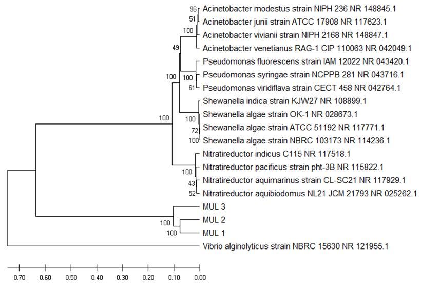
Phylogenetic tree analysis. The analysis of phylogenetic tree formation was carried out
by looking at the similarities that exist in the genetic isolates of MUL_1, MUL_2 and
MUL_3 bacteria with a database of the results of alignment of nucleotide sequences in
GenBank using the Mega X 10.0.5 WIN 64 application. Phylogenetic trees analysis used
the UPGMA (Unweighted Pair Group Method with Arithmetic Average), a clustering
method based on the pairwise distance arithmetic means. Phylogenetic analysis used 17
sequences with 3 sample sequences and 14 comparison sequences, the alignment
process used 1000 bootstraps and the Kimura 2 model parameters. Branches that
connect the dots (nodes) are taxonomic units, such as species or genes and tree roots
are the ancestors for all organisms. The dendogram is presented in Figure 5.
Figure 5 shows that the bacteria MUL_1, MUL_2 and MUL_3 are in 1 cluster and
that they are separated from the type of control bacteria, which means thatMUL_1 and
MUL_2 have close relationships, tending even to be of the same species or to belong to
the same subspecies, while MUL_3 bacteria are from the same genus. Based on the the
branching point’s location, it could be considered that the three isolates were closely
related to Nitratireductor and Shewanella.
According to the calculation results of the genetic distance (using Mega X 10.0.5
WIN 64), it was found that the distances between species varied, ranging from 0.00 to
1.6659. The smallest genetic distance, of 0.2005, was found between bacteria MUL_1
and MUL_2, while between MUL_1 and MUL_3 it was of 0.2081. The genetic distances
found were the following: 1.2531 between bacteria MUL_1 and Acinetobacter venetianus
(GenBank: NR.042049.1), 1.2955 between MUL_2 and Shewanella algae (GenBank:
NR117771.1) and 1.2468 between MUL_3 and Nitratireductor aquimarinus (GenBank:
117929.1). According to Tallei et al (2016), the smaller the genetic distance between two
organisms, the closer their kinship.
AACL Bioflux, 2021, Volume 14, Issue 1.
http://www.bioflux.com.ro/aacl
589Figure 5. Phylogenetic tree dendrogram of isolates MUL_1, MUL_2 and MUL_3 with 16
comparative species.
Bauvet & Grimont (1986) stated that Acinetobacter (Isolate MUL_1) belongs to the
kingdom Acinetobacter, phylum Proteobacteria, class Gammaproteobacteria, order
Pseudomonadales, family Moraxellaceae, genus Acinetobacter. In general, these bacteria
are rod-shaped, gram-negative, non-motile, catalase positive and oxidase negative, live
aerobically, and are opportunistic. Beleneva & Maslennikova (2004) and Soslau et al
(2011) reported that the Acinetobacter bacteria were found to be associated with several
invertebrates such as mollusks, sea cucumbers, sea urchins, starfish and crustaceans.
Shewanella bacteria (Isolate MUL_2) belong to the Bacteria kingdom, the
Proteobacteria phylum, the Gamma Proteobacteria class, the Alteromonadales order, the
family Shewanellaceae and the genus Shewanella. The special characteristics of
Shewanellae are their ability to capture electrons in oxygen deficient conditions and their
ability to survive in various habitats (Gralnick & Newman 2007). The genus Shewanella is
a gram-negative rod-shaped proteobacteria, 2-3 μm long and 0.4-0.7 μm in diameter.
These bacteria are facultative anaerobic bacteria that are commonly found in marine
sediments and can swim with the help of a single polar flagellum (Venkateswaran et al
1999). Shewanella is a genus of metal reducing bacteria found in marine environments,
freshwater, lakes, land or terrestrial, rivers, Arctic and Antarctic oceans, corroded oil
pipes and aquifer environments contaminated with uranium. These bacteria are widely
used for bioremediation or environmental cleaning of pollutants such as chlorinated
compounds, radionuclides and other environmental pollutants (Venkateswaran et al
1999).
According to Labbe et al (2004), Nitratireductor (Isolate MUL_3) belongs to the
kingdom Bacteria, phylum Proteobacteria, class Alpha Proteobacteria, order Rhizobiales,
family Phyllobacteriaceae and genus Nitratireductor. Nitratireductor bacteria isolated from
sea water in the Indian Ocean and marine sediments in the Pacific Ocean have the ability
to reduce ammonia, are rod-shaped and form white bacterial colonies. The optimum
temperature for their growth is 25-35⁰C and pH 5-12.
AACL Bioflux, 2021, Volume 14, Issue 1.
http://www.bioflux.com.ro/aacl
590Water quality. The results of observations on temperature, pH, dissolved oxygen (DO),
TAN, nitrite, (NO2) and nitrate (NO3) in all treatments during the study are presented in
Table 4.
Table 4
Average temperature, pH, dissolved oxygen (DO), total ammonia nitrogen (TAN), nitrite
(NO2) and nitrate (NO3) in all treatments during the study
Number of bioball of each filter container
Parameters
0 35 45 55 65
Temp. (0C) 28.6-29.1 28.4-29.1 28.7-29.1 28.6-29.1 28.6-29.1
pH 7.5-7.7 7.6-7.7 7.6-7.7 7.6-7.8 7.6-7.7
DO (mg L-1) 6.2-6.5 6.2-6.6 6.2-6.6 6.3-6.7 6.2-6.6
Salinity (g L-1) 15 15 15 15 15
TAN (mg L-1) 0.61.-1.58 0.28-1.59 0.18-1.56 0.06-1.57 0.12-1.56
Nitrite (mg L-1) 0.624-0.953 0.209-0.863 0.170-0.875 0.120-0.861 0.129-0.874
Nitrate (mg L-1) 0.820-1.12 0.98-1.40 0.96-2.15 0.93-3.07 0.98-1.42
Table 4 shows that all water parameters, namely: temperature, pH, dissolved oxygen
(DO), ammonia (NH3) and nitrite (NO2) are relatively similar in all treatments, except for
the nitrate content. The highest nitrate content was found in the treatment with 55
bioball filters per container. In general, temperature, pH, DO, TAN, nitrite and nitrate are
in a good range for the growth of T. blochii.
SNI 7901.4 (2013) and Ashari et al (2014) state that the optimal temperature for
the growth of T. blochii ranges from 28 to 32°C, and the pH ranges from 6.8 to 8.4.
According to Ezraneti et al (2019), T. blochii fish can live optimally at a salinity of 14-19
ppt. SNI 7901.4 (2013) requires a dissolved oxygen level of at least 5 mg L-1 for the
rearing of T. blochii seeds. Royan et al (2019) reported that the lethal concentration
(LC50) of TAN ranges from 1.10 to 22.8 mg L-1 for invertebrates and from 0.56 to 2.37
mg L-1 for fish, within 24-96 hours after exposure. The nitrite level for the maintenance
of T. blochii seeds is of maximum 1 mg L-1 (SNI 7901.4. 2013).
The highest nitrate concentration was found in the treatment with 55 bioball filters
containers-1 for a volume of 9.8 L, optimal for nitrification bacterial culture media. The
higher the number of bacteria, the faster the nitrification process, resulting in an increase
in the concentration of nitrate in the media. According to Lampert & Sommer (2007),
bacteria have proteolytic enzymes that can break down organic nitrogen into nitrates,
which are nutrients needed by phytoplankton and other aquatic microflora. Furthermore,
Nelvia et al (2015) obtained the almost the same optimal number of bioballs for the
growth of goldfish, namely 50 bioball filters per container of 9.8 L. Nitrate levels obtained
during the study were still in accordance with the quality standards referred to in the
Government Regulation No. 82 of 2001, namelyoxidation process of TAN, which is toxic to fish, into non-toxic nitrate through the
nitrification process.
Table 5
Linear regression analysis between the number of bioballs with temperature, pH, DO,
TAN, NO2 and NO3
Linear regression Correlation Coefficient of
Parameter
equation coefficient (r) determination (R2)
Temperature Y=289.72–0.038 X -0.767 0.589
pH Y=759.51+0.067 X 0.444 0.197
Dissolved oxygen (DO) Y=634.56+0.196 X 0.650 0.423
TAN Y=58.29–0.83 X -0.965 0.931
Nitrite (NO2) Y=587.55–8.345 X -0.949 0.900
Nitrate (NO3) Y= 94.73+1.75 X 0.664 0.441
The value of r=0 indicates that there is no correlation between the two variables (number of bioballs and water
quality), r0.25 and 0.5 and
0.75 andReferences
Acharya T., 2012 Oxidase test: principle procedure and oxidase positive organisms.
https://microbeonline.com/oxidase-test-principle-procedure-and-oxidase-positive-
organisms/
Adithiya D. S., Feliatra F., Afrizal T., 2017 [Using of bacteria heterotrophic as an anti-
bacterial againts pathogenic bacteria isolated from sea water in Dumai city,
Riau Province]. Jurnal Online Mahasiswa Perikanan dan Kelautan 4(2):1-17. [In
Indonesian].
Alfia A. R., Arini E., Elfitasari T., 2013 [The effect of different densities on livelihoods and
growth of tilapia (Oreochromis niloticus) in the recirculation system with bioball
filters]. Journal of Aquaculture Management and Technology 2(3):86-93. [In
Indonesian].
Ashari S. A., Rusliadi, Putra I., 2014 [Growth and livelihoods of silver pompano fish
(Trachinotus blochii, Lacepede) with different stocking densities reared in floating
net cages]. https://media.neliti.com/media/publications/199307-none.pdf. [In
Indonesian].
Azaldin M., Syawal H., Lukistyowati I., 2020 [Sensitivity of pineapple peel (Ananas
comosus) extract against Edwardsiella tarda bacteria]. Jurnal Ruaya 8(1):53-59. [In
Indonesian].
Bauvet P. J. M., Grimont P. A. D., 1986 Taxonomy of the genus Acinetobacter with the
recognition of Acinetobacter baumannii sp. nov., Acinetobacter haemolyticus sp.
Nov., Acinetobacter johnonii sp. nov., Acinetobacter junii sp. nov., and emended
description of Acinetobacter calcoaceticus and Acinetobacter iwoffii. International
Journal of Systematic Bacteriology 36(2):228-240.
Beleneva I. A., Maslennikova E. F., 2004 Spread of bacteria of the genus Acinetobacter in
the hydrobios of the bay of peter the great (the Sea of Japan). Journal of
Microbiology, Epidemiology and Immunobiology 3:88-90.
Dancourt M., Bollet C., Carlioz A., Martelin R., Gayral J. P., Raoult D., 2000 16S
ribosomal DNA sequence analysis of a large collection of environmental and clinical
unidentifiable bacterial isolates. Journal Of Clinical Microbiology 38:3623-3630.
Djokosetiyanto D., Sunarma A., Widanarni, 2006 [Changes of ammonia, nitrite and
nitrate at recirculation system of red tilapia (Oreochromis sp.) rearing]. Jurnal
Akuakultur Indonesia 5(1):13-20. [In Indonesian].
Dwyana Z., Murniati, 2020 [Sensitivity test of probiotic bacteria against Vibrio harveyi in
vitro causes of vibriosis]. Journal of Natural and Environmental Sciences 11(2):15-
23. [In Indonesian].
Effendi H., 2014 [Study of water quality for management of aquatic environmental
resources]. Kanasius, Yogyakarta, Indonesia, 258 p. [In Indonesian].
Ezraneti R., Adhar S., Alura A. M., 2019 [Effect of salinity on physiological conditions in
silver pompano fish seeds (Trachinotus blochii)]. Acta Aquatica: Aquatic Sciences
Journal 6(2):52-57. [In Indonesian].
Fadhil R., Endan J., Taip F. S., Ja’afar M. S. H., 2010 [Recirculating aquaculture system
technology to increase inland fishery production in Aceh: an overview]. Aceh
Development International Conference, Auditorium Hall, Faculty of Engineering,
Universiti Putra Malaysia, pp. 826–833. [In Indonesian].
Fahri M., 2008 [Pathogenic bacteria of Vibrio alginolyticus in aquaculture]. MSc Thesis,
Mahasiswa Program Pascasarjana, Brawijaya University, Malang, Indonesia, 66 p.
[In Indonesian].
Gralnick J. A., Newman D. K., 2007 Extracellular respiration. Molecular Microbiology
65:1–11.
Hagström A., Pinhassi J., Zweiefel U. L., 2000 Biogeoghraphycal diversity among marine
bacterioplankton. Aquatic Microbial Technology 21:231-244.
Hartini S., Sasanti A. D., Taqwa F. H., 2013 water quality, survival rate and growth of
snakehead (Channa striata) maintained in media with addition of probiotics. Jurnal
Akuakultur Rawa Indonesia 1(2):192-202.
Hartoko A., 2010 [Oceanography and marine fisheries resources in Indonesia]. UNDIP
AACL Bioflux, 2021, Volume 14, Issue 1.
http://www.bioflux.com.ro/aacl
593PRESS, Semarang, 466 p. [In Indonesian].
Jannata R. H., Gunadi A., Ermawati T., 2014 [Antibacterial activity of manalagi apple peel
(Malussylvestris mill.) extract on the growth of Streptococcus mutans]. Jurnal
Pustaka Kesehatan 2(1):1-9. [In Indonesian].
Labbe N., Parent S., Villemur R., 2004 Nitratireductor aquibiodomus gen. nov., sp. nov.,
a new alpha-proteobacterium isolated from the marine denitrification system of the
montreal biodome (Canada). International Journal of Systematic and Evolutionary
Microbiology 54:269-273.
Lampert W., Sommer U., 2007 Limnoecology: the ecology of lakes and streams. 2nd
edition. Oxford University Press, New York, 324 p.
Lekang O. I., 2008 Aquaculture engineering. Blackwell Publising Ltd., Oxford, UK, 340 p.
Madigan M. T., Martinko J. M., Stahl D. A., Clark D. P., 2012 Brock biology of
microorganisms. Pearson, San Fransisco, 8 p.
Mo Y., 2017 [Looking at the astrology potential of silver pompano]. www.isw.co.id. [In
Indonesian].
Mulyadi, Pamukas N. A., 2011 [Optimalisation of water for nursery and rearing of catfish
(Mystus nemurus C.V)]. Proceeding 2nd International Seminar of Fisheries and
Marine Science, Faculty of Fisheries and Marine Science, University of Riau, 107-118.
[In Indonesian].
Nelvia L., Elfrida, Basri Y., 2015 [Addition of bioball to the filter media on growth and
survival of goldfish seeds (Carassius auratus)]. Jurnal Manajemen Universitas Bung
Hatta 7(1): 1-12. [In Indonesian].
Nurhidayat, Nirmala K., Djokosetyanto D., 2012 [Performance effectiveness of biofilter
media in the recirculation system on water quality for growth and survival of red
rainbow fish (Glossolepis incises Weber)]. Jurnal Riset Akuakultur 7(2):279–292. [In
Indonesian].
Prahadi Y. Y., 2015 [Production of silver pompano is targeted to grow 31.5%]. Swa.
co.id./swa/trends/management. [In Indonesian].
Prayogo, Rahardja B. S., Manan A., 2012 [Exploration of indigenized bacteria in a closed
recirculation system of African catfish (Clarias sp.) hatchery]. Jurnal Ilmiah
Perikanan dan Kelautan 4(2):193-198. [In Indonesian].
Putra I., Pamukas N. A., 2011 [Maintenance of selais (Ompok sp.) fish with recirculation,
aquaponic system]. Jurnal Perikanan dan Kelautan 16(1):125-131. [In Indonesian].
Rengpipat S., Rukpratanporn S., Piyatiratitivorakul S., Menasaveta P., 2000 Immunity
enhancement in black tiger shrimp (Penaeus monodon) by a probiont bacterium
(Bacillus S11). Aquaculture 191:271–288.
Royan M. R., Solim M. H., Santanumurti M. B., 2019 Ammonia-eliminating potential of
Gracilaria sp. and zeolite: a preliminary study of the efficient ammonia eliminator in
aquatic environment. IOP Conference Series: Earth and Environmental Science.
236(1):012002.
Said N. I., 2002 [Biofilter application for small industrial wastewater management].
Cetakan 1. BPPT, Jakarta, Indonesia, 18 p. [In Indonesian].
Sarwono J., 2012 Quantitative approach thesis research method using SPSS procedures
(1st edition). Jakarta: PT Elex Media Komputindo, 308 p. [In Indonesian].
Seprianto, Feliatra, Nugroho T. T., 2017 [Isolation and identification of probiotic bacteria
from the intestine of tiger shrimp (Penaeus monodon) based on 16S rDNA gene
sequences]. Jurnal Ilmiah Biologi Biogenesis 5(2):83-92.
Sidharta B. R., 2000 [Characteristics of marine bacteria; introduction to marine
microbiology]. Universitas Atmajaya, Yogyakarta, Indonesia, 13 p. [In Indonesian].
Soslau G., Russell J. A., Spotila J. R., Mathew A. J., Bagsiyao P., 2011 Acinetobacter sp.
HM746599 isolated from leatherback turtle blood. FEMS Microbiology Letters
322:166–171.
Steel R. G. D., Torrie J. H., 1996 Principles and procedures of statistics: A biometrical
approach subsequent edition. McGraw-Hill College, 672 p.
Stoica C., 2016 Biochemical test & method for bacterial identification; KOH-Test.
http://www.tgw1916.net/Tests/nitrates.html
Sugita H., Kawasaki J., Kumazawa J., Deguchi Y., 1996 Production of amylase by the
AACL Bioflux, 2021, Volume 14, Issue 1.
http://www.bioflux.com.ro/aacl
594intestinal bacteria of japanese coastal animals. Letter in Applied Microbiology 23:
174-178.
Sunarto, 2003 [The role of decomposition in the production process in marine
ecosystems]. http://www.rudyct.com/PPS702-ipb/07134/sunarto.pdf [In Indonesian].
Tallei T. E., Rembet R. E., Pelealu J. J., Kolondam B. J., 2016 Sequence variation and
phylogenetic analysis of Sansevieria trifasciata (Asparagaceae). Bioscience Research
13(1):01-07.
Ulqodry T. Z., Yulisman, Syahdan M., Santoso, 2010 [Characteristics and distribution of
nitrates, phosphates, and dissolved oxygen in the waters of Karimunjawa, Central
Java]. Jurnal Penelitian Sains 13(1):35-41. [In Indonesian].
Venkateswaran K., Moser D. P., Dollhopf M. E., Lies D. P., Saffarini D. A., Gregor B. J. M.,
Ringelberg D. B., White D. C., Nishijima M., Sano H., Burghardt J., Stackebrand E.,
Nealson K. H., 1999 Polyphasic taxonomy of the genus Shewanella and description
of Shewanella oneidensis sp. nov. International Journal of Systematic Bacteriology
49:705-724.
*** Standard National Indonesia, SNI, 2013 [Silver pompano (Trachinotus blochii,
Lacepede) - part 4: seed production]. National standardization agency for Indonesia.
Jakarta, Indonesia, 8 p. [In Indonesian].
Received: 16 November 2020. Accepted: 17 February 2021. Published online: 28 February 2021.
Authors:
Mulyadi, Riau University, Faculty of Fisheries and Marine Science, Department of Aquaculture, Jl. H.R. Subantas
KM 12.5, 28293 Pekanbaru, Indonesia, e-mail: mulyadibrian26@yahoo.com
Niken Ayu Pamukas, Riau University, Faculty of Fisheries and Marine Science, Department of Aquaculture, Jl.
H.R. Subantas KM 12.5, 28293 Pekanbaru, Indonesia, e-mail: nikenayupamukas@gmail.com
Usman Muhammad Tang, Riau University, Faculty of Fisheries and Marine Science, Department of Aquaculture,
Jl.H.R. Subantas KM 12.5, 28293 Pekanbaru, Indonesia, e-mail: usman_mt@yahoo.co.id
This is an open-access article distributed under the terms of the Creative Commons Attribution License, which
permits unrestricted use, distribution and reproduction in any medium, provided the original author and source
are credited.
How to cite this article:
Mulyadi, Pamukas N. A., Tang U. M., 2021 Density and types of probiotic bacterial filter media with different
number of bioballs in silver pompano (Trachinotus blochii) culture with recirculation system. AACL Bioflux
14(1):580-595.
AACL Bioflux, 2021, Volume 14, Issue 1.
http://www.bioflux.com.ro/aacl
595You can also read