Detecting and Measuring Surface Area of Skin Lesions - Pattern ...
←
→
Page content transcription
If your browser does not render page correctly, please read the page content below
Detecting and Measuring Surface Area of Skin
                      Lesions

    Houman Mirzaalian-Dastjerdi1,2 , Dominique Töpfer2 , Michael Bangemann3 ,
                                Andreas Maier1
         1
             Department of Computer Science 5, University of Erlangen-Nürnberg
                                2
                                  Softgate GmbH, Erlangen
                             6
                               Praxisnetz Nürnberg Süd e.V.
                               houman.mirzaalian@fau.de

        Abstract. The treatment of skin lesions of various kinds is a common
        task in clinical routine. Apart from wound care, the assessment of treat-
        ment efficacy plays an important role. Fully manual measurements and
        documentation of the healing process can be very cumbersome and im-
        precise. Existing technical solutions often require the user to delineate
        the lesion manually and rarely provide information on measurement pre-
        cision or accuracy. We propose a method for segmenting and measuring
        lesions using a single image. Surface area of lesions on bent surfaces
        is estimated based on a paper ruler. Only roughly outlining the region
        of interest is required. Wound segmention evaluation was performed on
        10 images, resulting in an accuracy of 0.98 ± 0.02. For surface mea-
        suring evaluation on 40 phantom images we found an absolute error of
        0.32 ± 0.27 cm2 and a relative error of 5.2 ± 4.3 %.

1     Introduction
Due to the prevalence of diseases such as diabetes, pressure ulcers etc., dealing
with skin lesions is a common and frequent task in clinical routine. For monitor-
ing treatment efficacy it is important to be able to measure the area of the skin
lesion precisely. As pure manual methods (measuring width and height and ap-
proximating the shape by a rectangle or ellipse) are imprecise and cumbersome, a
great deal of effort has gone into developing (semi-)automated techniques. In [1],
a transparent grid film is placed on the lesion and its contour is marked manu-
ally. This allows for a more precise determination of the surface area, however,
it is also time-consuming and due to direct contact of the film with the lesion,
it may hurt the patient. Modern techniques such as laser scanners are requir-
ing costly additional equipment [2]. That is why photographic techniques have
gained more and more attention. In addition to already commercially available
tools using manual delineation, there have been recent attempts to make use
of 3D reconstructions from multiple images. The 3D model provides geometric
information for measuring [3, 4]. The aim of this work is to investigate an easy,
fast and low cost segmentation tool with high measurement accuracy. We pro-
pose a new method for both lesion segmentation and surface measurement based
                                                                                    29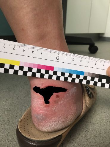
30 Mirzaalian-Dastjerdi et al.
on a single image taken with a commercial handheld digital camera or smart-
phone. From only one image, less information can be obtained than from a 3D
model, especially in case of the wound being on a curved surface. For helping to
estimate the local scale of the image even for curved surfaces, a flexible paper
ruler is used.
2 Material and methods
Our approach comprises two main steps, wound segmentation and lesion surface
area calculation. As only a single image is used, some conditions must be met
during image acquisition for keeping the measurement error as low as possible.
1) The camera shall be perpendicular to the wound surface. 2) The wound shall
be located in the center of the image (for minimizing lens distortion). 3) The
ruler has to be placed parallel to the largest wound diameter as close as possible
to it. Ideally, it should also reflect the curvature of the surface.
2.1 Wound segmentation
The user needs to provide a region of interest (ROI) including only wound and
skin by drawing a contour (Fig. 1(a)). For classifying the pixels in the ROI into
the classes skin and wound, a Random Forest (RF) classifier has been trained.
Feature vectors were generated by applying a filter bank consisting of Gaussian,
Difference of Gaussian, Sobel and Hessian filter using different values for the
standard deviation.
The output of the RF is a probability map defining how likely it is that a
single pixel belongs to wound or skin. Otsu’s thresholding is applied to generate
a binary mask. Finally, everything outside the ROI is discarded (Fig. 2).
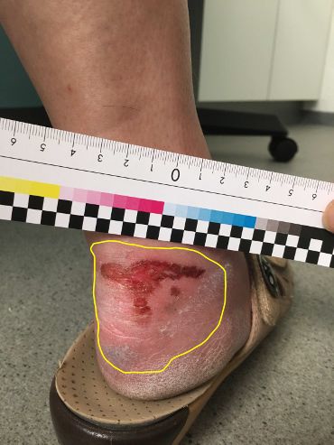
(a) (b)
Fig. 1. (a) Paper ruler and selected region of interest shown with yellow contour. (b)
The ground truth image obtained using the modified Random Walker.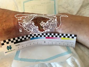
Measuring Skin Lesions       31

   To facilitate creating the ground truth masks for training the RF, the Ran-
dom Walker algorithm [5], with modified edge weight function based on Quater-
nion Color Curvature (QCC) [6] has been used (Fig. 1(b)).

2.2   Surface area measurement
For surface area calculation, the ruler should be first detected. As shown in
(Fig. 3(a)), the ruler contains a checkboard pattern with known size for easy
detection. The Structure Tensor filter is used for dividing the image into uniform
regions, corners, and edges according to the tensor’s eigenvalues [7] for each
pixel. The high eigenvalues roughly correspond to the ruler skeleton shown in
(Fig. 3(b)) and the low eigenvalues to corner points (Fig. 3(c)). Identification
of checkboard points is based on comparing intensities in a region around the
points. Fitting a curve to the local maxima of the distance transform of the
points seperates them to upper and lower points (Fig. 3(d)). Moving along this
curve allows finding pairs of corresponding points by detecting intensity changes
across checkerboard edges. (Fig. 3(c)).
    For obtaining the local measurement parameters, a heuristical approach is
used: Consider two corresponding points (p1 , p2 ) as shown in (Fig. 3(c)). Along
the line defined by p1 and p2 , points p3 , p4 , . . . are placed equidistantly using
d = kp1 − p2 k2 . Extrapolating each pair of points, a grid pattern covering the
wound is obtained. For measuring, each quadrilateral in the grid is unwarped
from perspective distortion [8] and mapped to a square. As the “true” size of
the square is known from the definition of the ruler, measuring comes down to
counting the wound pixels covered by the square and using the formula
                                   Nw
                            Aw =      · area of square                           (1)
                                   N
where N is the total number of pixels and Nw is the number of wound pixels
in the square. Adding these results for all squares yields the surface area of the
lesion.

Evaluation We recieved 50 images of skin lesions from clinical routine. They
were not taken under controlled conditions and by different persons with dif-
ferent commercially available (smartphone) cameras. For training the RF, this

        (a)                  (b)                   (c)                  (d)

Fig. 2. (a) Original image. (b) Probability map (result of Random Forest classifier).
(c) Result of Otsu’s filter. (d) Segmentation result after applying the ROI.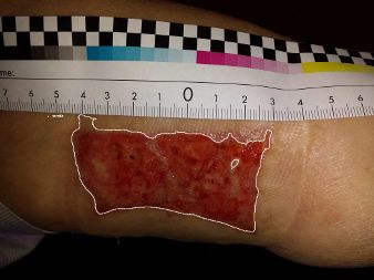
32      Mirzaalian-Dastjerdi et al.

set of images was split into a training set of 40 images and a test set of 10 im-
ages. Splitting was done manually as the set contained multiple images for some
patients. These images were all put in the training set.
    As we do not know the true size of the lesions in the clinical images, evaluation
of the measurements was performed using phantom images showing a geometric
shape with known surface area (Fig. 3(a)). Forty phantom images with different
geometric shapes of different sizes (ranging from 1.13–17.22 cm2 were taken with
an iPhone7’s camera. For simulating practical use we also bent the shapes
around cylindrical objects of different curvature and varied the angle of the
camera slightly.

3    Results

For wound segmentation, we found a mean accuracy, sensitivity and specificity
of 0.98 ± 0.02, 0.89 ± 0.13 and 0.99 ± 0.01, respectively, where accuracy is defined
as the proportion of correctly classified pixels according to the ground truth.
Fig. 4 shows a plot of these three values for each of the test images. Fig. 5
depicts three different results of segmented wounds.
    For the measurement evaluation based on the phantom images we found an
absolute error of 0.32 ± 0.27 cm2 and a relative error of 5.2 ± 4.3 %. Tab. 1 shows
the results grouped into flat images and images with lower and higher curvature.
    Among these 40 images, 16 images consisted of eight pairs showing the same
shape with slightly different size (differences ranging from 0.43 to 1.22 cm2 and 7

             (a)                         (b)                         (c)

                           (d)                         (e)

Fig. 3. (a) Phantom image. (b) Result of structure tensor filter containing high eigen-
values. (c) Result of structure tensor filter containing low eigenvalues. Two corre-
sponding corner points p1 and p2 are highlighted (red). (d) Distance transform, local
maxima shown with white points. (e) Obtained grid pattern and “wound segmentation”
(black circle).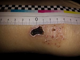
Measuring Skin Lesions       33

        1
                                                                       Fig. 4. The accuracy (red),
      0.95                                                             sensitivity (green) and speci-
       0.9                                                             ficity (blue) parameters of
      0.85
                                                                       wound segmentation of 10
       0.8
                                                                       patient wound images (bars
      0.75

       0.7
                                                                       were truncated for better
      0.65
                                                                       visualization).
       0.6

                                                        accuracy
      0.55
                                                        sensitivity
                                                        specificity
       0.5
             1     2   3   4   5     6   7     8   9      10

Table 1. Results for ruler-based measurements. Values for absolute and relative error
are given as mean ± standard deviation.

Type of Image                      absolute error          relative error   Min Area     Max Area
                                   (cm2 )                  (%)              (cm2 )       (cm2 )
Flat (N=20)                        0.23±0.21               4.54±4.86        2.80         17.22
Low Curvature (N=12)               0.42±0.34               5.03±2.98        1.13         9.73
High Curvature (N=8)               0.39±0.23               7.28±3.63        2.80         8.98
All (N=40)                         0.32±0.27               5.24±4.29        1.13         17.22

to 15 %). The true mean of surface areas in the group of smaller shapes was µs =
5.57 cm2 compared to µl = 6.33 cm2 for the group of larger shapes. Even though
the measurements reflect this difference (µ̄s = 5.73 cm2 and µ̄l = 6.52 cm2 ), a
paired, one-tailed t-test did not yield a significant difference.

3.1     Discussion and conclusion

We presented first results of our method for segmenting and measuring skin
lesions from a single image with acceptable error also for lesions on a bent surface.
A limitation of this study is the small number of data. For segmentation we
only could test our method on 10 images and a proper cross-validation was not
possible due to having multiple images of the same lesion. Skin lesions are greatly
varying in appearance (color and texture), so the approach must be tested using

                 (a)                                   (b)                             (c)

Fig. 5. (a) and (b) Examples of good segmentations. (c) A weak segmentation result
(the red region is misclassified as wound).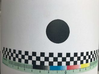
34      Mirzaalian-Dastjerdi et al.

more training and test images. Also color correction (which was not addressed
in this article) may help stabilizing the results [4].
    For measuring the area, our method is still lacking the possibility to reject
images not taken perpendicular to the wound surface. If the angle is only slightly
changed, the errors can quickly increase. It was beyond the scope of this study,
addressing this problem more systematically. Also here the sample size was quite
small, which also could explain the non-significant result for detecting changes
in surface area. We have also studied a different approach for extrapolating the
ruler points based on the cross ratio [9]; as we quickly found this approach to
be less stable and to have greater error, we did not pursue it further. In [3], the
announced measurement error is approximately 10% for available photographic
techniques and the precision may vary with wound size. We found comparable
results for our method, however, so far we have only addressed evaluation based
on phantom images. Also, a systematic precision analysis was out of the scope
of this work, due to the small amount of clinical images that contained the ruler.
    Providing a method for rejecting images taken from great angles and tools for
correcting failed segmentations could improve our approach. Further research
with more data is necessary for validating this method.

References
1. Foltynski P, Ladyzynski P, Wojcicki JM. A new smartphone-based method for
   wound area measurement. Artific Organs. 2014;38(4):346–352.
2. Liu X, Kim W, Schmidt R, et al. Wound measurement by curvature maps: a
   feasibility study. Phys Measure. 2006;27(11):1107.
3. Treuillet S, Albouy B, Lucas Y. Three-dimensional assessment of skin wounds using
   a standard digital camera. IEEE Trans Med Imaging. 2009;28(5):752–762.
4. Wannous H, Lucas Y, Treuillet S. Enhanced assessment of the wound-healing
   process by accurate multiview tissue classification. IEEE Trans Med Imaging.
   2011;30(2):315–326.
5. Grady L. Random walks for image segmentation. IEEE Trans Pattern Anal Machine
   Intell. 2006;28(11):1768–1783.
6. Shi L, Funt B, Hamarneh G. Quaternion color curvature. In: Color and Imaging
   Conference. vol. 2008. Society for Imaging Science and Technology; 2008. p. 338–341.
7. Baghaie A, Yu Z. Structure tensor based image interpolation method. AEU Int J
   Electronic Comm. 2015;69(2):515–522.
8. Jagannathan L, Jawahar C. Perspective correction methods for camera based docu-
   ment analysis. In: Proc. First Int. Workshop on Camera-based Document Analysis
   and Recognition; 2005. p. 148–154.
9. Lei G. Recognition of planar objects in 3-D space from single perspective views
   using cross ratio. IEEE Trans Robotic Automat. 1990;6(4):432–437.
You can also read