Diagnosis and Medical Management of Spondylolysis & Spondylolisthesis - William Primos, MD, FAAP Northeast Georgia Physicians Group Sports Medicine
←
→
Page content transcription
If your browser does not render page correctly, please read the page content below

Diagnosis and Medical Management of
    Spondylolysis & Spondylolisthesis
                        William Primos, MD, FAAP
                Northeast Georgia Physicians Group
                                   Sports Medicine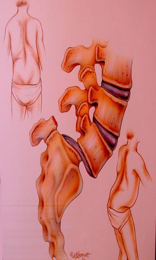
Disclosures
I have no financial interests, relationships, or potential conflicts of interest relative to this
presentation
Lower Back Pain in Young
Athletes
• Incr. sports participation in young people has led
  to back pain becoming more common
• 17.8% reported episode during 2 year period
• Most case are muscular
• Unlike in adults – often a specific diagnosis
Lower Back Pain Evaluation
• Obtain a history
  ▪ When? Onset, duration

  ▪ Why? Reason for pain, injury

  ▪ Where? Location of pain

  ▪ Describe pain. Type, severity, local or general, constant or intermittent
Lower Back Pain Evaluation
History (cont.)
  • Neurological Symptoms

  • Bowel / bladder incontinence

  • Aggravation/alleviation

  • Medication

  • Associated symptoms

  • Nighttime awakening

  • Family history
RED FLAGS

   ❑ Watch for these in a patient with low back pain

   ❑ May indicate a more serious condition as a cause of the
     back pain
RED FLAGS
❖ Significant trauma (fracture)

❖ Disabling pain –stops pleasurable
  activities (fracture, disc)

❖ Nighttime awakening (tumor, infection)

❖ Neurological Deficit / Radiating
  symptoms (disc, tumor)
RED FLAGS
❖ Unexplained weight loss (tumor)

❖ Fever or constitutional symptoms (infection

❖ Young patient (
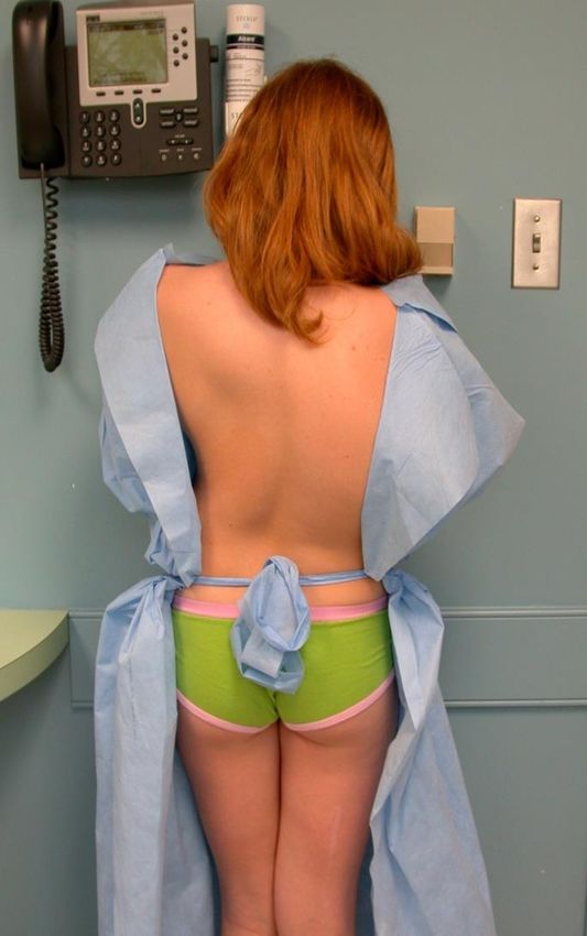
Physical Exam of the Spine
INSPECTION
  ▪ Gait
  ▪ Symmetry
  ▪ Posture
  ▪ Skin lesions/abnormalities
  ▪ Pelvis level
  ▪ Leg length comparison
Physical Exam of the Spine
Range of Motion – Is there pain?
  • Flexion
  • Extension
  • Rotation
  • Lateral bend
  • Hamstring flexibility
  • Hip motion
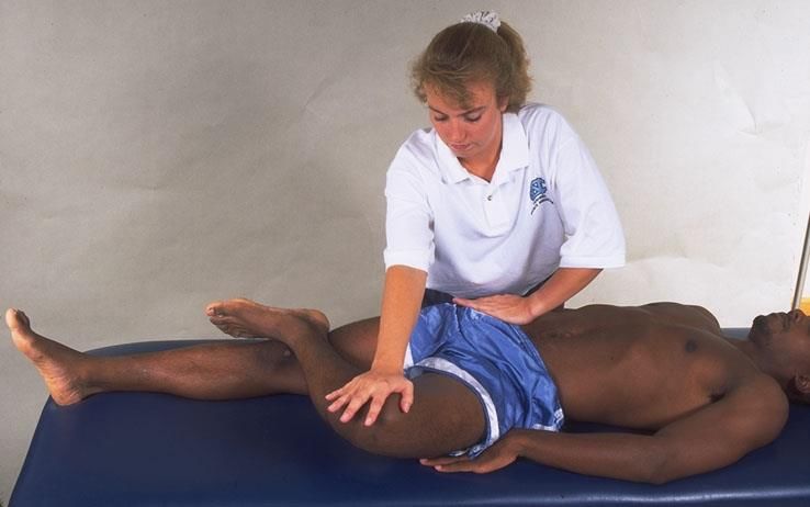
Physical Exam of the Spine
❑ Palpate – localize tenderness
❑ Straight leg raise
❑ FABER TEST – SI joint
❑ Neurological exam
  • Reflexes
  • Sensation
  • Strength
Imaging for Back Pain
❖Get plain films

❖AP/lateral views

❖Possibly oblique views
Causes of back pain in adolescents
➢ Spondylolysis/Spondylolisthesis
➢ Acute Lumbosacral Strain
➢ Mechanical Low Back Pain
➢ Degenerative Disc / Discogenic pain
➢ Scheurmann’s Kyphosis
➢ Infection
➢ Fracture
➢ Tumor
SPONDYLOLYSIS
❖ Anatomical defect of the pars
  interarticularis of the vertebral arch.

❖ May be unilateral or bilateral

❖ Most common- L5 (85-95%), then L4
  (5-15%)
SPONDYLOLYSIS
❑ Most common bony cause of back pain in young athletes

❑ Studies show spondylolysis causes 30-40% of low back pain in adolescent
  athletes

❑ In adolescent athletes, 8-14% have spondylolysis seen on x-rays

❑ Asymptomatic in majority

❑ Incidence ratio of 2:1 male to female
SPONDYLOLYSIS
❑ Develops during ambulatory activity

❑ Studies have shown spondylolysis is absent at birth or in non-ambulatory
  individuals

❑ Studies show prevalence of 4.4% in 6 years old

❑ Heredity plays a factor since it occurs more commonly in relatives than in
  general population
Spondylolysis Pathophysiology
❑May occur suddenly as an acute injury

❑But usually develops gradually due to overuse or repetitive
 hyperextension
  • Repetitive forces cause minute damage to bone

  • When the rate of damage overcomes the ability of the bone to repair itself

  • Then a stress fracture results.
Spondylolysis Pathophysiology
❑ Abnormalities that may increase risk
• Inflexibility due to rapid skeletal growth during adolescence
• Poor physical condition / core weakness
• Spina bifida occulta (posterior spinal fusion anomaly)
• Hyper-lordosis of the spine
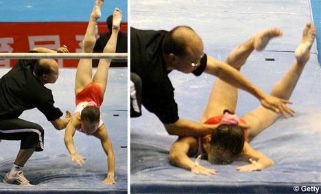
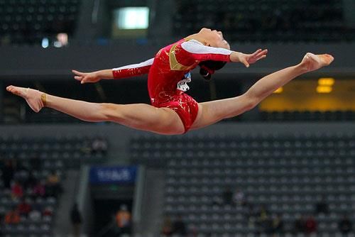
• ScoliosisSPONDYLOLYSIS Occurs more frequently in certain sports such as gymnastics, weight-lifting, baseball, soccer, football lineman
Pathophysiology
❑ Spondylolysis may progress
  to spondylolisthesis

❑ Displacement or slip of a
  vertebra on the other
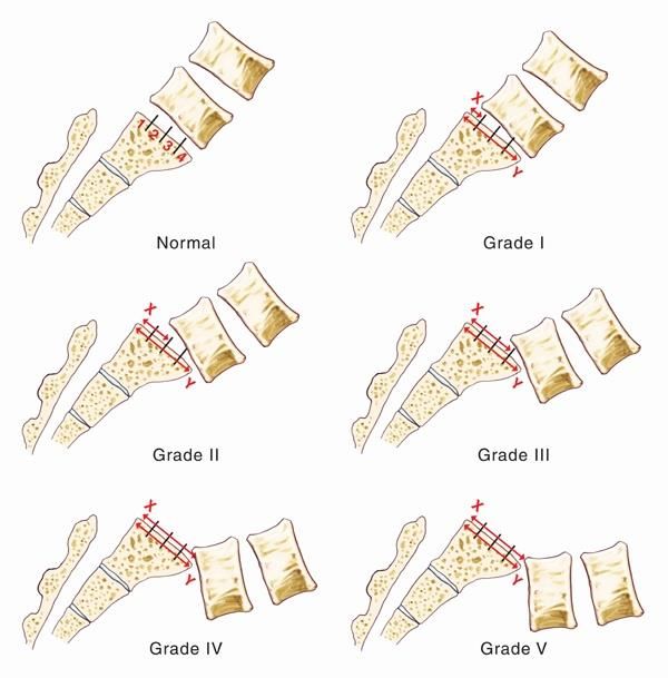
Spondylolisthesis
Graded by amount of displacement
  I : 1-25%

  II : 26-50%

  III : 51-75%

  IV : 76-100%

  V : > 100%
Categories of Spondylolisthesis
❖ Type I
  o Dysplastic- congenital deformity with abnormal rounding of superior aspect of S1
     vertebral body that allows L5 vertebra to slip

❖ Type II
  o Isthmic- stress or acute fracture in the pars interarticularis that leads to the slip

❖ Type III
  o Degenerative- slip occurs due to instability of the vertebra because of arthritis
Categories of Spondylolisthesis
❖ Type IV
o Traumatic- acute fracture from high energy trauma to spine results in the slip
❖ Type V
o Pathological- bone disease, tumor, or infection that causes weakness and the
slippage
❖ Type VI
o Iatrogenic- potential sequelae of spinal surgery which weakened the spineSPONDYLOLYSIS
History
• Back pain often begins after an increase
in training
• Pain worsens with activity, especially
extension of back
• Often unable to continue their sport due
to pain
• Pain near lower lumbar spine, may be
either right or left of spine or in midlineSPONDYLOLYSIS
Study by Hirano showed association of pain with lumbar hyperextension and
spondylolysis
▪ 100 young athletes with lower back pain
▪ 69% had pain with hyperextension
▪ All patients had x-rays, then CT if x-rays negative
▪ 42 had spondylolysis
o 34 of 42 (81%) had pain with hyperextension
▪ 58 did not have spondylolysis
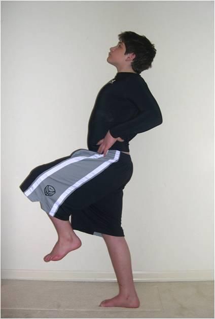
o 35 of 58 (60%) had pain with hyperextensionPHYSICAL EXAM ✓ Tenderness over lower spine ✓ Pain / stiffness with extension and rotation of back ✓ Usually, no pain with flexion ✓ Worsens with the stork test ✓ Negative straight leg raise test ✓ Hamstring inflexibility often present ✓ May feel step-off in spondylolisthesis
SPONDYLOLYSIS
Radiographs:

▪ May be seen with just AP/LAT views

▪ Obliques not always ordered
SPONDYLOLYSIS
Radiographs:
• May show “Scottie dog
  collar” sign on oblique view
SPONDYLOLYSIS
IMAGING- further studies to get if suspect :
• CT Scan

• MRI

• SPECT Bone scan
SPONDYLOLYSIS
IMAGING:
• SPECT Scan – very sensitive but
  not specific

• Radiation exposure
SPONDYLOLYSIS
IMAGING:
❑ CT Scan
 •   Effective in evaluating for fx and determining acuteness

 •   Radiation exposure
SPONDYLOLYSIS
IMAGING:
❑ MRI
 • Improving effectiveness
 • No radiation
 • Order with thin cuts and oblique views to improve
   sensitivity
SPONDYLOLYSIS
▪ Differing recommendations

▪ Most agree plain radiographs are reasonable screening tool

▪ SPECT bone scan, then CT provides anatomical and physiological
  info needed

▪ MRI potentially shows the info and also does not expose pt. to
  radiation
IMAGING
(Congeni)
  Radiographs : AP/LAT/OBL
    • Positive- no more studies and treat
    • Negative
      o SX greater than 6 weeks- get MRI
      o SX less than 6 weeks- get SPECT scan
IMAGING
(Gregory, et al)
Radiographs
• Positive: treat
• Negative: get SPECT Scan
o Positive SPECT : get CT scan
o Negative SPECT :get MRISPONDYLOLYSIS TREATMENT ❑ No definite standard of care ❑ Numerous opinions on proper treatment
SPONDYLOLYSIS TREATMENT
Controversial questions

 •   Is fracture healing possible?

 •   How long to rest from sports?

 •   Is a brace necessary?
SPONDYLOLYSIS TREATMENT
Is fracture healing possible? Depends on how recently
occurred.

 •   Chronic fracture (> 6 months ago ) unlikely to heal

 •   Recent fracture (
SPONDYLOLYSIS TREATMENT
 Chronic fracture (> 6 months )
  • Rest until pain-free
  • Then start rehab and progressive RTP

 Recent fracture (
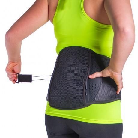
SPONDYLOLYSIS TREATMENT
 Brace or not?
  •   In the past, brace recommended to immobilize the
      spine to allow healing

  •   Recent studies show bracing may not be needed and
      improvement occurs with just rest and PT
SPONDYLOLYSIS TREATMENT Brace – even though constant bracing not needed for healing it can help relieve pain and also control activity
SPONDYLOLYSIS TREATMENT
Physical Therapy
 •   Core strengthening

 •   Hamstring stretches

 •   Spine range of motion
SPONDYLOLYSIS TREATMENT

❑ Low-intensity pulsed ultrasound may help bony
  healing

❑ Has been shown to speed healing time and
  improve success of treatment
SPONDYLOLISTHESIS TREATMENT
❑Grades 1 and II spondylolisthesis -usually treated conservatively,
 same as in spondylolysis

❑Grades II, IV, V spondylolisthesis –possible will need surgical
 treatment
Factors that may lead to surgery
▪ Progressive slippage
▪ Spondylolisthesis Stages III, IV, V
▪ Instability of the spine
▪ Neurological findings
▪ Cauda equina syndrome
▪ Severe, unremitting pain
▪ Failure to improve with conservative
  treatment for 6 months
References
• Gagnet P, Kern K, Andrews K, Elgafy H, Ebraheim N. Spondylolysis and spondylolisthesis: a review of the
  literature. J Orthop 2018;15(2): 404-407.

• Gregory P, Batt M, Kerslake R, Scammell B, Webb J. The value of combining single photon emission
  computerised tomography and computerised tomography in the investigation of spondylolysis. Eur Spine J
  2004: 13:503-509.

• Congeni J. Evaluating spondylolysis in adolescent athletes. J Musculoskel Med 2000; 17: 123-129.

• Hirano A, Takebayashi T, Yoshimoto M. Characteristics of clinical and imaging findings in adolescent
  lumbar spondylolysis associated with sports activities. J Spine. 2012; 1:124.

• Arima H, Suzuki Y, Togawa D, Mihara Y, Murata H, Matsuyama Y. Low-intensity pulsed ultrasound is
  effective for progressive-stage lumbar spondylolysis with MRI high-signal change. Eur Spine J 2017:1-7.
Thank You!
You can also read