Diverse novel phleboviruses in sandflies from the Panama Canal area, Central Panama
←
→
Page content transcription
If your browser does not render page correctly, please read the page content below
RESEARCH ARTICLE
Marklewitz et al., Journal of General Virology
DOI 10.1099/jgv.0.001260
Diverse novel phleboviruses in sandflies from the Panama Canal
area, Central Panama
Marco Marklewitz1,2,3,†, Larissa C. Dutari4,5,6,†, Sofia Paraskevopoulou1, Rachel A. Page3, Jose R. Loaiza3,4,6 and Sandra
Junglen1,2,*
Abstract
The genus Phlebovirus (order Bunyavirales, family Phenuiviridae) comprises 57 viruses that are grouped into nine species-
complexes. Sandfly-transmitted phleboviruses are found in Europe, Africa and the Americas and are responsible for febrile
illness and infections of the nervous system in humans. The aim of this study was to assess the genetic diversity of sandfly-
transmitted phleboviruses in connected and isolated forest habitats throughout the Panama Canal area in Central Panama.
In total, we collected 13 807 sandflies comprising eight phlebotomine species. We detected several strains pertaining to
five previously unknown viruses showing maximum pairwise identities of 45–78 % to the RNA-dependent RNA polymerase
genes of phleboviruses. Entire coding regions were directly sequenced from infected sandflies as virus isolation in cell
culture was not successful. The viruses were tentatively named La Gloria virus (LAGV), Mona Grita virus (MOGV), Peña
Blanca virus (PEBV), Tico virus (TICV) and Tres Almendras virus (TRAV). Inferred phylogenies and p-distance-based analy-
ses revealed that PEBV groups with the Bujaru phlebovirus species-complex, TRAV with the Candiru phlebovirus species-
complex and MOGV belongs to the proposed Icoarci phlebovirus species-complex, whereas LAGV and TICV seem to be
distant members of the Bujaru phlebovirus species-complex. No specific vector or habitat association was found for any of
the five viruses. Relative abundance of sandflies was similar over habitat types. Our study shows that blood-feeding insects
originating from remote and biodiverse habitats harbour multiple previously unknown phleboviruses. These viruses should
be included in future surveillance studies to assess their geographic distribution and to elucidate if these viruses cause
symptoms of disease in animals or humans.
Introduction phlebotomine sandflies have been implicated as vectors of
Neotropical sandflies are a diverse group of insects in the Leishmania parasites, the causing agent of American Cuta-
order Diptera, family Psychodidae, subfamily Phlebotominae, neous Leishmaniasis (ACL) [3–5], and sandflies of some of
which has undergone notable taxonomic revision recently [1]. these taxa are also suspected of transmitting phleboviruses
Historically, phlebotomine sandflies have been recognized as to a broad range of animals and humans [6–9]. Sandflies
vectors of various disease-causing pathogens to humans [2]. of the six species Psychodopygus panamensis, Nyssomyia
In Panama, anthropophilic species of at least six species of trapidoi, Lutzomyia gomezi, Nyssomyia ylephiletor, Lutzomyia
Received 13 February 2019; Accepted 21 March 2019; Published 03 May 2019
Author affiliations: 1Institute of Virology, Charité - Universitätsmedizin Berlin, corporate member of Free University Berlin, Humboldt-University Berlin,
and Berlin Institute of Health, Berlin, Germany; 2German Center for Infection Research (DZIF), Berlin, Germany; 3Smithsonian Tropical Research Institute,
Panama City, Republic of Panama; 4Centro de Biodiversidad y Descubrimiento de Drogas, Instituto de Investigaciones Científicas y Servicios de Alta
Tecnología (INDICASAT-AIP), Panama City, Republic of Panama; 5Biotechnology Department, Acharya Nagarjuna University, Guntur, India; 6Programa
Centroamericano de Maestría en Entomología, Vicerrectoría de Investigación y, Postgrado, Universidad de Panamá, Panama City, Republic of Panama.
*Correspondence: Sandra Junglen, sandra.junglen@charite.de
Keywords: Bunyavirales; Phlebovirus; species-complex; Neotropics; Panama; Phlebotominae.
Abbreviations: blast, Basic Local Alignment Search Tool; CDC, U.S. Center for Disease Control; CF, complement fixation; CHGV, Chagres virus; CL,
American Cutaneous Leishmaniasis; DFG, German Science Foundation; GPC, glycoprotein precursor protein; HI, hemagglutination inhibition; ICTV,
International Committee on Taxonomy of Viruses; JOAV, Joá virus; LAGV, La Gloria virus; MAFFT, multiple alignment using fast Fourier transform;
MEGA, Molecular Evolutionary Genetics Analysis; MOGV, Mona Grita virus; N, nucleocapsid; NGS, next-generation sequencing; NIQV, Nique virus; NPV,
Ntepes virus; ORF, open reading frame; PCR, polymerase chain reaction; PEBV, Peña Blanca virus; PTV, Punta Toro virus; RdRp, RNA-dependent RNA
polymerase; RT-PCR, reverse transcriptase polymerase chain reaction; RVDB, Reference Viral Database; SENACYT, National Secretariat of Science,
Technology and Innovation of Panama; SPP, Priority Program; TICV, Tico virus; TRAV, Tres Almendras virus; URIV, Uriurana virus.
Sequences of LAGV, MOGV, PEBV, TICV and TRAV have been deposited in GenBank under accession number MK524329-MK524350.
†These authors contributed equally to this work
001260 © 2019 The Authors
This is an open access article under the terms of the Creative Commons Attribution 4.0 International License, which permits unrestricted use, distribution and reproduction in any medium, provided
the original author and source are credited. Downloaded from www.microbiologyresearch.org by
IP: 200.46.27.89
1
On: Tue, 07 May 2019 20:06:24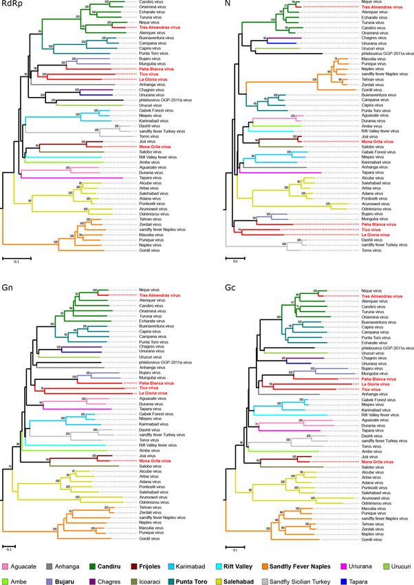
Marklewitz et al., Journal of General Virology 2019
sanguinaria and Psychodopygus thula are widespread across landscapes differing in their level of anthropogenic distur-
forested areas of Panama, feeding opportunistically on bance, (i) in continuous tropical lowland forest at the shore
animals of various species depending on habitat quality, host of Gatún Lake (continuous forests), (ii) in small forest frag-
availability and biomass [10]. ments (1.5–51 ha) surrounded by agricultural fields (forest
fragments) and (iii) in isolated forested islands (5.2–17.5 ha)
Sandflies almost exclusively transmit viruses of the genus
(forested islands) that are remnants of the original continuous
Phlebovirus (family Phenuiviridae, order Bunyavirales).
forest and emerged after flooding the Panama Canal in 1914.
According to the International Committee on Taxonomy of
In each of the three landscape types, five replicate sites were
Viruses (ICTV) members of the genus Phlebovirus are clas-
sified into nine virus species that comprise 57 viruses [11]. selected resulting in 15 sampling sites in total [24].
In addition, more than 15 putative phlebovirus species have Adult sandflies were collected using CDC (U.S. Centers
been described that are waiting to be classified by the ICTV. for Disease Control) miniature light traps (John W. Hock
The classified sandfly-transmitted viruses of the New World Company, Gainesville, FL, USA). The following sampling
belong to the species-complexes Punta Toro, Candiru, Chilibre design was used for each sampling site. In total, nine traps
and Frijoles phlebovirus. Infections with sandfly-borne phle- were placed along two parallel transects while two traps were
boviruses can cause unspecific symptoms in humans often placed in the canopy at approximately 10 m high. Traps oper-
misdiagnosed as dengue fever, malaria or influenza [7, 12]. ated at night for 12 h. Sandfly specimens were collected after
Clinical symptoms are ranging from high fever, severe head- sunrise, identified using female taxonomic keys, and stored
ache, muscle ache and aseptic meningitis to mild or severe in liquid nitrogen until further processing [25, 26].
meningoencephalitis [13]. A recent study in Panama has
shown that Punta Toro virus (PTV) causes febrile illness in
Phlebotomine sandfly community metrics
humans with symptoms similar to infections with dengue
viruses [12]. Between 1964 and 2009 various members of Phlebotomine sandfly species diversity was assessed at each
the PTV species-complex were isolated from phlebotomine landscape type using Shannon and Simpson Alpha diversity
sandflies, humans and sentinel hamsters in Panama and indexes [27, 28]. Species relative abundance was measured
Colombia [7]. In addition, Chagres virus (CHGV) has been and compared among landscape types using one-way
reported as being responsible for outbreaks of febrile illness Kruskal–Wallis nonparametric test as implemented in the
in humans [14]. Other phleboviruses detected in Panama- PAST3 program (available at https://folk.uio.no/ohammer/
nian sandflies in the 1950s are Aguacate, Cacao, Frijoles and past/).
Nique viruses [6, 15–17]. More recent contributions to the
expansion of the genetic diversity of phleboviruses of the New RNA-extraction and PCR screening
World have been provided by sequencing of viruses isolated Sandflies were pooled according to species, collection site
from sandflies (Phlebotominae spp.), spiny rats (Proechimys and date by individuals of ten. Specimens containing blood
sp.) and the Southern two-toed sloth (Choloepus brasiliensis) were processed individually. Pools were homogenized in
that was collected in Brazil between 1962 and 1985 [18], and 500 µl L-15 medium (Thermo Fisher Scientific, Waltham,
2014–2015 [19]. It is unknown if these viruses are pathogenic USA) using ceramic beads and a SpeedMill Plus (Analytik
for humans. The highest diversity of phleboviruses in Panama Jena, Germany). The suspensions were cleared by centrifu-
was detected in phlebotomine sandflies and human samples gation at 2500 r.p.m. for 10 min. RNA was extracted from
from the Central Panama region and the Darien province 50 µl supernatant using the QIAamp Viral RNA Mini Kit
[7, 20]. Both regions show increasing levels of habitat frag- (Qiagen, Hilden, Germany) according to the manufacturer's
mentation due to land-use change for cattle and crop farming instructions. cDNA was synthesized using the SuperScript
[21, 22]. Despite their impact on human and veterinary III RT System (Invitrogen, Karlsruhe, Germany) and random
health, phleboviruses have been insufficiently studied in hexamer primers according to the instructions from the
Panama [7, 12].
manufacturing company. Sandfly pools were tested for
The aim of this study was to investigate the genetic diversity phleboviruses in a PCR assay using Platinum Taq DNA Poly-
of phleboviruses in sandflies of forested areas of the Panama merase (Thermo Fisher Scientific, Waltham, USA) and first
Canal area. The lowland tropical rainforest of the Panama round primers F 5'-TCAARAAGAMNCAACATGGTGG, R
Canal area harbours a large diversity of phlebotomine sandfly 5'-TATGCCYTGCATCATYCCWG followed by nested round
species of the New World. This region has a long history of primers Fn 5'-GGACTTAGAGAGATYTAYGTNTTGG and
arbovirus outbreaks. For example, during the construction of Rn 5'-ACATGRTGACCYTGRTTCCA. Thermocycler condi-
the Panama Canal at the end of the nineteenth century tens of tions were as follows: 10 cycles of 95 °C for 3 min, 94 °C for
thousands of people died from yellow fever [23]. 15 s, 55 °C for 20 s and 72 °C for 30 s, then 30 cycles from
95 °C to 15 s, 50 °C to 20 s, 72 °C to 30 s and a final extension
step of 72 °C for 5 min. PCR products were visualized on
Methods 2 % agarose gels and positive samples were Sanger sequenced
Sandfly collection (Seqlab, Göttingen, Germany). Obtained sequences were
Fieldwork was conducted in the Panama Canal area, Central analysed using Geneious R9 [29] and compared to GenBank
Panama, between 2013 and 2014. Sandflies were collected in database (www.ncbi.nlm.nih.gov/genbank/).
Downloaded from www.microbiologyresearch.org by
IP: 200.46.27.89
2
On: Tue, 07 May 2019 20:06:24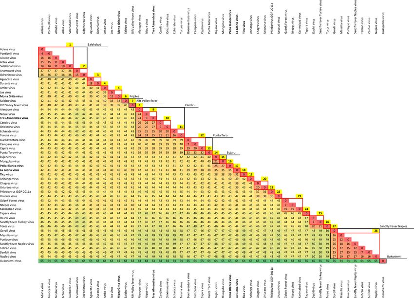
Marklewitz et al., Journal of General Virology 2019
Table 1. Sandfly species abundance and diversity across study sites
Abbreviations are as follows: number of specimens (n), percent (%), Shannon diversity (H), Simpson diversity (λ).
Species Continous forests Forest fragments Forested islands
n % -pi*ln(pi) pi² n % -pi*ln(pi) pi² n % -pi*ln(pi) pi²
Psychodopygus 4010 43.9 0.36 0.19 512 34.36 0.37 0.12 1152 38.51 0.37 0.15
panamensis
Nyssomyia 812 8.89 0.22 0.01 282 18.92 0.32 0.04 213 7.12 0.19 0.01
trapidoi
Lutzomyia gomezi 899 9.84 0.23 0.01 164 11.1 0.24 0.01 69 2.3 0.09 0.00
Bichromomyia 61 0.66 0.00 0.00 – – – – 14 0.46 0.00 0.00
olmeca sp.
Psychodopygus 292 3.19 0.11 0.00 20 1.34 0.06 0.00 290 9.69 0.23 0.01
thula
Nyssomyia 97 1.06 0.05 0.00 21 1.34 0.06 0.00 46 1.53 0.06 0.00
ylephiletor
Pressatia 321 3.51 0.12 0.00 7 0.46 0.03 0.00 40 1.34 0.06 0.00
dysponeta
Phlebotominae 2523 27.62 0.36 0.08 469 31.47 0.36 0.10 916 30.7 0.36 0.09
spp.
Total 9133 100 H=1.49 λ=0.29 1490 100 H=1.48 λ=0.27 2991 100 H=1.56 λ=0.26
Genome sequencing ATCTAGCGACCTCCACACACAAAG. Products were
RNA derived from phlebovirus-positive sandfly pools was subsequently Sanger sequenced.
used to sequence full genomes on an Illumina MiSeq next-
generation sequencing (NGS) platform. Library preparation Genome analysis
and Illumina MiSeq sequencing was carried out by using Full genomes were analysed regarding sizes, putative coding
the SuperScript One-Cycle cDNA Kit, the Nextera XT DNA regions and 5'/3' noncoding regions using Geneious R9 [29].
Library Preparation Kit, and V3 chemistry (2×300 bp) N-glycosylation sites were identified using NetNGlyc v1.0
according to the manufacturers’ instructions. The individual Server (www.cbs.dtu.dk/services/NetNGlyc/), prediction of
samples were normalized, pooled and sequenced on an
transmembrane domains was done using TMHMM Server
Illumina MiSeq instrument, with a designated yield of ~25
v2.0 (www.cbs.dtu.dk/services/TMHMM/) and posttransla-
million paired-end reads. After demultiplexing the obtained
tional GPC processing products were identified using Pfam
raw reads were subjected to two filtering steps using Lighter
v31.0 (https://pfam.xfam.org/) as implemented in Geneious
v1.1.0 [30] for correcting sequencing errors and bwa v0.7.17
[29].
[31] to subtract host-originated background information
using the mitochondrial reference genome of Phlebotomus
papatasi as no genome of a phlebotomine sandfly species Phylogenetic analysis
closer related to the species used in this study is available. Translational alignments of N, Gn, Gc and L genes were
The remaining reads were de novo assembled into contigs with performed using the MAFFT E-INS-I algorithm [34]. Model-
the genome assembler software Spades v3.11.1 [32]. An initial test was performed as implemented in mega7 to identify the
blastx search against the Reference Viral Database (RVDB) most suitable evolutionary model [35]. Phylogenies were
[33] was performed, followed by a second blastx search inferred using PhyML with the WAG substitution model as
against two custom databases containing only the S and the implemented in Geneious R9, with 1000 bootstrap replicates
M segment sequences of Phenuiviridae. Reference mapping [36].
of NGS reads to the respective PCR fragment was performed
using Geneious mapper [29]. Genome ends were amplified
by conventional semi-nested RT-PCR (reverse transcription Virus isolation
PCR) using Platinum Taq DNA Polymerase (Thermo Fisher Grivet (Chlorocebus aethiops) (VeroE6/7) and Lutzomyia
Scientific, Waltham, USA), segment-specific primers (Table 1) longipalpis (LL-5) cells were used to perform virus isola-
and a primer containing the terminal conserved nucleotides tion trails from sandfly homogenates as described previously
of phleboviruses fused to an anchor sequence PTN 5'-GACC [37].
Downloaded from www.microbiologyresearch.org by
IP: 200.46.27.89
3
On: Tue, 07 May 2019 20:06:24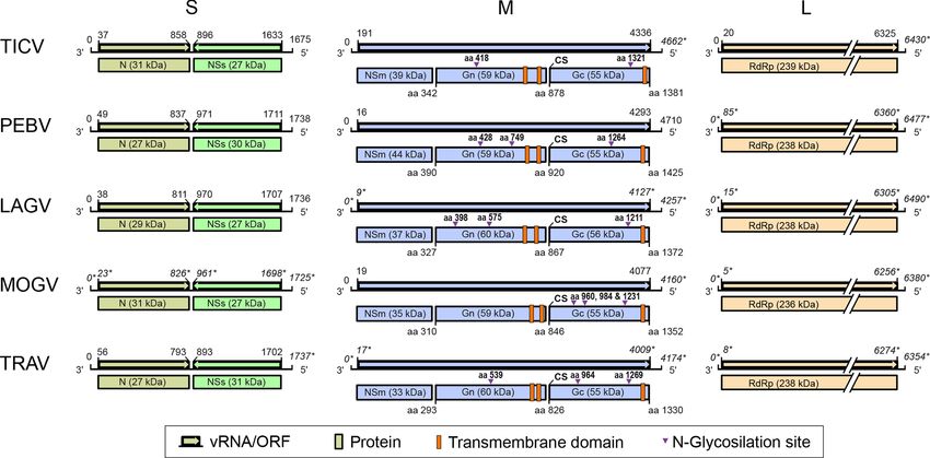
Marklewitz et al., Journal of General Virology 2019

Table 2. Virus names, designated strains, locality of sandfly specimen collection, sex and species of viruspositive sandflies
Abbreviation of localities are as follows: forest fragment (F), continuous forest (C), forested island (I).

  Virus                                                Strain                          Locality               Sex           Sandfly species

  Tico virus                                      SP0157/PA/2013                          F                    f           Phlebotominae spp.

  Peña Blanca virus                               SP1681/PA/2013                          C                    f           Phlebotominae spp.
                                                  SP1683/PA/2013                          C                    f       Psychodopygus panamensis
                                                  SP1684/PA/2013                          C                    f           Nyssomyia trapidoi

  La Gloria virus                                 SP0535/PA/2013                          F                   f            Nyssomyia trapidoi
                                                  SP0538/PA/2013                          F                   f            Phlebotominae spp.
                                                  SP0543/PA/2013                          F                   m            Phlebotominae spp.
                                                  SP0584/PA/2013                          F                   f        Psychodopygus panamensis
                                                  SP1070/PA/2013                          C                   f            Nyssomyia trapidoi
                                                  SP1075/PA/2013                          C                   f            Nyssomyia trapidoi

  Mona Grita virus                                SP0260/PA/2013                          I                    f           Nyssomyia trapidoi

  Tres Almendras virus                            SP0412/PA/2013                          I                    f       Psychodopygus panamensis

Results                                                                            between the relative abundance of phlebotomine sandflies
Sandfly species composition and diversity                                          among landscape types.
In total, 13 692 phlebotomine sandflies were collected in
continuous tropical lowland forest (continuous forests), in                        Virus detection and genome analyses
fragmented forest surrounded by agricultural fields (forest                        Sandflies were tested for phleboviruses by generic RT-PCR.
fragments) and in isolated forest on islands in the Panama                         Sequences with 45–78 % nt identity to the RNA-dependent
Canal area, Central Panama (forested islands). Eight different                     RNA polymerase (RdRp) gene of phleboviruses were iden-
phlebotomine sandfly species were identified, with Psycho-                         tified in 12 pools (Table 3). The sequences formed five
dopygus panamensis as the predominating species (41.6 %)                           groups with intra-group pairwise nt identities ranging from
(Table 2). Due to damage of specimens, 28.7 % of the sandflies                     96–100 % and inter-group pairwise nt identities ranging from
could not be identified to species level and were summarized                       67–74 %, suggesting the detection of several strains pertaining to
as Phlebotominae spp..                                                             five phlebovirus species. Attempts to isolate the viruses in verte-
                                                                                   brate (VeroE6/7) and sandfly (LL-5) cells were not successful.
Great differences were detected in the number of sandflies per
                                                                                   Complete coding regions of one strain from each putative
landscape type (sandfly abundance). Highest abundance was
                                                                                   species were obtained by NGS and semi-nested RT-PCR from
found in continuous forests (n=9,133, 67 %). Abundance was
                                                                                   homogenates of infected sandflies. The viruses were tentatively
much lower in forest fragments surrounded by agricultural
                                                                                   named after the geographic location where the sandflies have
fields (n=1,490, 15 %) and on forested islands in the Panama
                                                                                   been collected, namely La Gloria virus (LAGV), Mona Grita
Canal (n=2,991, n=18 %). Surprisingly, species richness
                                                                                   virus (MOGV), Peña Blanca virus (PEBV), Tico virus (TICV)
was similar across the different landscape types, with eight
                                                                                   and Tres Almendras virus (TRAV).
species found in continuous forests and on forested islands
in the Panama Canal, and a slightly lower species richness                         All viruses had a tripartite genome organization similar to
of seven species found in forest fragments surrounded by                           phleboviruses, comprising a large (L), middle (M) and small
agricultural fields (Table 2). Except Bichromomyia olmeca                          (S) segment (Fig. 1). The transduced L segment ORFs of LAGV,
sp. which was not present in forest fragments, all of the                          MOGV, PEBV, TICV and TRAV showed the highest similari-
other seven phlebotomine sandfly species were found in all                         ties of 68–91 % to phlebovirus RdRp proteins suggesting the
landscape types. Accordingly, no great differences in species                      identification of five previously unknown viruses. Bunya-
diversity entropy (H) and evenness (EH) across landscape                           viruses, including phleboviruses, carry the conserved palm
types were found, continuous forests H=1.49 and EH=0.51,                           motifs Pre-A and A through E on the L segment. These motifs
forest fragments H=1.48 and EH=0.55, and forested islands                          were detected in all five viruses (Fig. 2a). The conserved
H=1.56 and EH=0.54. The results from the Kruskal-Wallis H                          N-terminal motifs H...D...PD...ExT…K (cation-binding
test (χ2(2)=5.527, p=0.063) suggest that there is no difference                    residues and catalytic lysine) of the endonuclease domain of
                                                  Downloaded from www.microbiologyresearch.org by
                                                                 IP: 200.46.27.89
                                                                        4
                                                          On: Tue, 07 May 2019 20:06:24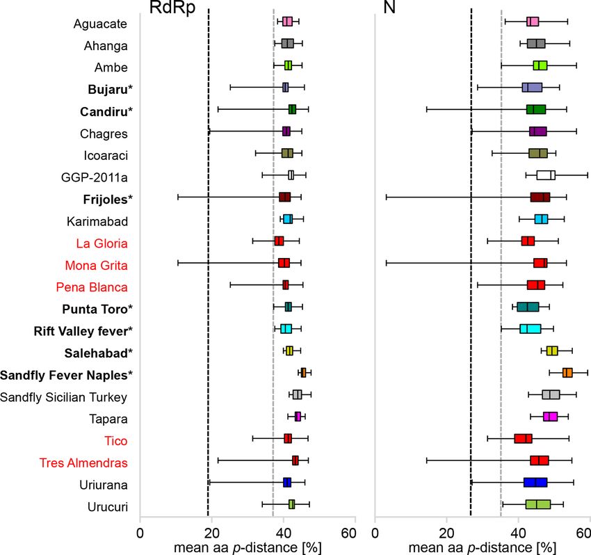
Marklewitz et al., Journal of General Virology 2019
Table 3. Segment-specific primers used to amplify genome ends
Abbreviations are as follows: Tico virus (TICV), Peña Blanca virus (PEBV), La Gloria virus (LAGV), Mona Grita virus (MOGV) and Tres Almendras virus
(TRAV).
Virus Segment end Primer sequences
First round PCR Nested round PCR
TICV M-5' 5'-TGCAGGTTGATCTCAGTTGGG 5'-TTTGAGACCTGGCCGAGTTG
L-5' 5'-CCACCTTCAGCCCACTATTC 5'-TGGAGGAAGAAATCTGGCAAC
PEBV M-5' 5'-ACTGATGGCTCGTGAATGGG 5'-ATAACTTGGCTGGCGACTCC
M-3' 5'-GATTTCGTTCAGTCAGGCGG 5'-CTCATGGATGGGTGGTCCTG
LAGV S-5' 5'-ACTGAACTGGCAGCTCCTTC 5'-GGTGGCTCTTGTCTTTGTCG
S-3' 5'-TCACAGGCTTATTCCCACGAG 5'-AGAGCCAGCACAATCATCTTC
M-5' 5'-TCACCTGATGTCCCGATCTG 5'-TGTCTTAACACTCTGACGGGTG
L-5' 5'-GGACTGCTGATTAGTGTTGAGC 5'-ACTCAACTCTTAGACTTGATCCC
MOGV S-5' 5'-GCCTCTAGTCAGAGCGAGAAC 5'-GTCCTCTCTCTTGGACCAGTTG
M-5' 5'-TTCCACTCTGTTATTTATCCCTTGC 5'-AGTCTGATCCAGAGTAACTGCAC
TRAV S-5' 5'-CTTGTCTGACATCTTAGTCATCATC 5'-TGGTTTGTTTCCTCGAGTGAG
S-3' 5'-TGGTGCACTGTGATAGTTGTTG 5'-TTGCGATCAGTAGCACTTCTTG
M-5' 5'-TCCTGCATCCTGAGAGCTTG 5'-CTTCCCAGTAGTACACTAGCCC
L-5' 5'-TTGGACCAGACACCTTGTGC 5'-GTGCAGTCAGACGATCTGTTC
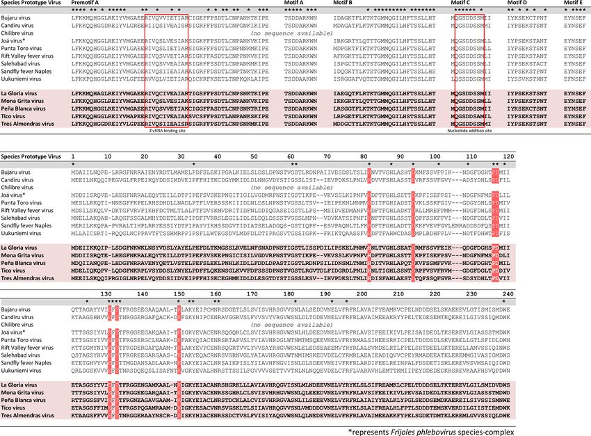
phleboviruses were present as well (Fig. 2b). This domain is (Fig. 1). The ORF closer to the 3′-end showed highest similari-
able to bind and to cleave RNA for transcription processes and ties of 64–81 % to the nucleocapsid gene (N) of phleboviruses.
is involved in the cap-snatching process typical for negative- The second ORF is encoded in ambisense, which is typical
sense RNA viral polymerases like in bunya- and orthomyxo- for phleboviruses, and most likely encodes the nonstructural
viruses [35, 38]. A second bunyavirus typical region, the motif protein NSs.
G (RY), was present between aa 668 and aa 675 in all protein
sequences of the novel viruses [39]. The conserved arginine Phylogenetic analyses and classification
is positioned in the polymerase active site to interact with the
The novel viruses consistently fall into three main clades in
priming nucleotide triphosphate.
the phylogenetic trees of RdRp, N, Gc and Gn proteins. TRAV
The M segments of LAGV, MOGV, PEBV, TICV and TRAV branches as a sister taxon to Nique virus (NIQV) within the
encode one single ORF of 3992–4277 nt in length with the Candiru phlebovirus species-complex (Fig. 3). MOGV is part
highest similarity (40–86 %) to glycoprotein precursor genes of the Frijoles phlebovirus species-complex as it is placed as
(GPC) of phleboviruses (Fig. 1). The translated GPC is a sister taxon to Joá virus (JOAV). PEBV is a novel lineage
predicted to be posttranslationally cleaved into the glycopro- in basal phylogenetic relationship to the Bujaru phlebovirus
teins Gn and Gc, and the nonstructural protein NSm. Protein species-complex, which is defined by Bujaru and Munguba
sequence searches using the pfam database revealed a similar viruses. TICV and LAGV share a common ancestor and are
predicted molecular weight of 59–60 kDa for both Gn and Gc. placed as two deep rooting lineages in basal position to the
The predicted molecular weights of the NSm proteins were Bujaru phlebovirus species-complex.
more variable, ranging from 33 to 44 kDa.
In order to further analyse if the novel viruses may pertain
The S segments of the five putative novel viruses LAGV, to Phlebovirus species-complexes, the range of intra-species
MOGV, PEBV, TICV and TRAV contained two ORFs pairwise distances (p-distance) was analysed based on
Downloaded from www.microbiologyresearch.org by
IP: 200.46.27.89
5
On: Tue, 07 May 2019 20:06:24Marklewitz et al., Journal of General Virology 2019
Fig. 1. Schematic genome organization of phleboviruses sequenced in this study. Dark lines represent genome segments, arrows
represent coding sequences and boxes represent translated proteins.
Fig. 2. Phleboviral polymerase motifs and endonuclease domain. Sequence alignments of highly conserved motifs within the phleboviral
RdRp protein of species-complex prototype viruses and viruses sequenced in this study. Residues conserved throughout all taxa are
marked with an asterisk. Highly conserved endonuclease residues are highlighted in red.
Downloaded from www.microbiologyresearch.org by
IP: 200.46.27.89
6
On: Tue, 07 May 2019 20:06:24Marklewitz et al., Journal of General Virology 2019
Fig. 3. Phylogenetic analyses of phleboviruses. Maximum-likelihood phylogenies of selected phleboviruses (>5 % aa distance in RdRp
proteins) inferred from translated RdRp, N, Gn and Gc genes. Coloured branches represent established (bold type) and suggested
species-complexes. Viruses described in this study are shown in red.
Downloaded from www.microbiologyresearch.org by
IP: 200.46.27.89
7
On: Tue, 07 May 2019 20:06:24Marklewitz et al., Journal of General Virology 2019
Fig. 4. Mean pairwise distance among established and suggested phlebovirus species-complexes and phleboviruses sequenced in this
study. Mean aa p-distance comparison of RdRp and N protein among established (bold, asterisk) and suggested Phlebovirus species-
complexes and viruses sequenced in this study (red). Whiskers represent the data range with the whisker top representing the highest
and the whisker bottom the lowest values. Dashed lines represent cut-off value based on projected serological cross reactivity to genetic
level using established (grey) and including suggested species-complexes (black).
complete RdRp and N protein sequences among established proteins, respectively). LAGV (p-distance of 33.7 and 35.2
and suggested species-complexes. The grouping based on in RdRp and N proteins, respectively) and TICV (p-distance
genetic distances confirms the phylogeny-based grouping of 36.1 and 36.4 in RdRp and N proteins, respectively) show
(Fig. 4). The analyses of p-distances for established species- borderline values to be grouped within the Bujaru phlebovirus
complexes revealed 38.4 and 37.6 % as the lowest p-distance species-complex.
values among RdRp and N proteins respectively, agreeing
with the currently existing genetic cut-off value for members To further test if LAGV and TICV group with other described
of established species-complexes (shown as a grey dashed line phleboviruses, we extended our analysis and included the
in Fig. 4). Using these cut-off values, three of the novel viruses sequences of more than 15 putative novel phlebovirus species
fall clearly into the diversity of established species-complexes, that have not yet been officially classified by the ICTV. These
while two of them show borderline values to be grouped into viruses are suggested to form 11 novel phlebovirus species-
established species-complexes. MOGV, TRAV and PEBV complexes. Among these, the closely related CHGV and
group with members of the Frijoles phlebovirus (p-distances Uriurana virus (URIV) have been suggested to establish
of 10.6 and 3.4 % in RdRp and N proteins, respectively), distinct virus species-complexes as they do not show sero-
Candiru phlebovirus (p-distances of 21.7 and 14.5 % in RdRp logical cross reactivity [14, 18]. Our analyses compare CHGV
and N proteins, respectively), and Bujaru phlebovirus species- and URIV to established and suggested species complexes,
complexes (p-distances of 25.2 and 28.6 % in RdRp and N and reveal p-distances of 19.5 and 27.1 % in the RdRp and N
Downloaded from www.microbiologyresearch.org by
IP: 200.46.27.89
8
On: Tue, 07 May 2019 20:06:24Marklewitz et al., Journal of General Virology 2019
Fig. 5. Distance matrix of phleboviruses. Distance matrix of the RdRp protein of selected phleboviruses (Marklewitz et al., Journal of General Virology 2019
species composition and habitat type are not significantly trapidoi. JOAV was isolated from an unidentified species of
associated (χ2(2)=5.527, p=0.063, df=16). phlebotomine sandflies in Brazil [18]. The vector of TICV
could not be not identified.
Discussion Due to the huge expansion of sequence information from
phleboviruses, which were not isolated in cell culture, there
Insects originating from biodiverse tropical forests have
is an urgent need to establish genetic-based classification
been shown to be infected with genetically highly diverse
criteria. Until recently, the classification of phleboviruses into
viruses. While the majority of viruses is found in mosqui-
species-complexes was only based on the presence or absence
toes [17, 37, 40, 41] other blood-feeding arthropods such as
of serological cross-reactivity. Distinct species were differ-
sandflies are largely unexplored for viral infections. Here, we
entiated by fourfold difference in neutralization tests, which
collected sandflies in remote forest areas of the Panama Canal
requires the availability of reference viruses and antisera, as
area and screened them for infections with phleboviruses.
well as high laboratory safety standards. To circumvent this,
We detected and genetically characterized five previously
haemagglutination inhibition (HI) and complement fixation
unknown phleboviruses found in Psychodopygus panamensis
(CI) tests have been widely used to test for serological cross-
and Nyssomyia trapidoi species. Although a comparatively
reactivity, leading to the establishment of species-complexes
large number of sandflies was analysed (n=13 807), which
as these tests cannot differentiate between serologically closely
have been collected within 1 year, we did not detect any of
related viruses. In 2015, the ICTV has started an extensive
the sandfly-associated viruses known to occur in Panama.
reorganization of the bunyavirus taxonomy, including the
The latter viruses are mainly found in urban areas and their
genus Phlebovirus. The genomes of nearly all phlebovirus
prevalence may be low in forested areas with comparatively
isolates that had only been characterized by serology were
little landscape modification.
sequenced and classified into distinct viruses (designated
Continuous forests showed the highest abundance of sandflies member ‘virus’ by the ICTV), which were assigned to species-
overall, which may be due to the extensive presence of leaf- complexes (designated ‘species’ by the ICTV). This classifica-
litter with decomposing organic matter for larval feeding, plus tion largely reflects the previous serology-based classification
abundant burrows and crevices that serve as optimal breeding [11]. Although, the genus Phlebovirus currently contains
and resting sites for adult phlebotomine sandflies in more nine species-complexes, which comprise 57 recognized
humid and undisrupted forest environments [25]. We did viruses, no genetic-based species demarcation criteria have
not find differences in phlebotomine sandfly species richness been defined yet. In addition, the grouping of viruses into
among landscape types, suggesting that phlebotomine sand- species-complexes seems to be a rather rough classification,
flies are probably more resilient to forest fragmentation than as for example the Salehabad phlebovirus species-complex is
other blood-feeding dipterans [42]. However, this could also extremely diverse and includes viruses with distances of up
be an artefact of our sampling strategy as CDC light traps tend to 37 % within their RdRp proteins. Like in orthonairo- and
to over select for certain species. Hence, community metrics orthohantaviruses, the members of the genus Phlebovirus are
might be better estimated using a combination of sampling not classified in species but assigned to species-complexes.
strategies instead of using a single trapping method. Bichro- Our analyses of the intraspecies genetic diversity of 154
momyia olmeca sp. was the only taxon exclusively found in RVFV strains yielded a maximum distance of 2 % distance
continuous forest. The absence of Bichromomyia olmeca sp. in RdRp protein sequences. Thus, a species demarcation
from other landscape types is in line with findings by Fair- criterion of 5 % aa distance between RdRp protein sequences
child and Theodor [43]. Our results, in terms of phlebotomine would represent a rather conservative cut-off value. Such a
sandfly species richness and overall species abundance, are criterion would be in line with the recently discovered novel
very similar to those reported by Azpurua and colleagues who sandfly-borne Ntepes virus (NPV), that groups with other
also investigated sandfly community structure and pathogen members of the so far unclassified ‘Karimabad phlebovirus’
transmission in the Panama Canal area using CDC light traps species-complex [44]. NPV is at least 7 % distant in the RdRp
[5]. However, like Azpurua et al., we only surveyed for phle- protein from other phleboviruses (Fig. 5) and did not react
botomine sandflies at night, therefore diurnal species were with related viruses in neutralization assays suggesting that it
not part of our analysis. Further studies will have to corrobo- defines a distinct species [45]. Furthermore, distinct species
rate whether diurnal species fit the community outcomes in a related bunyavirus genus, the genus Orthobunyavirus,
presented here. shall be differentiated by at least 4 % distance in their RdRp
proteins [38]. Taken together, these data suggest that cut-off
PEBV, TRAV and LAGV were detected in Psychodopygus
values of 4–5 % aa distance within RdRp proteins are appro-
panamensis, which is a known vector of NIQV in Panama
priate in a biological sense as species demarcation criteria.
[17]. NIQV belongs to the Candiru phlebovirus species-
complex and is the closest relative to TRAV. Some members Our phylogenetic analyses differed from the one presented by
of the Candiru phlebovirus species-complex have been shown Nunes-Neto and colleagues [18]. In their analyses, Uriurana
to infect humans [17]. Whether TRAV can also infect humans virus groups together with Tapara virus and the authors
and cause symptoms of disease needs further investigation. propose to classify both viruses into a single species-complex.
MOGV groups as a sister taxon to JOAV within the Frijoles In contrast, our phylogenetic analysis places Uriurana virus
phlebovirus species-complex and was detected in Nyssomyia as a sister taxon to Chagres virus (Fig. 3), a topology which
Downloaded from www.microbiologyresearch.org by
IP: 200.46.27.89
10
On: Tue, 07 May 2019 20:06:24Marklewitz et al., Journal of General Virology 2019
has also been found by others [19]. Chagres virus has been 3. Telford SR, Herrer A, Christensen HA. Enzootic cutaneous leish-
isolated from a human in 1960 from Panama [14] and maniasis in eastern Panama. Annals of Tropical Medicine & Parasi-
tology 2016;66:173–179.
Uriurana virus was isolated in 1985 from Phlebotominae
4. Christensen HA, Herrer A. Development of a Panamanian strain of
sp. collected in Brazil [18]. Despite their long history, both Leishmania mexicana in co-indigenous Lutzomyia sanguinaria and
viruses are not officially classified by the ICTV. According to Lu. gomezi (Diptera: Psychodidae). J Med Entomol 1980;17:188–189.
their genetic distance of 20 % in their RdRp protein sequences 5. Azpurua J, De La Cruz D, Valderama A, Windsor D. Lutzomyia sand
(Fig. 5), Uriurana virus and Chagres virus seem to be two fly diversity and rates of infection by Wolbachia and an exotic Leish-
distinct species of a single species-complex, tentatively named mania species on Barro Colorado Island, Panama. PLoS Negl Trop
Dis 2010;4:e627.
‘Chagres phlebovirus’ species-complex.
6. Palacios G, da Rosa AT, Savji N, Sze W, Wick I et al. Aguacate virus,
The detection and genomic characterization of five novel a new antigenic complex of the genus Phlebovirus (family Bunya-
viridae). J Gen Virol 2011;92:1445–1453.
viruses identified in Panamanian phlebotomine sandflies
7. Palacios G, Wiley MR, Travassos da Rosa APA, Guzman H,
species show that the taxonomic diversity of phleboviruses Quiroz E, Ladner JT, Tesh RB et al. Characterization of the Punta
is larger than what has been reported before in the Panama Toro species complex (genus Phlebovirus, family Bunyaviridae).
Canal area. In this study, Psychodopygus panamensis and J Gen Virol 2015;96:2079–2085.
Nyssomyia trapidoi were the most prevalent species showing 8. Tesh RB, Chaniotis BN, Peralta PH, Johnson KM. Ecology of viruses
the highest overall proportions regardless of landscape type. isolated from Panamanian phlebotomine sandflies. Am J Trop Med
Hyg 1974;23:258–269.
The fact that both species have been previously incriminated
9. Tesh RB, Boshell J, Young DG, Morales A, Ferra de Carrasquilla C,
as vectors of human pathogens in Panama, combined with De Rodriguez C, Travassos da rosa APA et al. Characterization of
their considerable abundance, is very suggestive of their five new phleboviruses recently isolated from sand flies in tropical
involvement in the transmission of LAGV, MOGV, PEBV, America. Am J Trop Med Hyg 1989;40:529–533.
TICV and TRAV. However, future studies will have to inves- 10. Dutari LC, Loaiza JR. American cutaneous leishmaniasis in
tigate if these sandfly species are competent vectors, as well Panama: a historical review of entomological studies on anthropo-
philic Lutzomyia sand fly species. Parasit Vectors 2014;7:218.
as if these viruses infect mammals. Our findings underline
11. Maes P, Adkins S, Alkhovsky SV, Avšič-Županc T, Ballinger MJ
the importance to extend surveillance to blood-feeding
et al. Taxonomy of the order Bunyavirales: second update 2018.
arthropods other than mosquitoes, including hard ticks, Arch Virol 2019;164:927–941.
biting-midges and sandflies. 12. Gundacker ND, Carrera J-P, Castillo M, Díaz Y, Valenzuela J, Valen-
zuela J, Pascale JM, Tamhane A et al. Clinical manifestations of
Punta Toro virus species complex infections, Panama, 2009. Emerg
Funding information Infect Dis 2017;23:872–874.
This research was funded by the German Science Foundation (DFG) 13. Baldelli F, Ciufolini MG, Francisci D, Marchi A, Venturi G et al.
and is part of the DFG Priority Program SPP 1596 Ecology and Unusual presentation of life-threatening Toscana virus menin-
Species Barriers in Emerging Infectious Diseases (grant JU 2857/3-1 goencephalitis. Clin Infect Dis 2004;38:515–520.
and JU 2857/3-2 to S.J.). Larissa C. Dutari was supported by the 14. Peralta PH, Shelokov A, Brody JA. Chagres virus: a new human
National Secretariat of Science, Technology and Innovation of Panama isolate from Panama. Am J Trop Med Hyg 1965;14:146–151.
(SENACYT), through the international internship scholarship with code
APY-GC-2014-031 (Convocations for the Generation of Scientific and 15. Lambert AJ, Lanciotti RS. Consensus amplification and novel
Technological Capabilities 2014 - 2015). multiplex sequencing method for S segment species identifi-
cation of 47 viruses of the Orthobunyavirus, Phlebovirus, and
Acknowledgements Nairovirus genera of the family Bunyaviridae. J Clin Microbiol
We thank Gloria Marklewitz and Bastian Sauer for their support in 2009;47:2398–2404.
sandfly collection. For granting us permission to perform field-work 16. Xu F, Liu D, Nunes MRT, DA Rosa APAT, Tesh RB, DA ROSA APAT,
on their terrains we are grateful to the private landowners. We also et al. Antigenic and genetic relationships among Rift Valley fever
thank Stefan Brändel for his help with the research permit applications. virus and other selected members of the genus Phlebovirus
For excellent technical assistance we would like to acknowledge Pascal (Bunyaviridae). Am J Trop Med Hyg 2007;76:1194–1200.
Trippner and Tobias Bleicker. Finally, we thank the Ministry of Environ-
17. Palacios G, Tesh R, Travassos da Rosa A, Savji N, Sze W et al.
ment of Panama (MiAmbiente) for granting the permits for collection
Characterization of the Candiru antigenic complex (Bunyaviridae:
and export of biological samples.
phlebovirus), a highly diverse and reassorting group of viruses
Author contributions affecting humans in tropical America. J Virol 2011;85:3811–3820.
S. J. conceived and designed the study. M. M. and R. A. P. coordinated 18. Nunes-Neto JP, Souza WMde, Acrani GO, Romeiro MF, Fumagalli M
and M. M. performed field work. L. C. D. identified the sandfly species. M. et al. Characterization of the Bujaru, frijoles and Tapara antigenic
M. and L. C. D. performed the experiments. M. M. analysed the data. M. complexes into the sandfly fever group and two unclassified phle-
M. and L. C. D. wrote the first draft of the manuscript. S. P. performed the boviruses from Brazil. J Gen Virol 2017;98:585–594.
bioinformatic analyses. All authors played a vital part in the preparation 19. de Carvalho MS, de Lara Pinto AZ, Pinheiro A, Rodrigues JSV,
and revision of the final version of the manuscript. Melo FL et al. Viola phlebovirus is a novel Phlebotomus fever sero-
Conflicts of interest group member identified in Lutzomyia (Lutzomyia) longipalpis from
The authors declare that there are no conflicts of interest. Brazilian Pantanal. Parasit Vectors 2018;11:405.
20. Xu F, Chen H, Travassos da Rosa APA, Tesh RB, Xiao S-Y. Phylo-
References genetic relationships among sandfly fever group viruses (Phlebo-
1. Shimabukuro PHF, de Andrade AJ, Galati EAB. Checklist of Amer- virus: Bunyaviridae) based on the small genome segment. J Gen
ican sand flies (Diptera, Psychodidae, Phlebotominae): genera, Virol 2007;88:2312–2319.
species, and their distribution. Zookeys 2017;660:67–106. 21. Rompré G, Robinson WD, Desrochers A. Causes of habitat loss in
2. Ready PD. Biology of phlebotomine sand flies as vectors of disease a Neotropical landscape: the Panama canal corridor. Landsc Urban
agents. Annu Rev Entomol 2013;58:227–250. Plan 2008;87:129–139.
Downloaded from www.microbiologyresearch.org by
IP: 200.46.27.89
11
On: Tue, 07 May 2019 20:06:24Marklewitz et al., Journal of General Virology 2019
22. Condit R, Robinson WD, Ibáñez R, Aguilar S, Sanjur A et al. The 34. Katoh K, Misawa K, Kuma K-ichi, Miyata T. MAFFT: a novel method
status of the Panama canal watershed and its biodiversity at the for rapid multiple sequence alignment based on fast Fourier trans-
beginning of the 21st century: long-term ecological studies reveal form. Nucleic Acids Res 2002;30:3059–3066.
a diverse flora and fauna near the Panama canal, harbored within 35. Reguera J, Malet H, Weber F, Cusack S. Structural basis for
a corridor of forest stretching from the Caribbean to the Pacific, encapsidation of genomic RNA by La Crosse Orthobunyavirus
but deforestation, land degradation, erosion, and overhunting nucleoprotein. Proceedings of the National Academy of Sciences
remain threats. Bioscience 2001;51:389–398. 2013;110:7246–7251.
23. Panama Canal Authority. 2010. A history of the Panama canal: 36. Guindon S, Gascuel O. A simple, fast, and accurate algorithm to
French and American construction efforts. http://www.pancanal. estimate large phylogenies by maximum likelihood. Syst Biol
com/eng/history/history/index.html [accessed 12 February 2003;52:696–704.
2019]. 37. Marklewitz M, Zirkel F, Kurth A, Drosten C, Junglen S. Evolutionary
24. Schmid J, Rasche A, Eibner G, Jeworowski L, Page RA et al. and phenotypic analysis of live virus isolates suggests arthropod
Ecological drivers of Hepacivirus infection in a neotropical rodent origin of a pathogenic RNA virus family. Proc Natl Acad Sci U S A
inhabiting landscapes with various degrees of human environ- 2015;112:7536–7541.
mental change. Oecologia 2018;188:289–302. 38. Reguera J, Gerlach P, Rosenthal M, Gaudon S, Coscia F et al.
25. Young DG, Duncan MA. Guide to the identification and geographic Comparative structural and functional analysis of bunyavirus
distribution of Lutzomyia sand Flies in Mexico, the West Indies, and arenavirus Cap-Snatching endonucleases. PLoS Pathog
Central and South America. Gainesville, Florida, USA: Associated 2016;12:e1005636.
Publishers; 1994. 39. Gerlach P, Malet H, Cusack S, Reguera J. Structural insights into
26. Chaniotis BN. Use of external characters for rapid identifica- bunyavirus replication and its regulation by the vRNA promoter.
tion of phlebotomine sandflies in vector studies. J Med Entomol Cell 2015;161:1267–1279.
1974;11:501–501. 40. Hermanns K, Zirkel F, Kopp A, Marklewitz M, Rwego IB et al.
27. Shannon CE. A mathematical theory of communication. Bell Syst Discovery of a novel alphavirus related to Eilat virus. J Gen Virol
Tech J 1948;27:623–656. 2017;98:43–49.
28. Simpson EH. Measurement of diversity. Nature 1949;163:688. 41. Carrera J-P, Guzman H, Beltrán D, Díaz Y, López-Vergès S et al.
Mercadeo virus: a novel mosquito-specific flavivirus from Panama.
29. Kearse M, Moir R, Wilson A, Stones-Havas S, Cheung M et al. Am J Trop Med Hyg 2015;93:1014–1019.
Geneious basic: an integrated and extendable desktop software
platform for the organization and analysis of sequence data. Bioin- 42. Loaiza JR, Dutari LC, Rovira JR, Sanjur OI, Laporta GZ et al. Distur-
formatics 2012;28:1647–1649. bance and mosquito diversity in the lowland tropical rainforest of
central Panama. Sci Rep 2017;7:7248.
30. Song L, Florea L, Langmead B. Lighter: fast and memory-effi-
cient sequencing error correction without counting. Genome Biol 43. Fairchild GB, Theodor O. On Lutzomyia flaviscutellata (Mangabeira)
2014;15:509. and L. Olmeca (Vargas and Diaz-Najera) (Diptera: Psychodidae).
J Med Entomol 1971;8:153–159.
31. Li H, Durbin R. Fast and accurate long-read alignment with
44. Tchouassi DP, Marklewitz M, Chepkorir E, Zirkel F, Agha SB et al.
Burrows-Wheeler transform. Bioinformatics 2010;26:589–595.
Sand fly-associated phlebovirus with evidence of neutralizing
32. Bankevich A, Nurk S, Antipov D, Gurevich AA, Dvorkin M et al. antibodies in humans, Kenya. Emerg Infect Dis 2019;25:681–690.
SPAdes: a new genome assembly algorithm and its applications to
45. Lambert AJ, Adkins S, Alkhovsky SV, Beer M, Blair CD et al.
single-cell sequencing. J Comput Biol 2012;19:455–477. Thirty-eight new species within the genus Orthobunyavirus.
33. Goodacre N, Aljanahi A, Nandakumar S, Mikailov M, Khan AS Pending Proposal awaiting ICTV ratification. https://talk.ictvon-
et al. A reference viral database (RVDB) to enhance bioinformatics line.o rg/f iles/p roposals/a nimal_d srna_a nd_s srna-_v iruses/
analysis of high-throughput sequencing for novel virus detection. m/animal_rna_minus_ec_approved/7656 2019 [accessed 2
mSphere 2018;3:00069–18. December 2019].
Five reasons to publish your next article with a Microbiology Society journal
1. The Microbiology Society is a not-for-profit organization.
2. We offer fast and rigorous peer review – average time to first decision is 4–6 weeks.
3. Our journals have a global readership with subscriptions held in research institutions around
the world.
4. 80% of our authors rate our submission process as ‘excellent’ or ‘very good’.
5. Your article will be published on an interactive journal platform with advanced metrics.
Find out more and submit your article at microbiologyresearch.org.
Downloaded from www.microbiologyresearch.org by
IP: 200.46.27.89
12
On: Tue, 07 May 2019 20:06:24You can also read