DNA-stable isotope probing DNA-stable isotope probing (DNA-SIP) identifies marine sponge-associated bacteria actively utilizing dissolved organic ...
←
→
Page content transcription
If your browser does not render page correctly, please read the page content below
Environmental Microbiology (2021) 00(00), 00–00                                                        doi:10.1111/1462-2920.15642

DNA-stable isotope probing DNA-stable isotope probing
(DNA-SIP) identifies marine sponge-associated bacteria
actively utilizing dissolved organic matter (DOM)

Sara Campana ,1* Kathrin Busch ,2                                    application of DNA-SIP to detect substrate incorporation
Ute Hentschel ,2 Gerard Muyzer 1 and                                 into a marine holobiont with a complex associated bacte-
Jasper M. de Goeij 1,3                                               rial community and provide new experimental evidence
1
  Department of Freshwater and Marine Ecology, Institute             that links the identity of diverse sponge-associated bac-
for Biodiversity and Ecosystem Dynamics, University of               teria to the consumption of DOM.
Amsterdam, P.O. Box 94240, 1090 GE Amsterdam,
Netherlands.                                                         Introduction
2
  Department of Marine Ecology, Research Unit Marine
Symbioses, GEOMAR Helmholtz Centre for Ocean                         Sponges are among the oldest extant metazoans and a
Research Kiel, Düsternbrooker Weg 20, 24105 Kiel,                    unique model system to study early metazoan–microbe
Germany.                                                             interactions because of their exceptionally diverse asso-
3
  CARMABI Foundation, Piscaderabaai z/n, P.O. Box                    ciated microbial communities (Thomas et al., 2016;
2090, Willemstad, Curaçao.                                           Moitinho-Silva et al., 2017a). Sponge holobionts, con-
                                                                     sisting of the sponge host and its associated microbiota,
                                                                     are important aquatic ecosystem drivers because of their
Summary                                                              efficient filtering system, their interactions with surround-
Sponges possess exceptionally diverse associated                     ing organisms and within the holobiont itself in terms of
microbial communities and play a major role in (re)                  nutrient cycling and providing habitat (Webster and
cycling of dissolved organic matter (DOM) in marine eco-             Taylor, 2012; Webster and Thomas, 2016; de Goeij
systems. Linking sponge-associated community struc-                  et al., 2017; Pita et al., 2018). In the oceans, primary pro-
ture with DOM utilization is essential to understand                 ducers, such as phytoplankton, macroalgae, and dinofla-
host–microbe interactions in the uptake, processing,                 gellates in corals release the product of their
and exchange of resources. We coupled, for the first                  photosynthetic activity in the form of dissolved organic
time, DNA-stable isotope probing (DNA-SIP) with 16S                  matter (DOM), which is the main pool of organic matter in
rRNA amplicon sequencing in a sponge holobiont to                    seawater and an important food source for sponges
identify which symbiotic bacterial taxa are metabolically            (Tanaka et al., 2011; de Goeij et al., 2013;
active in DOM uptake. Parallel incubation experiments                Thornton, 2014; Hansell and Carlson, 2015; Rix
with the sponge Plakortis angulospiculatus were                      et al., 2017; Bart et al., 2020). In fact, dissolved organic
amended with equimolar quantities of unlabelled (12C)                carbon (DOC) – a component of the DOM pool – can
and labelled (13C) DOM. Seven bacterial amplicon                     contribute to 60%–97% of the daily carbon intake of
sequence variants (ASVs), belonging to the phyla                     sponges (Yahel et al., 2003; de Goeij et al., 2008; Mueller
PAUC34f, Proteobacteria, Poribacteria, Nitrospirae, and              et al., 2014; Hoer et al., 2018; McMurray et al., 2018).
Chloroflexi, were identified as the first active consumers              Sponges consume DOM and release particulate organic
of DOM. Our results support the predictions that
                                                                     matter as detritus, which is readily available to higher tro-
PAUC34f, Poribacteria, and Chloroflexi are capable of
                                                                     phic levels (de Goeij et al., 2013) or stored as biomass,
organic matter degradation through heterotrophic car-
                                                                     which is hypothesized to be predated upon by
bon metabolism, while Nitrospirae may have a potential
                                                                     spongivores (McMurray et al., 2018). This so-called
mixotrophic metabolism. We present a new analytical
                                                                     sponge loop is of high ecological relevance in tropical
                                                                     coral reefs where sponges are estimated to cycle DOC in
                                                                     the same order of magnitude as the primary production
Received 10 February, 2021; revised 11 June, 2021; accepted 11
June, 2021. *For correspondence. E-mail s.campana@uva.nl; Tel.       rates of the entire ecosystems (de Goeij et al., 2013). In
(+31) 0 205256408, Fax: (+31) 020 525 7832.                          the deep-sea, sponges are also very abundant, forming
© 2021 The Authors. Environmental Microbiology published by Society for Applied Microbiology and John Wiley & Sons Ltd.
This is an open access article under the terms of the Creative Commons Attribution-NonCommercial-NoDerivs License, which permits
use and distribution in any medium, provided the original work is properly cited, the use is non-commercial and no modifications or
adaptations are made.
2 S. Campana et al.
large sponge-dominated ecosystems called sponge gro-               creates a density gradient along which the cellular com-
unds (Maldonado et al., 2017), or as part of cold-water            ponents labelled by the heavy isotope can be isolated
coral reefs (Cathalot et al., 2015). The processing of             into multiple fractions and sequenced, thus linking the
DOM by deep-sea sponges has been experimentally                    identity of the organisms to the uptake of specific sub-
confirmed (Rix et al., 2016; Leys et al., 2018; Maier               strates or to specific metabolic functions (Radajewski
et al., 2020; Bart et al., 2020; Bart et al., 2021), but the       et al., 2000; Neufeld et al., 2007a). In the marine environ-
ecological relevance is not yet established.                       ment, SIP has been used to identify bacterioplankton
   Organic and inorganic nutrient uptake (from here on             taxa incorporating readily bioavailable DOM as opposed
collectively called ‘nutrients’) is believed to occur through      to the refractory component of DOM, which generally
a symbiotic relationship within the sponge holobiont with          comprises 75%–90% of the DOM pool (Nelson and
reciprocal translocation of resources: the sponge micro-           Carlson, 2012; Bryson et al., 2017; Liu et al., 2020). SIP
biota can benefit by a supply of nutrients, such as ammo-           is therefore a promising approach to track direct incorpo-
nia, released by the host, while the host can profit from           ration of DOM into the biomass of responding microbial
the internal nutrient supply performed by the diverse              populations and to understand DOM metabolism in the
microbial metabolism (Taylor et al., 2007; Hentschel               sponge microbiota. Here, we conducted for the first time
et al., 2012; Webster and Taylor, 2012; Zhang                      a DNA-SIP experiment in the sponge Plakortis
et al., 2019). Although the proposed symbiotic functions           angulospiculatus (Porifera, Homoscleromorpha) to inves-
are extensive and mostly described for photoautotrophic            tigate which of the associated bacterial taxa can actively
nutrient    cycling    within     the     sponge    holobiont      incorporate DOM. The species P. angulospiculatus was
(Wilkinson, 1983; Steindler et al., 2002; Erwin and                chosen because it is widespread across the Caribbean
Thacker, 2008; Weisz et al., 2010; Freeman                         and contains a high abundance of associated microbes
and Thacker, 2011; Fiore et al., 2013), examples of het-           (Hudspith et al., 2021). Seven bacterial amplicon
erotrophic interaction (e.g., the uptake and cycling of            sequence variants (ASVs) belonging to the phyla
organic matter) between sponge host cells and microbial            PAUC34f, Proteobacteria, Poribacteria, Nitrospirae, and
symbionts are scarce. Recent nanoscale secondary ion               Chloroflexi were identified as active DOM processors in
mass spectrometry (nanoSIMS) studies have visualized               P. angulospiculatus, therefore providing new experimen-
and quantified DOM assimilation by the sponge host cells            tal evidence of their ecological roles in the sponge
and associated symbionts (Achlatis et al., 2019; Rix               holobiont.
et al., 2020) along with the transfer of carbon and nitro-
gen within the holobiont (Hudspith et al., 2021). Both
sponge holobionts with high and low abundances of                  Experimental procedures
microbial symbionts were found to assimilate DOM, with
                                                                   Collection and maintenance of sponges
different relative contributions of host and symbionts.
Whereas microbial symbionts extensively assimilated                Individuals of the sponge Plakortis angulospiculatus
DOM, the filter cells (choanocytes) of the sponge host              were collected by SCUBA diving from between 5 and
also readily take up DOM (de Goeij et al., 2009; Achlatis          30 m water depth on the fringing reef close to Piscadera
et al., 2019; Rix et al., 2020) and even translocation of          Bay on Curaçao (12 12’ N, 68 96’ W). All experimental
the assimilated DOM from the host cells to the symbionts           work was conducted at the CARMABI Research Station
was found (Hudspith et al., 2021). However, the identity           between May and August 2018. Collection and experi-
of the microbial groups actively involved in DOM uptake            ments were performed under the research permit
and processing within the sponge holobiont remains                 (#2012/48584) issued by the Curaçaoan Ministry of
unresolved.                                                        Health, Environment and Nature (GMN) to the CARMABI
   In recent years, environmental microbiology has taken           Foundation. After collection, sponge individuals were
a step further in linking metabolic activity with the phylo-       trimmed to sizes of 8–10 cm3 with at least two functioning
genetic identity of uncultivated microorganisms through            oscula (i.e., outflow opening; active pumping tested with
the development of stable isotope-based techniques                 fluorescent dye) and cleared from epibionts. The spon-
coupled with next generation sequencing. The principle             ges were immediately transported without air exposure to
of stable isotope probing (SIP) is to amend an experi-             the CARMABI aquarium facilities and kept in 100 l flow-
mental incubation with a substrate labelled with a heavy           through aquaria, with a flow rate of 3 l min1, supplied
stable isotope (e.g., 13C or 15N) and track the labelled           with seawater pumped directly from the reef at 10 m
compounds into cellular biomass components, like DNA               water depth. All individuals were allowed to recover from
or RNA (Radajewski et al., 2000; Dumont and                        tissue damage caused by the initial trimming and to accli-
Murrell, 2005; Whiteley et al., 2007; Neufeld                      matize in the aquaria up to 4 weeks prior to incubation
et al., 2007a). A long ultracentrifugation step (36–65 h)          experiments (Alexander et al., 2015). Only visually
          © 2021 The Authors. Environmental Microbiology published by Society for Applied Microbiology and John Wiley & Sons Ltd.,
                                                                                                       Environmental Microbiology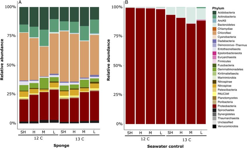
DNA-SIP application in a sponge holobiont    3
healthy individuals (actively pumping, with open oscula)            Stable isotope analysis
were used in the experiments.
                                                                    Sponge tissue samples for bulk stable isotope analysis
                                                                    were weighted before (wet weight) and after freeze-
                                                                    drying (dry weight). Dry tissue was milled with a mortar
SIP incubations                                                     and pestle. For each sample, a subset of thoroughly
                                                                    mixed tissue powder was decalcified by acidification with
Two different treatments were amended: 12C-unlabelled
                                                                    a few drops of fuming HCl until effervescence ceased
DOM and 13C-labelled DOM. Per treatment, three sponge
                                                                    and freeze-dried again. Duplicate (10–30 mg) acidified
replicates and one seawater control without sponge were
                                                                    and non-acidified samples were transferred to tin cups
incubated. The added tracer-DOM was extracted from
                                                                    for bulk ∂13C isotope analysis. The carbon content and
1 g of unlabelled (12C) or 1 g of labelled (13C) lyophilized
                                                                    the 13C/12C isotopic composition was measured simulta-
algal cells (>98% atom purity, ULM-2177 and CLM-2065
                                                                    neously with a high-temperature combustion element
Agmenellum quadruplicatum, Cambridge Isotope Labo-
                                                                    analyser (Vario Isotope cube, Elementar GmbH,
ratories). Briefly, the algal cells were resuspended in
                                                                    Langenselbold, Germany) coupled with an isotope ratio
Milli-Q water, sonicated for 15–20 min and then cen-
                                                                    mass spectrometer (BioVision, Elementar, Stockport,
trifuged for 10 min at 8000g. Subsequently, the superna-
                                                                    UK). A two-point calibration curve was used to correct
tant was first filtered over a 0.7-μm GF/F grade filter and
                                                                    the isotopic data. The 13C values were measured against
then over a 0.2-μm polycarbonate membrane filter to
                                                                    caffeine (IAEA-600, ∂13C = 27.77‰) and UL-D-glucose
remove possible algal cell remnants from the DOM solu-
                                                                    (IAEA-309B, ∂13C = 535.3‰), used as low and high
tion. This process was repeated with the pellet left after
                                                                    internal stable-isotope standards, respectively. The 13C
the centrifugation step, in order to extract most of the
                                                                    stable-isotope data were calculated as previously
DOM. The filtered DOM was freeze dried and analysed
                                                                    described (Rix et al., 2020; Bart et al., 2020; for details
by elemental analyser–isotope ratio mass spectrometry
                                                                    see Supporting Information). Isotope data from the incu-
(EA-IRMS) to determine the C and N content and isotopic
                                                                    bations are corrected for: the background ∂13C values
composition. The freeze-dried DOM was then solubilized
                                                                    (measured in the sponges incubated with unlabelled
in ultrapure water (18.2 MΩ-cm type I, Elga Purelab Clas-
                                                                    DOM), the sponge dry weight, the incubation time, and
sic UV) at CARMABI Research Station prior to the incu-
                                                                    the enrichment of the DOM source used as substrate.
bations. Experimental incubations were carried out in 3 l
                                                                    Isotope-tracer-DOM assimilation rates are expressed as
incubation chambers for 6 h. The sponges were trans-
                                                                    μmol Ctracer mmol Csponge1 h1.
ferred, without air exposure, to air-tight, stirred, incubation
chambers (de Goeij et al., 2013), which were filled with
0.7-μm filtered seawater (FSW). The resuspended DOM
                                                                    DNA extractions
was added at the beginning of the incubation using a
sterile syringe (at a final concentration of 120 μM DOC).            DNA was extracted from the sponge tissue samples
One FSW control (without a sponge) was also incubated               using the DNeasy® Blood & Tissue Kit (Qiagen). The tis-
per treatment under the same conditions to exclude pos-             sue was first homogenized in 180 μl Buffer ATL and 20 μl
sible feeding of the sponge on enriched seawater bacter-            proteinase K using a small sterile pestle that fits in a
ioplankton. All incubations were conducted in the dark              1.5 ml microcentrifuge tube. Afterwards, all tissue sam-
and dissolved oxygen concentrations were monitored                  ples were incubated overnight at 56 C. After the incuba-
continuously with an optical probe (OXY-4; PreSens,                 tion, 200 μl of Buffer AL were added to each sample,
Regensburg, Germany). Incubation chambers were                      followed by 200 μl of ethanol [100% (v/v)] and mixed thor-
placed in a flow-through aquarium (water pumped up                   oughly by vortexing. Before adding the homogenate onto
from 10 m water depth from the nearby reef) to ensure               the DNeasy mini spin column, all samples were cen-
near in situ temperatures. At the end of each incubation,           trifuged at 6000g for 2 min to precipitate unlysed mate-
the sponges were rinsed in non-labelled 0.7-μm FSW                  rial. The supernatant was then transferred onto the spin
and dipped in ultrapure water to remove salts before                column and the protocol was further followed as
sampling. Each sponge was cut in half, one half was kept            described by the manufacturer. DNA from the Sterivex fil-
for 13C bulk tissue (i.e. holobiont tissue) stable isotope          ters was also extracted using DNeasy® Blood & Tissue
analysis, and the other half was immediately snap-frozen            Kit (Qiagen). The volumes of Buffer ATL and proteinase
and stored at 80 C until DNA extraction. The seawater             K were doubled as deviation from the original manufac-
from each control incubations (3 l) was collected and fil-           tory protocol and pipetted into the filter cartridge after
tered over a Sterivex filter (GP 0.22 μm) using a peristal-          which the whole filter was incubated in an oven at 55 C
tic pump. The Sterivex filters were stored at 80 C until           with a sample rocker. After the incubation, 400 μl of
DNA extraction.                                                     Buffer AL were pipetted into the filter cartridge through
© 2021 The Authors. Environmental Microbiology published by Society for Applied Microbiology and John Wiley & Sons Ltd.,
Environmental Microbiology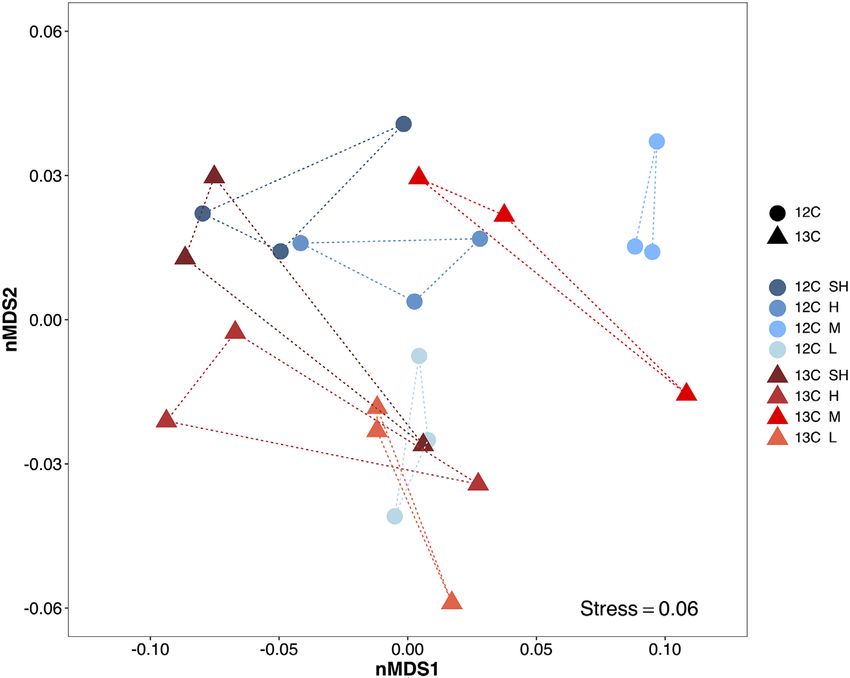
4 S. Campana et al.
the luer-lock side and the filter was incubated for another         heaviest fraction) and fraction 15 (the lightest fraction)
20 min at 70 C to deactivate the proteinase K. The entire         contain mostly CsCl or sterile water they were excluded
volume was extracted from the cartridge using a sterile            for further analysis.
3 ml syringe by taking it up into the syringe and trans-
ferred to a 2 ml microcentrifuge tube. Then, 400 μl of
                                                                   16S amplicon sequencing and data analyses
100% (v/v) ethanol was added and after mixing by
vortexing the entire sample was loaded onto the spin               Pooled fractions of the CsCl gradient were submitted for
column. The protocol was further followed as described             16S amplicon sequencing. The V3-V4 region of the 16S
by the manufacturer. The concentration and purity of               rRNA gene was amplified using the primer pair 341f/806r
the extracted DNA were checked with a NanoDrop                     (dual-barcoding approach; Kozich et al., 2013; primer
1000™ spectrophotometer (Thermo Fischer Scientific,                 sequences: 5’-CCTACGGGAGGCAGCAG-30 and 5’-GG
MA, USA).                                                          ACTACHVGGGTWTCTAAT-3’). The PCR conditions
                                                                   were as follows: initial denaturation step for 30 s at 98 C,
                                                                   30 cycles of denaturation for 9 s at 98 C, annealing for
Density gradient centrifugation and fractionation
                                                                   30 s at 55 C and extension for 30 s at 72 C, followed by
For DNA separation, caesium chloride (CsCl) gradients              a final extension step of 10 min at 72 C. The product
were prepared as described by Neufeld et al. (2007a).              quality and quantity were checked using gel electropho-
Briefly, around 2 μg of DNA was mixed with 4.8 ml of                resis. PCR products were normalized (SequalPrep nor-
7.163 M CsCl solution and the appropriate gradient buffer          malization plate kit; Thermo Fisher Scientific, Waltham,
(GB) (0.1 M Tris, 0.1 M KCl and 1 mM EDTA) volume to               USA) and pooled. Amplicon libraries were sequenced
obtain a final density of 1.725 g ml1. The solution was            using the MiSeq v3 chemistry sequencing kit
transferred to a sterile 4.9 OptiSeal polypropylene centri-        (2  300 bp) on an Illumina MiSeq platform (MiSeq FGx;
fuge tube (Beckham Coulter, CA, USA) and centrifuged               Illumina, San Diego, USA). Raw sequences were quality
in an Optima L-90 K ultracentrifuge (Beckham Coulter)              filtered and trimmed based on quality scores. Amplicon
fitted with a VTi 90 rotor (Beckham Coulter), for 40 h at           sequence variants (ASVs) were computed with the
177 000g and 20 C, with the vacuum on, maximum                    DADA2 algorithm within QIIME2 (version 2018.11). To
acceleration and deceleration without brake. After centri-         train the error model, 1 million reads were used. Chloro-
fugation the gradients were recovered dropwise from the            plasts and mitochondrial sequences were removed from
bottom of the tube by injecting ultrapure water with 0.2%          further analyses. Phylogenetic ASV trees were generated
(v/v) of loading dye on top of the tube using a LEGATO®            with the FastTree2 plugin. A primer-specific trained Naive
100 syringe pump (KD Scientific, MA, USA) with a speed              Bayes taxonomic classifier was used to classify repre-
of 0.5 ml min1. A total of 15 fractions of 330 μl each            sentative ASVs based on the Silva 132 99% OTUs 16S
were collected per sample. The refractive index (nD-TC)            database. Weighted UniFrac distances were calculated
of each gradient fraction was measured using an AR200              and sample separation in ordination space visualized by
digital refractometer (Reichert, NY, USA) and converted            non-metric multidimensional scaling (nMDS) in RStudio
to buoyant density based on a standard curve of nD-TC              Version 1.2. Permutational multivariate analyses of vari-
versus GB-CsCl solution. The DNA from all fractions was            ance (PERMANOVAs) were performed with 999 permuta-
precipitated with 1 μl of glycogen (20 μg) and two vol-            tions to determine whether bacterial communities were
umes of polyethylene glycol (PEG) 6000 solution. Pel-              statistically significantly different between treatments.
leted DNA was washed with 70% (v/v) ethanol and                    Furthermore, significant enrichment between treatments
resuspended in nuclease-free water. The DNA concen-                and density fractions was determined and ranked using
tration of each fraction was measured with Qubit® dsDNA            the linear discriminant analysis effect size (LEfSe) algo-
HS Assay Kit and Qubit® 2.0 Fluorometer (Invitrogen,               rithm, with a P value of 0.05 and LDA threshold of
CA, USA). DNA was stored at 20 C until further analy-            2 (Segata et al., 2011). Microbiome Analyst software
sis. For each experimental treatment, a DNA density pro-           (Chong et al., 2020; www.microbiomeanalyst.ca) was
file was created (Fig. 1). Equal density ranges were                used to perform PERMANOVAs and LEfSe analyses.
selected for the sponge and the seawater control profiles,          Among the significantly enriched taxa (P < 0.05, LEfSe)
based on the observed differences in the amount of DNA             in the sponge-associated bacterial community, substrate
between labelled and unlabelled treatments. DNA frac-              incorporation was validated based on two criteria: (i) the
tions were grouped in the following density ranges: super          increasing relative abundance of a taxon in the 13C treat-
heavy (fractions 2, 3, 4), heavy (fractions 5, 6, 7),              ment occurs in at least two consecutive density fractions,
medium (fractions 8, 9, 10, 11) and light (fractions 12, 13,       with highest abundance in the heaviest of the two frac-
14), and the fractions within each range were pooled vol-          tions, and ii) the relative abundance of the taxon in the
umetrically (5 μl of each fraction). Because fraction 1 (the       13
                                                                      C treatment is greater than its relative abundance in
          © 2021 The Authors. Environmental Microbiology published by Society for Applied Microbiology and John Wiley & Sons Ltd.,
                                                                                                       Environmental Microbiology
DNA-SIP application in a sponge holobiont 5
Fig 1. Distribution of the DNA in the
CsCl gradients of (A) sponge DNA
(n = 3) and (B) seawater bacter-
ioplankton DNA (n = 1). The line charts
show the percentage of total gradient
DNA contained in each fraction of 12C
treatment (blue line) and 13C treatment
(red line) (data shown as mean
% SD). Density ranges constituting
each pooled fraction are separated by
the dotted grey arrow and indicated as
Super Heavy, Heavy, Medium, and
Light. [Color figure can be viewed at
wileyonlinelibrary.com]
the 12C treatment in both density fractions. Because only respectively), showing a mean carbon assimilation rate of
one seawater sample was collected per treatment, the 0.46 0.07 μmol Ctracer mmol Csponge1 h1 (mean SD).
seawater bacterial community comparisons were only
made between treatments, using the density fractions as
technical replicates, and thus allowing only conclusion on Density gradient centrifugation and fractionation
taxa enrichment between treatments, but not between The density of all fractions ranged between 1.68 and
density fractions. Taxa detected only in one of the two 1.77 g ml1, with a linear trend decreasing from the bot-
treatment were removed from the enrichment analysis in tom to the top, indicating proper gradient formation
order to exclude possible artefacts of the methodology (Supporting Information Fig. S1). DNA extracted from the
itself. The sequence data have been deposited in the sponge samples did not show a clear distinction in distri-
NCBI database under BioProject ID PRJNA698441. bution when looking at the light fractions; however, in the
medium and heavy fractions (see magnification panel in
Fig. 1A), an increase in the DNA concentration was visi-
Results ble in the 13C treatment as compared to the 12C treat-
ment (Fig. 1A). The distinction in DNA distribution was
Stable isotope analysis
evident in the DNA extracted from the seawater controls,
Sponge tissue was enriched with tracer-DOM in the 13C where the highest peak of DNA abundance shifted from
treatment as compared to the 12C treatment (210.97 the light fraction of the 12C treatment to the medium frac-
36.62 and 18.57 0.47 ∂13C ‰ mean SD, tion of the 13C treatment (Fig. 1B).
© 2021 The Authors. Environmental Microbiology published by Society for Applied Microbiology and John Wiley & Sons Ltd.,
Environmental Microbiology6 S. Campana et al.
16S amplicon sequencing There was a significant difference in the sponge-
associated bacterial community among the density frac-
After filtering and quality control, 908 906 bacterial
tions of the 13C-labelled and 12C-unlabelled DOM treat-
sequences were obtained from 32 samples, resulting in an
ments (PERMANOVA, F-value = 1.647; R2 = 0.42;
average frequency of 28 403 sequence reads per sample.
P = 0.048). A total of 10 sponge-associated bacterial
We identified 2102 bacterial amplicon single nucleotide
ASVs resulted significantly enriched (P < 0.05, LEfSe), of
variants (ASVs) affiliated to 23 bacterial phyla. Non-metric
which seven in the 13C treatment and three in the 12C
multidimensional scaling (nMDS) based on weighted
treatment (Fig. 4 and Table 1). Sponge-associated bacte-
UniFrac distances clearly separated the sponge-associated
rial ASVs significantly enriched in the 13C treatment
bacterial community and the seawater bacterioplankton
super heavy fraction were a PAUC34f bacterium and a
community (Supporting Information Fig. S2). An nMDS
Proteobacteria of the genus Endozoicomonas (Fig. 4A
ordination plot of the sponge-associated bacterial commu-
and B) and in the heavy fraction a Poribacterial ASV, a
nity alone showed a slight separation between the density
Nitrospira genus and three ASVs belonging to the
fractions along the horizontal axis with heavier ones
Chloroflexi SAR202 clade (Fig. 4C–G). The three
(darker shaded ‘SH’ and ‘H’) clustering to the left and ligh-
sponge-associated ASVs significantly enriched in the 12C
ter ones (lighter shaded ‘M’ and ‘L’) to the centre-right
super heavy fraction were an Entotheonellaeota bacte-
(Fig. 2). Overall, the three replicates of each fractions of
rium, a PAUC26f Acidobacterium and an uncultured
the 13C treatment were more dispersed compared with the
Chloroflexi bacterium (Fig. 4H–J). LEfSe analysis
more closely clustered replicates of the 12C treatment.
showed that four of these sponge-associated ASVs were
Taxonomic assignment revealed that in the sponge-
significantly enriched at all taxonomical levels (from phy-
associated bacterial community the predominant phyla
lum to ASVs), namely, the Poribacteria, PAUC34f, and
were Chloroflexi (35.9%), Proteobacteria (22.7%),
Nitrospirae taxa in the 13C treatment and the
Acidobacteria (14.2%), Actinobacteria (10.2%),
Entotheonellaeota ASV in the 12C treatment (Table 1).
Gemmatimonadetes (4.9%), Nitrospirae (3.8%), Spiro-
A significant change was also detected between the 13C
chaetes (1.8%), Entotheonellaeota (1.7%), and PAUC34f,
and 12C treatment in the seawater bacterioplankton com-
Poribacteria, Nitrospinae, and Dadabacteria, each
munity composition (PERMANOVA, F-value = 32.129;
accounting for 0.9% (Fig. 3A). The seawater bacter-
R2 = 0.41; P = 0.028). Specifically, 48 ASVs were found
ioplankton community, instead, consisted mostly of Prote-
to be significantly enriched (P < 0.05, LEfSe) in one of the
obacteria accounting for 94.2% on average (Fig. 3B).
two treatment, and 42 of them at all taxonomical levels
Fig 2. Sponge-associated bacterial
community composition visualized by
a non-metric multidimensional scaling
plot on weighted UniFrac distances at
the ASV level. Each marker is one
bacterial community, with symbols and
colours indicating the combination of
treatment (12C circles in shades of
blue and 13C triangles in shades of
red) and density fractions (SH: Super
Heavy, H: Heavy, M: Medium, and L:
Light). [Color figure can be viewed at
wileyonlinelibrary.com]
© 2021 The Authors. Environmental Microbiology published by Society for Applied Microbiology and John Wiley & Sons Ltd.,
Environmental MicrobiologyDNA-SIP application in a sponge holobiont 7
Fig 3. Relative abundance of bacterial phyla among the pooled density fraction of the 12C and 13C treatments in (A) the sponge-associated bacte-
rial community and (B) the seawater control. Density fractions are indicated as SH: Super Heavy, H: Heavy, M: Medium, and L: Light. [Color fig-
ure can be viewed at wileyonlinelibrary.com]
(Table 1). The Bacteroidetes phylum was enriched in the Cellvibrionaceae. Among the Alphaproteobacteria, nine
13
C treatment (9.8%) compared to 12C treatment (0.4%) ASVs were enriched in the labelled treatment, including the
(Supporting Information Fig. S3a). Within this phylum, the genera Thalassospira, Nautella, Pelagibaca, Ruegeria,
enriched genera were Tenacibaculum, Marinoscillum, and Thalassobius, while 15 other alphaproteobacterial
Roseivirga, and Pontibacter. The Proteobacteria phylum ASVs were enriched in the 12C treatment, encompassing
was overall enriched in the 12C treatment (98.9%) com- the genera Ponticaulis, Shimia, Thalassobius, Tropici-
pared to 13C treatment (89.6%), however when looking at bacter, a Sphingomonadaceae bacterium, and seven
the class level a division was clear between the other Rhodobacteraceae. Exiguobacterium of the phylum
Alphaproteobacteria, which abundance increased in the Firmicutes was also enriched in the seawater 12C treat-
12
C treatment (27.35% vs 54.4%, 13C vs 12C treatment, ment (Supporting Information Fig. S3d).
respectively; Supporting Information Fig. S3b), and the
Gammaproteobacteria, which abundance instead
increased in the 13C treatment (46.1% vs 34.5%;
Discussion
Supporting Information Fig. S3c). Ten gam-
maproteobacterial ASVs were enriched in the seawater DNA-SIP has been widely used in environmental microbi-
13
C treatment and belonged to the genera Vibrio, ology, but there are only few studies that applied this
Alteromonas, Thalassotalea, Litoricola, Neptuniibacter, technique in vivo to understand the role of microbes that
and Reinekea. One of the Alteromonas ASVs that resulted live in symbiotic association with a host (Shao
enriched in the seawater bacterial community was also et al., 2014; Alonso-Pernas et al., 2017). To the best of
enriched in the sponge-associated bacterial community our knowledge, this is the first study applying DNA-SIP to
from the order to the ASV level in the 13C treatment. The a marine holobiont. Here we linked the identity of
other eight Gammaproteobacterial ASVs were enriched in sponge-associated bacterial taxa to their ability of incor-
the seawater 12C treatment and belonged to the genera porating DOM – a complex mixture of low- and high-
Oleibacter, Vibrio, Acinetobacter, Pseudomonas, molecular weight biomolecules, including proteins, lipids,
Marinobacter, Pseudoalteromonas, and an uncultured and carbohydrates (Nebbioso and Piccolo, 2012) –,
© 2021 The Authors. Environmental Microbiology published by Society for Applied Microbiology and John Wiley & Sons Ltd.,
Environmental Microbiology8 S. Campana et al.
Fig 4. Legend on next page.
© 2021 The Authors. Environmental Microbiology published by Society for Applied Microbiology and John Wiley & Sons Ltd.,
Environmental MicrobiologyDNA-SIP application in a sponge holobiont 9
which is the largest source of organic matter in the the relative low abundances of the ASVs that resulted
oceans (Hansell and Carlson, 2015) and is largely inac- significantly enriched in the sponge-associated bacterial
cessible to other heterotrophic marine metazoans. community. In fact, the total difference in relative abun-
dance between the labelled and unlabelled treatments of
the significantly enriched ASVs was around 4%. Further-
Stable isotope analysis, density gradient centrifugation
more, in these samples, the extracted DNA contained
and fractionation
both the DNA of the sponge host and its associated sym-
The challenge of DNA-SIP experiments is employing suf- bionts and we hypothesize that the host DNA was not
ficient substrate (e.g., DOM) concentration and incuba- enriched in 13C as much as the DNA of the associated
tion time, but staying as close as possible to the in situ symbionts. For instance, in another sponge with high
ambient conditions (Dumont and Murrell, 2005; Neufeld abundance of associated microbes, the percent contribu-
et al., 2007b). Too short incubation times may cause tion of host sponge cells to bulk uptake of algal tracer
insufficient above-background enrichment for the suc- DOM was only 35%, with the remaining 65% assigned to
cessful isolation of 13C-labelled DNA (Teng-xiang the symbiont microbes (Rix et al., 2020). Most likely, the
et al., 2016), whereas too long incubation times could density shift of the bacterial DNA may have been masked
cause enrichment bias due to cross-feeding by second- by the presence of unlabelled host DNA. One option to
ary consumers on the metabolic by-products released by avoid this effect could be to perform a priori cell separa-
the initial consumers (Neufeld et al., 2007b). Experimen- tion of the sponge tissue before DNA extraction (Fieseler
tal conditions become more unpredictable in holobionts et al., 2006; Thomas et al., 2010; Freeman et al., 2013;
with complex microbial communities compared to pure Astudillo-García et al., 2018; Engelberts et al., 2020). A
bacterial cultures. couple of studies have applied cell separation techniques
The treatment conditions used in our experiment were with results reaching up to 95%–99% pure microbial frac-
suitable to detect isotopic enrichment in the labelled sam- tions (Freeman et al., 2013; Rix et al., 2020) but do not
ples compared to the unlabelled ones. Furthermore, our report the number and composition of host and symbiont
approach resulted in isotopic enrichment and DOM incor- cells lost in the numerous washing steps during the pro-
poration rates similar to those observed in previous stud- cess. Therefore, using separated microbial cell fractions
ies where diatom or cyanobacterial DOM was used as a for microbial community composition studies may not be
labelled substrate for estimating DOM uptake and release representative of the overall microbial diversity because
by sponges and their associated microbes (de Goeij bacterial cells lost during the washing steps could result
et al., 2013; Achlatis et al., 2019; Rix et al., 2020; Bart in a biased relative abundance of some bacterial taxa or
et al., 2020; Hudspith et al., 2021; Campana et al., 2021). in the complete disappearance of less abundant taxa
The suitability of our treatment conditions was also (Fieseler et al., 2006). Cell separation can be nonethe-
proven by the clear shift in DNA distribution visible in the less useful in (meta)genomic studies where sufficient
seawater bacterioplankton, indeed the highest peak of coverage of given microbial lineages is required (Fieseler
DNA abundance shifted from the light fraction in the 12C et al., 2006; Bayer et al., 2018; Mori et al., 2018; Schorn
treatment to the medium fraction in the 13C treatment et al., 2019; Moeller et al., 2019). Another option to
after a 6 h incubation time (Fig. 1B). This shift indicates reduce host DNA contamination could be the use of
an increase in the density of the DNA caused by the methylation-based approaches, such as the NEBNext
incorporation of the heavier (13C) isotope by the active Microbiome DNA Enrichment Kit and the Molzym MolYsis
microbial groups, as observed previously in a DNA-SIP Basic kit, to enrich microbial DNA after extraction
study on DOM uptake by Sargasso Sea bacterioplankton (Thoendel et al., 2016). However, similarly to the cell sep-
(Liu et al., 2020). aration method, there are some considerations to be
Unlike the bacterioplankton, the DNA distribution pro- made regarding this approaches. The NEBNext micro-
file of the sponge bacterial symbionts did not show a biome DNA enrichment kit has been used on one sponge
clear and general shift towards heavier fractions in the species for the preparation of metagenomic libraries
13
C treatment as compared to the 12C treatment. Only (Burgsdorf et al., 2019), but its application has not been
the medium to heavier fractions became slightly heavier yet optimized for the use on other sponge species. Fur-
in the 13C treatment (Fig. 1A). This could be explained by thermore, it is unknown whether this techniques may or
Fig 4. Relative abundance profiles of significantly enriched sponge-associated ASVs across treatments and density fractions (n = 3). A–G:
Enrichment in the 13C treatment. H–J: Enrichment in the 12C treatment. Blue dots represent unlabelled fractions (12C amendments). Red trian-
gles represent labelled fractions (13C amendments). Density fractions are indicated as SH: Super Heavy, H: Heavy, M: Medium, and L: Light.
Asterisks (*) indicate in which density fraction the significant enrichment (LefSE, P < 0.05) was found for the sponge-associated taxa. [Color fig-
ure can be viewed at wileyonlinelibrary.com]
© 2021 The Authors. Environmental Microbiology published by Society for Applied Microbiology and John Wiley & Sons Ltd.,
Environmental Microbiology13 12
Table 1. Bacterial taxa that were significantly enriched in either the C or C treatment in the sponge-associated (n = 3) or in the seawater bacterial communities (n = 1, four technical
replicates).
Taxa Activity LefSE (P < 0.05)
Phylum Class Order Family Genus ASV Treatment Sponge Seawater
Sponge-associated
13
PAUC34f _ _ _ _ c21208a04e6186513386c2c482345edc C SH *
13
Proteobacteria Gammaproteobacteria Oceanospirillales Endozoicomonadaceae Endozoicomonas a2478cc476af4056defe87a849618323 C SH **(A)
10 S. Campana et al.
13
Poribacteria _ _ _ _ 64b6cef452f5e253205a0543df547dab CH *
13
Nitrospirae Nitrospira Nitrospirales Nitrospiraceae Nitrospira 658a5a79e887116ab355ee0a52e3dd0f CH *
13
Chloroflexi Dehalococcoidia SAR202clade Uncultured Uncultured 60bdd0e2fee3b5ff24c4bf975df23c58 CH *(F,G,A)
Chloroflexibacterium Chloroflexibacterium
13
Uncultured sponge Uncultured sponge aac86c50ed061422950976fe61a5e949 CH *(F,G,A)
symbiont PAWS52f symbiont PAWS52f
13
_ _ a2478cc476af4056defe87a849618323 CH *(A)
12
Entotheonellaeota Entotheonellia Entotheonellales Entotheonellaceae _ c4682f3866a514d67f9fb8f721f208f5 C SH *
12
Acidobacteria Acidobacteriia Solibacterales Solibacteraceae PAUC26f 12f08292a37324271780e278b29d091a C SH *(C,O,F,
(Subgroup3) G,A)
12
Chloroflexi TK30 uncultured Uncultured bacterium Uncultured bacterium 52ced10f6a64091cc72afd91c25f3827 C SH *(C,O,F,
bacterium G,A)
Seawater
13
Bacteroidetes Bacteroidia Flavobacteriales Flavobacteriaceae Tenacibaculum f21f749f2b1805136d34383f8c97ece4 C *
eff75c879b746cdcedf6afb6760e97ae
24ca9fa104b19f14339d1a7311813089
13
Cytophagales Cyclobacteriaceae Marinoscillum 7275d37e008a936d8d5bc763e6bb4bc4 C *
13
Roseivirga 38d7233bd6197cf2336e07a3048db6cb C *
13
Hymenobacteraceae Pontibacter e97fb331fff682a80370cee733bd1d78 C *
12
Proteobacteria Alphaproteobacteria Caulobacterales Hyphomonadaceae Ponticaulis a813833b5bf150261ffc393184d30d44 C *
40d8840a3be2a4fe8dde5eda0476ad31
12
uncultured 6de8b46c22914de5a20a9ee515922538 C *
12
Sphingomonadales Sphingomonadaceae _ 561ccfc9d063356c8373a600171733ec C *
12
Rhodobacterales Rhodobacteraceae Shimia 206c94637323740ba5098d13f68ca613 C *
12
Thalassobius 6dcfa855474bb1059df1cb23148cb7a7 C *
12
Tropicibacter 9f2a8d79acd772807cef2413a7eec54f C *
12
_ 01a15b2901a87b10a9c86180308bed9f C *
23213ccd4beac18b2bc709906fd88799
f9b669c4bc34d832d53e35560fa82bb3
07d7253127dd990ba7624c7583eae864
d465efd32b6588a04333c4ecf8cb4b12
a3db2151875b48c2902e982d93392359
ce1f4d53c75557d9f75dea813b22abf1
13
Nautella ac400ce27d3fc3110fc2470116a8ed5b C *
13
Pelagibaca 9f90f4404e5d2136a0a3e6c7b338728d C *
13
Ruegeria 64eb1f3c8c959ed10dcf396ed914c6f0 C *
13
Thalassobius 9bd0341d1fa5ed1bc0b72992262bc0ce C *
1238794ef18ee8cb8a3d859c49baff1b
13
_ 1ce2fdedd72ccc2531126fdddfc0a17e C *
5e812dd01970e02d58df8803208c4ccf
29fbbbc6e3ca28a22617dd162afcfa20
Environmental Microbiology
© 2021 The Authors. Environmental Microbiology published by Society for Applied Microbiology and John Wiley & Sons Ltd.,
13
Rhodospirillales Thalassospiraceae Thalassospira 0bb177fe74aef34be459e2e44c6db8aa C *13
Gammaproteobacteria Vibrionales Vibrionaceae Vibrio 0d03338e4efc93449bc6fd3ff698df17 C *(A)
12
Vibrionales Vibrionaceae Vibrio d236227c6c2e37e2a2e29320e7511b4a C *(A)
88f99b7a436d728ba7dba26a1e76e946
12
Pseudomonadales Moraxellaceae Acinetobacter ddcd85a2958ed67c1d62118ca454acec C *(A)
12
Pseudomonadaceae Pseudomonas 70bc0124178ab46e0f1ebc57a31d814e C *(A)
12
Cellvibrionales Cellvibrionaceae uncultured 822420f29aeac1825f5e8da048d2a70b C *(A)
12
Alteromonadales Marinobacteraceae Marinobacter 91ec927d4b3dd369fccbf441b0797054 C *
12
Pseudoalteromonadaceae Pseudoalteromonas 4706b0f83ab177d31fead600620e5d4a C *
13
Alteromonadaceae Alteromonas 31ea5a141edff498d612057a56d03ee8 C *(O,F,G, *
A)
13
Environmental Microbiology
7d2144b913b1bbe6cb18a3c96aa6a8ef C *
13
_ a599a94b6f7d8247f7adb108b6f536aa C *
a69949ac60af0f6e6e35ca9f7f8d95d8
13
Colwelliaceae Thalassotalea 46f9eaa760044af4acffa4a79c577b82 C *
13
Oceanospirillales Litoricolaceae Litoricola 62425045241579aa2274efe55060a69b C *
73504068893298f99a4da53ea1bdef49
13
Nitrincolaceae Neptuniibacter 3b307f47731270021d0f8334d4185d0e C *
13
Saccharospirillaceae Reinekea ce54be95d7dcd5bc326bf8c67da61668 C *
12
Oleibacter 02ed0c05bcf4178b7d5bffa548811bc2 C *
12
Firmicutes Bacilli Bacillales FamilyXII Exiguobacterium f2a5b9165468b028bc5ad45d02ef0a67 C *
“*” represents significant enrichment at all listed taxonomical levels; *() represents significant enrichment only at the taxonomical level included in parenthesis where C = Class, O = Order,
F = Family, G = Genus and A = ASV; Density fractions are indicated as SH: Super Heavy, H: Heavy, M: Medium, and L: Light.
© 2021 The Authors. Environmental Microbiology published by Society for Applied Microbiology and John Wiley & Sons Ltd.,
DNA-SIP application in a sponge holobiont 1112 S. Campana et al.
may not introduce bias in relative abundance analysis, treatment of both sponge and seawater bacterial commu-
for example, due to the loss of some bacterial taxa. nities (Table 1). Therefore, we did not consider the
Alteromonas ASV as an indication of endosymbiotic
processing of DOM.
Bacterial taxa actively incorporating 13C-labelled DOM
To further relate the uptake of certain organic com-
To date, no clear or consistent shift in the sponge micro- pounds from the DOM pool to the enriched bacterial taxa
bial community composition was found in relation to rates in the 13C treatment, we can assess their reconstructed
of DOM uptake through the sponge holobiont (Gantt metabolic pathways as heterotrophic carbon consumers.
et al., 2019). Our study demonstrates that, at least, seven The metabolic reconstruction of two metagenome-
sponge-associated bacterial ASVs, belonging to the assembled genomes (MAGs) of PAUC34f, also known as
phyla PAUC34f, Proteobacteria, Poribacteria, Nitrospirae, sponge-associated unclassified lineage (SAUL), sug-
and Chloroflexi, are active incorporators of 13C-labelled gests that the members of this phylum possess genes
DOM. The relative abundance of these ASVs varied sig- involved in the tricarboxylic acid cycle (TCA), glycolysis,
nificantly among different density fractions. Higher rela- pentose phosphate pathway, Wood–Ljungdahl pathway,
tive abundance of a microbial taxon in the heavier and oxidative phosphorylation (Astudillo-García
fractions of the 13C-labelled treatment can indicate higher et al., 2018). Moreover, genes encoding glycoside hydro-
metabolic activity in incorporating a certain isotope-tracer lases (GH), glycoside transferases (GT), polysaccharide
substrate, i.e. DOM in our case. Therefore, the pattern lyases (PL) and carbohydrate esterases (CE) were also
observed in our results suggest that in the sponge present, suggesting the ability of PAUC34f to degrade
P. angulospiculatus a PAUC34f bacterium and an glycolipids, glycopeptides and glycoproteins, compounds
uncultured Proteobacteria of the genus Endozoicomonas typically found within the sponge mesohyl and as dis-
could be the first DOM consumers followed by a solved organic matter in seawater (Blunt et al., 2011;
Poribacteria, a Nitrospira and three Chloroflexi bacteria. Astudillo-García et al., 2018). Endozoicomonas bacteria
We cannot, however, exclude that other microbial taxa of the phylum Proteobacteria are globally distributed
present in P. angulospiculatus were also able to process endosymbionts and their genomes have been found
DOM to a certain extent. In fact, a NanoSIMS study on enriched in genes for carbon sugar transport, indicating a
the uptake of 13C and 15N-labelled DOM by the same potential role in the cycling of carbohydrates to their host
sponge species indicated that after a 3-h pulse, about (Neave et al., 2017). The Poribacteria phylum, originally
50% of the microbial symbiont cells were enriched in 13C discovered and described in marine sponges (Fieseler
(Hudspith et al., 2021). Our results rather provide direct et al., 2004), has a well-described genomic repertoire
evidence that the seven mentioned bacterial taxa had (Siegl et al., 2011; Giles et al., 2013; Kamke et al., 2014;
significant metabolic activity in the consumption and Slaby et al., 2017; Podell et al., 2018). The predicted
incorporation of the labelled carbon (13C) source into their functions of the central carbohydrate metabolism of
DNA within 6 h of incubation time. If other bacteria were Poribacteria include complete pathways for glycolysis,
able to take up the stable-isotope-enriched dissolved oxidative phosphorylation, the TCA cycle, branches of
organic carbon, but not yet incorporate it into their DNA, the pentose phosphate pathway, and carbon fixation via
the isotopic signal could be detected by NanoSIMS but the Wood–Ljungdahl pathway (Podell et al., 2018). Fur-
missed with the DNA-SIP method in the observed time thermore, polysaccharides and other complex carbohy-
frame. Longer DNA-SIP incubations may allow to identify drate degradation pathways can be carried out by
sponge-associated incorporating taxa with slower division polysaccharide lyases, and glycoside hydrolases (GH);
time, but cofounding results could occur. Hudspith the genes encoding these enzymes are abundantly pre-
et al. (2021) found that there is translocation of DOM sent in the genomes of Poribacteria (Kamke et al., 2013;
processed by sponge cells – predominantly the ‘choano- Podell et al., 2018; Robbins et al., 2021). In analogy with
cytes’ or sponge filter cells – to the microbial symbionts the PAUC34f and the Poribacteria, the metabolism of the
48 h after a 3-h pulse with 13C/15N-labelled DOM. It Chloroflexi SAR202 clade includes glycolysis, TCA cycle,
would be challenging to distinguish direct microbial pentose phosphate pathway and the respiratory chain as
processing of DOM versus translocated host metabolites. energy-producing pathways, additionally, autotrophic car-
The bacterial taxa enriched in the sponge-associated bon fixation via the reductive citrate acid cycle and the
bacterial communities differed from those enriched in the Wood–Ljungdahl pathway were also identified (Bayer
seawater control; therefore, we can exclude an enrich- et al., 2018). A relevant metabolic specialization in
ment bias caused by sponge feeding on the seawater SAR202 is the potential to degrade recalcitrant DOM,
bacterioplankton. The only sign of potential due to a large repertoire of oxidative enzymes that may
seawater bacteria ingestion by the sponge was the help in the oxidation of recalcitrant alicyclic ring structures
enrichment of the Alteromonas ASV in the 13C-labelled to more labile carboxylic acid (Landry et al., 2017; Bayer
© 2021 The Authors. Environmental Microbiology published by Society for Applied Microbiology and John Wiley & Sons Ltd.,
Environmental MicrobiologyDNA-SIP application in a sponge holobiont 13
et al., 2018). The reconstructed genome of a member of et al., 2019). DOM-uptake strategies within the sponge
the sponge-associated genus Nitrospira proposes an holobiont have been shown to vary between LMA and
autotrophic carbon metabolism via the reductive TCA HMA sponges: while the LMA sponge Dysidea avara
(rTCA) cycle (Moitinho-Silva et al., 2017b). Nonetheless, relied for more than 95% on DOM uptake by host cho-
there is some evidence of a possible mixotrophic lifestyle, anocyte cells, in the HMA sponge Aplysina aerophoba
indeed some Nitrospira from marine ecosystems or acti- the microbial symbionts accounted for the majority (65%)
vated sludge can use simple organic substrates, such as of DOM uptake (Rix et al., 2020). Since the microbial taxa
pyruvate, acetate, formate and glycerol for carbon assimi- enriched in P. angulospiculatus are indicator taxa of
lation, but pure heterotrophic growth has not been HMA sponges, our results suggests that in HMA sponges
observed yet (Lucker et al., 2010; Koch et al., 2015; DOM incorporation may depend on specific microbial
Pachiadaki et al., 2017). taxa rather than on host cell uptake. However, to validate
The Bacteroidetes phylum was the most active consumer the hypothesis that sponges with different quantities of
of DOM in the (0.7-μm filtered) seawater bacterioplankton. associated microbes have evolved different strategies to
The Proteobacteria phylum was overall enriched in the 12C exploit DOM in the ocean (Rix et al., 2020), a similar
treatment, but when looking at lower taxonomic level we DNA-SIP study in an LMA sponge would be necessary.
can see differential activities between members of the
Alphaproteobacteria and Gammaproteobacteria, with the
Conclusions
latter being the most active in the 13C treatment. It is well
recognized that Bacteroidetes (especially Cytophagales This manuscript, to best of our knowledge, represents the
and Flavobacteriales) and Gammaproteobacteria are able first application of a DNA-SIP experiment in combination
to mineralize organic aggregates, such as cellulose and chi- with 16S amplicon sequencing in a marine holobiont. We
tin, which are compounds that are often found in the high- identified sponge-associated bacterial taxa that are meta-
molecular mass (HMW) fraction of DOM (Kirchman, 2002; bolically active in the uptake and processing of DOM.
Edwards et al., 2010). Our bacterioplankton observation, Seven ASVs, belonging to the phyla PAUC34f, Prote-
however, serves here mainly as control to distinguish obacteria, Poribacteria, Nitrospirae and Chloroflexi were
sponge bacterial symbionts from seawater bacteria and significant incorporators of DOM in P. angulospiculatus.
indicate possible bacterioplankton feeding as opposed to PAUC34f, Poribacteria, and Chloroflexi have similar meta-
endosymbiont processing of DOM. The limited replication bolic capabilities and could be directly involved in the deg-
and the filtration over a 0.7-μm filter (that likely changed the radation of labile and recalcitrant organic matter through
ambient bacterioplankton community composition) of our heterotrophic carbon metabolism, while Nitrospirae may
seawater control prevents solid ecological conclusions from have an indirect role through carbon fixation of respired
the bacterioplankton treatment. Previous studies have organic carbon or a potential mixotrophic metabolism. The
assessed the uptake of different types of DOM by bacter- DNA-SIP technology identified the first DOM-processing
ioplankton community of the Sargasso Sea through DNA- taxa within the holobiont and could be used in the future to
SIP, showing that a wide variety of taxa (among which sev- trace the uptake of different types of DOM sources or
eral groups of the Proteobacteria, Flavobacteria, other non-DOM substances in sponge–microbe
Actinobacteria and Verrucomicrobia) are capable of incor- interactions.
porating DOM, and that the quality and type of DOM influ-
ences the response of these taxa in incorporating such
Author contributions
DOM (Nelson and Carlson, 2012; Liu et al., 2020). Like-
wise, DNA-SIP can become a powerful tool to understand S.C. and J.M.G. designed the experiments. S.C. performed
which sponge-associated microbial groups mediate the the experiments, analysed the samples and wrote the man-
uptake of DOM released by different primary producers pre- uscript. S.C. and K.B. analysed the data. All authors
sent on coral reefs, such as macroalgae, and corals. reviewed and contributed to the manuscript writing.
Most of the DOM incorporating taxa identified in
P. angulospiculatus are characteristic taxa of high micro-
Acknowledgements
bial abundance (HMA) sponges. In HMA sponges, the
bacteria can constitute up to 40% of their biomass and The authors want to thank Meggie Hudspith, Celine Demey,
have a distinctive community composition, while sponges Niklas Kornder, Benjamin Mueller, Mark Vermeij and the
staff of CARMABI for fieldwork and logistical support, Sus-
with low-microbial abundance of associated microbes,
anne Wilken for help with the DNA-SIP protocol develop-
i.e., low microbial abundance (LMA) sponges, generally
ment, Jorien Schoorl for IRMS sample analysis at the
possess a microbial community similar to that of the sea- University of Amsterdam and the Competence Centre for
water in both composition and abundance (Taylor Genomic Analysis (CCGA) at Kiel University for 16S
et al., 2007; Moitinho-Silva et al., 2017b; Gantt amplicon sequencing. This project has received funding from
© 2021 The Authors. Environmental Microbiology published by Society for Applied Microbiology and John Wiley & Sons Ltd.,
Environmental Microbiology14 S. Campana et al.
the European Research Council (ERC) under the European Cathalot, C., van Oevelen, D., Cox, T.J.S., Kutti, T.,
Union’s Horizon 2020 research and innovation programme Lavaleye, M., Duineveld, G., and Meysman, F.J.R. (2015)
(grant agreement #715513; personal grant to J.M. de Goeij). Cold-water coral reefs and adjacent sponge grounds: hot-
spots of benthic respiration and organic carbon cycling in
the deep sea. Front Mar Sci 2: 37.
Chong, J., Liu, P., Zhou, G., and Xia, J. (2020) Using micro-
References
biome analyst for comprehensive stastistical, functional,
Achlatis, M., Pernice, M., Green, K., de Goeij, J.M., and meta-analysis of microbiome data. Nat Protocols 15:
Guagliardo, P., Kilburn, M.R., et al. (2019) Single-cell visu- 799–821.
alization indicates direct role of sponge host in uptake of de Goeij, J.M., van den Berg, H., van Oostveen, M.M.,
dissolved organic matter. Proc Royal Soc B 286: 2019– Epping, E.H.G., and van Duyl, F.C. (2008) Major bulk dis-
2153. solved organic carbon (DOC) removal by encrusting coral
Alexander, B.E., Achlatis, M., Osinga, R., van der Geest, H. reef cavity sponges. Mar Ecol Prog Ser 357: 139–151.
G., Cleutjens, J. P. M., Schutte, B., & de Goeij, J.M. de Goeij, J.M., De Kluijver, A., Van Duyl, F.C., Vacelet, J.,
(2015). Cell kinetics during regeneration in the sponge Wijffels, R.H., De Goeij, A.F.P.M., et al. (2009) Cell kinet-
Halisarca caerulea: how local is the response to tissue ics of the marine sponge Halisarca caerulea reveal rapid
damage?. PeerJ, 3: e820. cell turnover and shedding. J Exp Biol 212: 3892–3900.
Alonso-Pernas, P., Bartram, S., Arias-Cordero, E., de Goeij, J.M., Lesser, M.P., and Pawlik, J.R. (2017) Nutrient
Novoselov, A.L., Halty-deLeon, L., Shao, Y., and fluxes and ecological functions of coral reef sponges in a
Boland, W. (2017) In vivo isotopic labeling of symbiotic changing ocean. In Climate Change, Ocean Acidification
bacteria involved in cellulose degradation and nitrogen and Sponges: Impacts across Multiple Levels of Organiza-
recycling within the gut of the forest cockchafer (Mel- tion, Carballo, J.L., and Bell, J.J. (eds). Cham: Springer
olontha hippocastani). Front Microbiol 8: 1970. International Publishing, pp. 373–410.
Astudillo-García, C., Slaby, B.M., Waite, D.W., Bayer, K., de Goeij, J.M., van Oevelen, D., Vermeij, M.J.A., Osinga, R.,
Hentschel, U., and Taylor, M.W. (2018) Phylogeny and Middelburg, J.J., de Goeij, A.F.P.M., and Admiraal, W.
genomics of SAUL, an enigmatic bacterial lineage fre- (2013) Surviving in a marine desert: the sponge loop
quently associated with marine sponges. Environ retains resources within coral reefs. Science 342:
Microbiol 20: 561–576. 108–110.
Bart, M.C., de Kluijver, A., Hoetjes, S., Absalah, S., Dumont, M.G., and Murrell, J.C. (2005) Stable isotope prob-
Mueller, B., Kenchington, E., et al. (2020) Differential ing - linking microbial identity to function. Nat Rev
processing of dissolved and particulate organic matter by Microbiol 3: 499–504.
deep-sea sponges and their microbial symbionts. Sci Rep Edwards, J.L., Smith, D.L., Connolly, J., McDonald, J.E.,
10: 17515. Cox, M.J., Joint, I., et al. (2010) Identification of carbohy-
Bart, M.C., Mueller, B., Rombouts, T., van de Ven, C., drate metabolism genes in the metagenome of a marine
Tompkins, G.J., Osinga, R., et al. (2021) Dissolved biofilm community shown to be dominated by
organic carbon (DOC) is essential to balance the meta- Gammaproteobacteria and Bacteroidetes. Genes 1:
bolic demands of four dominant North-Atlantic deep-sea 371–384.
sponges. Limnol Oceanogr 66: 925–938. Engelberts, J.P., Robbins, S.J., de Goeij, J.M., Aranda, M.,
Bayer, K., Jahn, M.T., Slaby, B.M., Moitinho-Silva, L., and Bell, S.C., and Webster, N.S. (2020) Characterization of a
Hentschel, U. (2018) Marine sponges as Chloroflexi hot sponge microbiome using an integrative genome-centric
spots: genomic insights and high-resolution visualization approach. ISME J 14: 1100–1110.
of an abundant and diverse symbiotic clade. mSystems 3: Erwin, P.M., and Thacker, R.W. (2008) Phototrophic nutrition
e00150–e00118. and symbiont diversity of two Caribbean sponge–
Blunt, J.W., Copp, B.R., Munro, M.H.G., Northcote, P.T., and cyanobacteria symbioses. Mar Ecol Prog Ser 362:
Prinsep, M.R. (2011) Marine natural products. Nat Prod 139–147.
Rep 28: 196–268. Fieseler, L., Horn, M., Wagner, M., and Hentschel, U. (2004)
Burgsdorf, I., Handley, K.M., Bar-Shalom, R., Erwin, P.M., Discovery of the novel candidate phylum ‘Poribacteria’ in
and Steindler, L. (2019) Life at home and on the roam: marine sponges. Appl Environ Microbiol 70: 3724–3732.
genomic adaptions reflect the dual lifestyle of an intracel- Fieseler, L., Quaiser, A., Schleper, C., and Hentschel, U.
lular, facultative symbiont. mSystems 4: e00057–e00019. (2006) Analysis of the first genome fragment from the
Bryson, S., Li, Z., Chavez, F., Weber, P.K., Pett-Ridge, J., marine sponge-associated, novel candidate phylum
Hettich, R.L., et al. (2017) Phylogenetically conserved Poribacteria by environmental genomics. Environ
resource partitioning in the coastal microbial loop. ISME J Microbiol 8: 612–624.
11: 2781–2792. Fiore, C.L., Jarett, J.K., and Lesser, M.P. (2013) Symbiotic
Campana, S., Husdpith, M., Lankes, D., de Kluijver, A., prokaryotic communities from different populations of the
Demey, C., Schoorl, J., Absalah, S., van der giant barrel sponge, Xestospongia muta. Micro-
Meer, M. T. J., Mueller, B., & de Goeij, J. M. (2021). biologyOpen 2: 938–952.
Processing of naturally sourced macroalgal- and coral- Freeman, C.J., and Thacker, R.W. (2011) Complex interac-
dissolved organic matter (DOM) by high and low microbial tions between marine sponges and their symbiotic micro-
abundance encrusting sponges. Front Mar Sci, 8: 452. bial communities. Limnol Oceanogr 56: 1577–1586.
© 2021 The Authors. Environmental Microbiology published by Society for Applied Microbiology and John Wiley & Sons Ltd.,
Environmental MicrobiologyYou can also read