Effective rational humanization of a PASylated anti galectin 3 Fab for the sensitive PET imaging of thyroid cancer in vivo
←
→
Page content transcription
If your browser does not render page correctly, please read the page content below
www.nature.com/scientificreports
OPEN Effective rational humanization
of a PASylated anti‑galectin‑3
Fab for the sensitive PET imaging
of thyroid cancer in vivo
Emanuel Peplau1, Francesco De Rose2, Andreas Eichinger1, Sybille Reder2,
Markus Mittelhäuser2, Giorgia Scafetta4, Markus Schwaiger2, Wolfgang A. Weber2,
Armando Bartolazzi3,4, Calogero D’Alessandria2 & Arne Skerra1*
The lack of a non-invasive test for malignant thyroid nodules makes the diagnosis of thyroid cancer
(TC) challenging. Human galectin-3 (hGal3) has emerged as a promising target for medical TC imaging
and diagnosis because of its exclusive overexpression in malignant thyroid tissues. We previously
developed a human-chimeric αhGal3 Fab fragment derived from the rat monoclonal antibody (mAb)
M3/38 with optimized clearance characteristics using PASylation technology. Here, we describe
the elucidation of the hGal3 epitope recognized by mAb M3/38, X-ray crystallographic analysis
of its complex with the chimeric Fab and, based on the three-dimensional structure, the rational
humanization of the Fab by CDR grafting. Four CDR-grafted versions were designed using structurally
most closely related fully human immunoglobulin VH/VL regions of which one—employing the acceptor
framework regions of the HIV-1 neutralizing human antibody m66—showed the highest antigen
affinity. By introducing two additional back-mutations to the rodent donor sequence, an affinity
toward hGal3 indistinguishable from the chimeric Fab was achieved (KD = 0.34 ± 0.02 nM in SPR). The
PASylated humanized Fab was site-specifically labelled with the fluorescent dye Cy7 and applied for
the immuno-histochemical staining of human tissue sections representative for different TCs. The
same protein was conjugated with the metal chelator Dfo, followed by radiolabelling with 89Zr(IV).
The resulting protein tracer allowed the highly sensitive and specific PET/CT imaging of orthotopic
tumors in mice, which was confirmed by quantitative analysis of radiotracer accumulation. Thus, the
PASylated humanized αhGal3 Fab offers clinical potential for the diagnostic imaging of TC.
Thyroid cancer (TC) generally occurs in the form of nodules in the thyroid parenchyma or of small sclerotic
areas with irregular borders. The prevalence of thyroid nodules is very high in geographic regions with iodine
deficiency and may reach up to 68% in the adult population according to recent s tudies1,2. However, as the over-
whelming majority of thyroid nodules is benign, the challenge lies in their distinction from malignant lesions3,4.
Currently, thyroid ultrasound (US) and fine needle aspiration biopsy (FNA) are the standard tools to diagnose
TC. However, in particular differentiated TC (DTC) with follicular structure expressing thyroglobulin is hard
to distinguish from benign nodules preoperatively, due to the common cyto-architectural features. Apart from
better diagnostic tools in general, there is also a medical need for improved therapies of poorly differentiated
TC (PDTC) and anaplastic TC (ATC). These two forms of TC are frequently fatal, exhibit rapid growth with
metastases in up to 75% of the patients, and they are often recognized and diagnosed late at advanced s tages5.
Both thyroid US imaging and FNA have contributed to consistently improving the diagnosis of thyroid
nodules, but both do not easily allow preoperative discrimination between benign and malignant lesions. As a
consequence, the surgical overtreatment of benign thyroid nodules is c ommon6,7 because so far only histology can
reliably differentiate between follicular TC and benign thyroid nodules. Thus, the preoperative characterization
of thyroid nodules is clinically highly relevant because it would facilitate selection of patients to be referred to
1
Lehrstuhl für Biologische Chemie, Technische Universität München, 85354 Freising (Weihenstephan),
Germany. 2Klinikum rechts der Isar, Nuclear Medicine Department, Technical University Munich, Ismaninger
Str. 22, 81675 Munich, Germany. 3Pathology Research Laboratory, Cancer Center Karolinska, Karolinska Hospital,
17176 Stockholm, Sweden. 4Pathology Research Laboratory, Sant’Andrea Hospital, University Sapienza, via di
Grottarossa 1035, 00189 Rome, Italy. *email: skerra@tum.de
Scientific Reports | (2021) 11:7358 | https://doi.org/10.1038/s41598-021-86641-0 1
Vol.:(0123456789)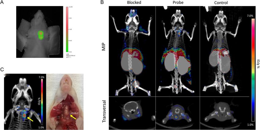
www.nature.com/scientificreports/
surgery, thus contributing to better TC follow-up as well as treatment optimization. In this regard, the overex-
pressed β-galactoside-binding lectin galectin 3 (Gal3) has shown promise as a molecular target for the diagnosis
of TC, as this phenotypic feature is absent in normal thyroid c ells8.
To exploit Gal3 overexpression for tumor imaging via positron emission tomography (PET) we recently
developed a 89Zr-labeled chimeric Fab-fragment derived from a well characterized rat monoclonal antibody
(mAb M3/38) which demonstrated high imaging contrast in m ice9. In accordance with earlier in vivo imaging
10,11
studies of other tumor targets , a Fab version with moderately prolonged plasma half-life utilizing PASyla-
tion technology turned out to result in excellent and highly specific uptake in orthotopic thyroid tumor models.
With the goal of clinical translation, further aspects beyond good tumor penetration and rapid clearance
have to be considered. While the preparation of a chimeric Fab—here comprising variable domains from rat
combined with human constant domains—considerably reduces the immunogenicity of the protein reagent
compared with a natural rodent antibody (also owing to the absence of the Fc portion), there are still reported
cases of an anti-drug antibody (ADA) response directed against the non-human V-regions12. A solution to this
problem is the humanization of the Fab via grafting of the complementarity determining regions (CDRs) from
rat onto a human immunoglobulin (Ig) framework as initially demonstrated more than 30 years a go13,14. In fact,
since the first market approval of trastuzumab for the treatment of HER2-positive metastasizing breast cancer,
CDR grafting has become a generally accepted strategy for biopharmaceutical mAb development up to n ow15,16.
However, a successful humanization campaign depends on a high structural similarity between donor and
acceptor Ig frameworks since even minute conformational changes in the paratope upon CDR transplantation
may critically affect the antigen-binding a ctivity17. Therefore, we have elucidated the crystal structure of the
chimeric Fab in complex with its antigen as a prerequisite for the informed choice of a suitable human Ig scaf-
fold. This enabled the functional CDR grafting via rational protein design, resulting in a humanized αGal3 Fab
that allowed sensitive PET imaging of orthotopic human thyroid tumors in mice.
Results
Gal3 epitope determination. Human Gal3 (hGal3) comprises a C-terminal carbohydrate recognition
domain (CRD) and a characteristic flexible N-terminal domain (ND) of 113 residues with several 9-amino acid
repeats: Pro-Gly-Ala-Tyr-Pro-Gly-Xaa-Xaa-Xaa18 (Fig. 1). Due to the inherent flexibility of the ND, hGal3 rep-
resents a challenging target for structural analysis. In fact, the crystal structure of full length Gal3 is unknown
to date, which prompted us to narrow down the epitope that is recognized by the mAb M3/38 or its chimeric
recombinant Fab9. On a western blot of both the full length recombinant hGal3 and a truncated version lack-
ing the ND only the complete antigen led to a signal with the fluorescence-labelled chimeric αhGal3-Fab-
PAS200-Cy5.59 (Fig. 1B), indicating that a linear epitope within the ND is recognized.
Thus, the SPOT technique19 was applied to identify the epitope using an array of consecutive synthetic hGal3
peptides that were immobilized on a hydrophilic membrane. To this end, a set of 45 8mer peptides, each shifted
by 2 residues and covering the sequence of the full ND (residues 1–112), was synthesized. Incubation with the
Cy5.5-labelled chimeric αhGal3-Fab-PAS200 led to prominent fluorescent binding signals for three peptide spots,
nos. 24, 25 and 29 (Fig. 1C), whereby the first two signals correspond to peptides with overlapping sequences. The
common minimal sequence motif of all three spots was: Ala-Pro-Pro-Gly-Ala-Tyr. Of note, this linear epitope
occurs in two of the 9-residue repeats within the hGal3 ND mentioned above.
Crystallographic analysis of the αhGal3‑Fab·peptide complex. The recombinant chimeric Fab was
functionally produced in E. coli and purified to homogeneity as previously described9, then mixed with the
N-terminally acetylated synthetic peptide Gln-Ala-Pro-Pro-Gly-Ala-Tyr-Pro-Gly. The protein crystallized in
the presence of 25% (w/v) PEG4000, 0.1 M HEPES/NaOH pH 7.0 in the space group C2 with one Fab·peptide
complex in the asymmetric unit. A synchrotron X-ray diffraction data set at 1.9 Å resolution was collected and
the structure of the complex was solved by molecular replacement (see “Materials and methods”). Residues
Asp(L1)–Cys(L214) of the light chain and Gln(H1)–Cys(H230) of the heavy chain (residue numbering accord-
abat20) as well as the complexed 9mer peptide were defined in the electron density map, with weaker den-
ing to K
sity for residues Cys(L214), Gly(H42), Lys(H43) and Cys(H230). In total 5 peptide bonds with cis-configuration
were found at typical positions of the Ig chains: Thr(L7)–Pro(L8), Phe(L94)–Pro(L95), Tyr(L140)–Pro(L141),
Phe(H148)–Pro(H149) and Glu(H150)–Pro(H151). Also, the five conserved disulfide bridges, including the
Val(L51) and Ser(H229) exhibit outlier ϕ/φ angles in the Ramachandran plot (Table 1) but are well defined in
one linking the light and heavy chains, Cys(L214)–Cys(H230) (with partial definition), were visible. Residues
the electron density.
The three-dimensional structure of the rat/human chimeric Fab exhibits the typical fold of a Fab fragment
as known from human and rodent Igs (Fig. 2). The paratope comprises a shallow cleft between the VH and VL
domains about 12 Å by 18 Å wide and 9 Å deep. Within this cleft the 9-residue hGal3 epitope peptide is bound
primarily by CDR-H3 and CDR-L3. The peptide shows an elongated, curved backbone conformation including a
short 310-helix formed by residues Pro(P4) to Ala(P6). A PISA a nalysis21 revealed that 61.4% of the total solvent-
accessible surface area of the peptide (647 of 1053 Å2) is buried in the binding site. Altogether 26 residues of the
Fab form van der Waals contacts to the bound peptide with a contact surface greater than 1 Å2, 17 arising from
the heavy chain and 9 from the light chain; of these, 10 are hydrophobic and 15 polar (Table 2). Furthermore, 4
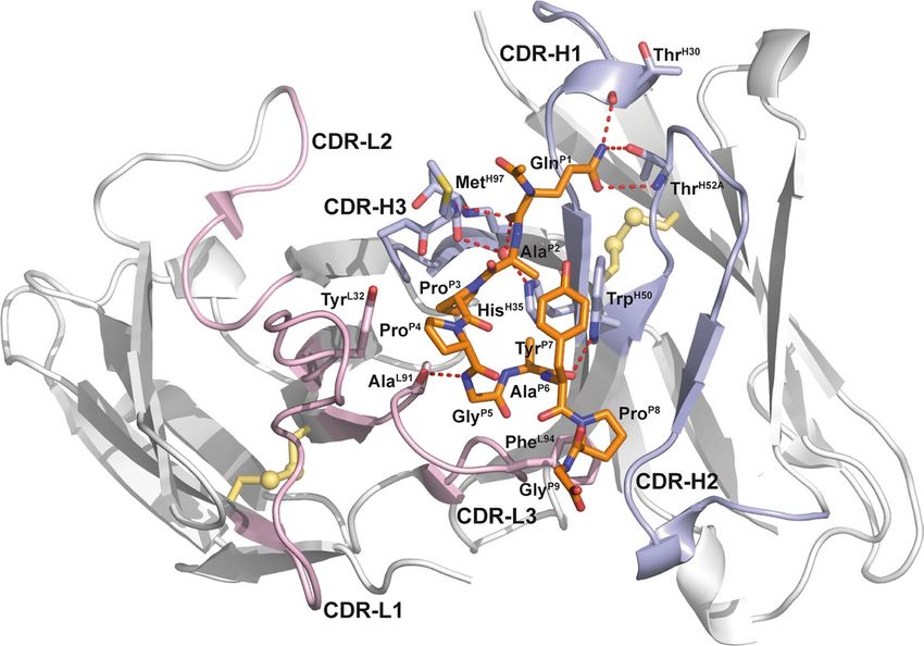
residues in the heavy chain and one in the light chain form hydrogen bonds to the peptide: Ala(L91), Thr(H30),
Trp(H50), Thr(H52A) and Met(H97). Apart from Trp(H47), all contacting residues are located within CDRs.
There are also some other relevant features of this antibody-antigen complex: Gln(P1) forms with its side
chain carboxamide group one and two hydrogen bonds, respectively, to residues Thr(H30) and Thr(H52A) as
well as with its main chain carbonyl oxygen hydrogen bonds to Met(H97) and to the only water molecule that is
Scientific Reports | (2021) 11:7358 | https://doi.org/10.1038/s41598-021-86641-0 2
Vol:.(1234567890)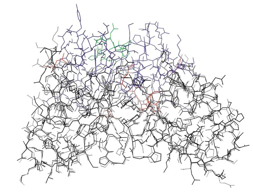
www.nature.com/scientificreports/
A C #1: M-A-D-N-F-S-L-H
#2: D-N-F-S-L-H-D-A
hGal3 hGal3 #3: F-S-L-H-D-A-L-S
full length CRD fragment #4: L-H-D-A-L-S-G-S
#5: D-A-L-S-G-S-G-N
#6: L-S-G-S-G-N-P-N
#7: G-S-G-N-P-N-P-Q
#8: G-N-P-N-P-Q-G-W
#9: P-N-P-Q-G-W-P-G
#10: P-Q-G-W-P-G-A-W
#11: G-W-P-G-A-W-G-N
C-terminal #12: P-G-A-W-G-N-Q-P
CRD #13: A-W-G-N-Q-P-A-G
#14: G-N-Q-P-A-G-A-G
#15: Q-P-A-G-A-G-G-Y
#16: A-G-A-G-G-Y-P-G
PGAYP(X) #17: A-G-G-Y-P-G-A-S
repeat #18: G-Y-P-G-A-S-Y-P
domain #19: P-G-A-S-Y-P-G-A
#20: A-S-Y-P-G-A-Y-P
#21: Y-P-G-A-Y-P-G-Q
#22: G-A-Y-P-G-Q-A-P
N-terminal #23: Y-P-G-Q-A-P-P-G
domain #24: G-Q-A-P-P-G-A-Y
#25: A-P-P-G-A-Y-P-G
#26: P-G-A-Y-P-G-Q-A
#27: A-Y-P-G-Q-A-P-P
#28: P-G-Q-A-P-P-G-A
#29: Q-A-P-P-G-A-Y-P
#30: P-P-G-A-Y-P-G-A
#31: G-A-Y-P-G-A-P-G
B #32: Y-P-G-A-P-G-A-Y
#33: G-A-P-G-A-Y-P-G
M 1 2 1 2 #34: P-G-A-Y-P-G-A-P
#35: A-Y-P-G-A-P-A-P
kDa #36: P-G-A-P-A-P-G-V
#37: A-P-A-P-G-V-Y-P
#38: A-P-G-V-Y-P-G-P
#39: G-V-Y-P-G-P-P-S
#40: Y-P-G-P-P-S-G-P
55 - #41: G-P-P-S-G-P-G-A
#42: P-S-G-P-G-A-Y-P
43 - #43: G-P-G-A-Y-P-S-S
#44: G-A-Y-P-S-S-G-Q
#45: Y-P-S-S-G-Q-P-S
34 - #46: S-S-G-Q-P-S-A-T
#47: G-Q-P-S-A-T-G-A
26 - #48: P-S-A-T-G-A-Y-P
#49: A-T-G-A-Y-P-A-T
#50: G-A-Y-P-A-T-G-P
#51: Y-P-A-T-G-P-Y-G
#52: G-P-Y-G-A-P-A-G
15 -
0.0 0.5 1.0
Em675 (normalized)
D 1
.
10
.
20
.
30
.
40
.
50
.
60
.
hGal3: MADNFSLHDALSGSGNPNPQGWPGAWGNQPAGAGGYPGASYPGAYPGQAPPGAYPGQAPPGAYPG--APG
mGal3: ...S...N...A.........Y........-........A.........................QAP.S
70 80 90 100 110
. . . . .
hGal3: AYPGAPAPGVYPGPPSGPGAYPSSGQPSATGAYPAT-GPYGAPAG
mGal3: ....PT...A....-TA.....GQPA.G.FPGQ.GAP.A.PQCS.
Figure 1. Elucidation of the hGal3 epitope recognized by the M3/38 Fab. (A) Schematic representation of the
full-length hGal3 (27 kDa) and its truncated CRD (residues 113–252; 14 kDa) which were investigated here.
(B) Western-blot analysis of the full length recombinant hGal3 and its CRD with the chimeric αhGal3-Fab-
PAS200-Cy5.5. Lanes: M, molecular size standard; 1, full length hGal3; 2, hGal3 CRD. Left: coomassie-stained
SDS-PAGE. Right: western-blot probed with the fluorescent chimeric αhGal3-Fab-PAS200-Cy5.5. (C) Linear
epitope scan of the ND of hGal3 using the SPOT technique and the chimeric αhGal3-Fab-PAS200-Cy5.5 for
direct detection by fluorescence at 675 nm (normalized to 1). The 8mer peptides cover the sequence of the ND
of hGal3 as shown in (D) with a two-residue offset. (D) Amino acid sequence alignment between the NDs of
human and murine Gal3 (sequence numbers are indicated for the human protein). The epitope of the αhGal3-
Fab seen in (C) is highlighted in bold. Graphics were prepared using Quant version 12.2 (https://totallab.com)
and Origin(Pro) version 2017 (https://www.originlab.com).
Scientific Reports | (2021) 11:7358 | https://doi.org/10.1038/s41598-021-86641-0 3
Vol.:(0123456789)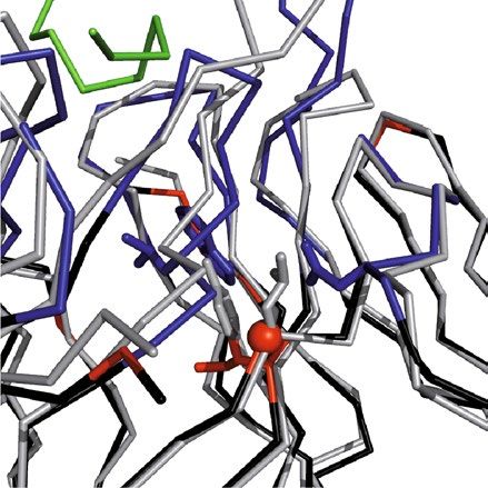
www.nature.com/scientificreports/
Fab M3/38·hGal3 peptide
Crystal Data:
Space group C2
Unit cell dimensions
a, b, c [Å] 94.1, 61.3, 80.9
α, β, γ [°] 90.0, 103.4, 90.0
Molecules per asym. unit 1 complex
Data collection
Wavelength [Å] 0.91840
Resolution range [Å]a 78.72–1.90 (2.00–1.90)
I/σ[I]a 5.7 (1.8)
Rmerge [%]a, b 8.8 (41.1)
Unique reflections 34,354
Multiplicitya 5.7 (5.9)
Completenessa 97.0 (97.9)
Refinement
Rcryst/Rfreec 20.3/24.3
Protein atoms 3367
Peptide atoms 64
Solvent atoms 201
Average B-factor [Å2]
Protein 27.3
Peptide 35.9
Water 32.3
Geometry
R.m.s.d. bond lengths, angles [Å, °] 0.007, 1.519
Ramachandran analysisd:
89.0, 9.9, 0.8, 0.3
core, allowed, generously allowed, disallowed [%]
Table 1. Crystallographic analysis
and refinement statistics.
a
Values in parentheses
are for the highest
b
resolution shell. Rmerge = h i |li (h) − �l(h)�|/ h i li (h) Rcryst = h ||Fo (h)| − |Fc (h)||/ h |Fo (h)|.
c 79
Rfree is Rcryst with 5% of the reflections that were randomly selected and excluded from r efinement .
d
Calculated with PROCHECK.
buried in the cleft (see Fig. 2). The side chain of Trp(H50) in CDR-H2 forms a hydrogen bond to peptide residue
Ala(P6) and also an edge-on contact to the aromatic side chain of Tyr(P7). In a similar way, the peptide residue
Pro(P4) contacts with its Cγ-atom the aromatic plane of the Tyr(L32) side chain in CDR-L1. The aliphatic ring
of Pro(P8), on the other hand, stacks against the side chain of Phe(L94) in CDR-L3.
Humanization of the mAb M3/38V‑regions. Basis of our humanization approach was a structural
alignment of the X-ray structure determined for the chimeric αhGal3-Fab against the AbDB, a specialized col-
lection of pre-numbered non-redundant antibody Fv portions of antibodies with known three-dimensional
structures22. For this alignment only a subset of those 695 Fv structures with a reported human origin was
chosen. The Cα-atom superposition was conducted with PDBeFold, which rates each match based on structural
similarity and paired amino acid sequence length23. The first 20 hits resulting from this search showed a narrow
distribution for the ranking factor Q, between 0.89 and 0.85 (Table 3).
A following literature search revealed an unclear origin or not fully human nature for 10 of these 20 candi-
dates, as some of them were already result of a previous humanization campaign, for example. The remaining 10
original human Fv structures were further assessed for their potential as an acceptor scaffold for CDR grafting.
This endeavour was guided by the plausible assumption that a close structural similarity between animal donor
and human acceptor framework regions will most likely retain the functional CDR conformations after grafting17.
First, a rational analysis of the Fv structures was performed using computer graphics. A Cα alignment of each
potential acceptor framework (residues L1–23, L35–49, L57–88, L98–107, H1–25, H36–49, H66–94, H103–111;
numbering according to Kabat20) with the crystal structure of the chimeric αhGal3-Fab described above allowed
us to spot critical regions that might influence the conformations of the grafted CDRs (i.e., residues L24–34,
L50–56, L89–97, H26–35, H50–65, H95–102; cf. Fig. 3). From this analysis it appeared that four Fv structures
derived from the PDB showed the least conflicts: PDB ID 3KYM, a mAb targeting the surface glycoprotein
LINGO-124, PBD ID 4KQ3, an anti-IL-17A antibody25, PDB ID 5I8C, the HIV-1 neutralizing Fab VRC34.0126,
and PDB ID 4NRY, the HIV-1 neutralizing antibody m 6627.
Structural inspection indicated that despite the high similarity between the rat framework amino acid
sequences of mAb M3/38 and each of the four human target Fv portions (Fig. 3), residues Leu(H71), Ala(H78)
Scientific Reports | (2021) 11:7358 | https://doi.org/10.1038/s41598-021-86641-0 4
Vol:.(1234567890)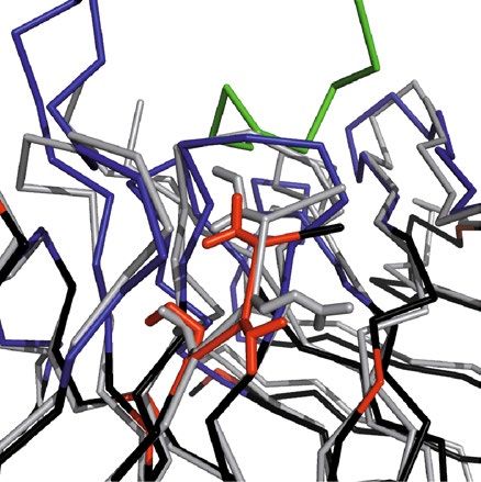
www.nature.com/scientificreports/
Figure 2. Crystal structure of the chimeric αhGal3-Fab fragment (Fv portion) in complex with the hGal3
epitope peptide, Ac-Gln-Ala-Pro-Pro-Gly-Ala-Tyr-Pro-Gly. The (N-acetylated) hGal3 peptide (residues P1-P9,
orange) is bound in a cleft between both variable domains. The secondary structure elements are representated
as cartoons and colored in light gray while disulfide bonds are shown yellow in ball-and-stick representation; the
CDRs of the light and heavy chains are colored in light pink and light blue, respectively. The mAb residues that
are engaged in aromatic contacts or form hydrogen bonds (red dotted lines) to the hGal3 peptide, together with
the two residues that form bridged hydrogen bonds through a bound water molecule (red sphere), are depicted
as sticks. Those residues that make direct hydrogen bonds to the epitope peptide are labeled (see Table 2).
Graphics were prepared using PyMOL version 1.30 (https://www.schrodinger.com).
Residue BSA [Å2] Interaction Residue BSA [Å2] Interaction
Thr(H30) 4.30 HB His(L27D) 22.26
Asp(H31) 13.70 Asp(L28) 5.77
Tyr(H32) 2.93 Tyr(L32) 27.40
Ala(H33) 20.88 Trp(L89) 2.50
His(H35) 18.81 (HB)a Ala(L91) 27.81 HB
Trp(H47) 19.95 Thr(L92) 8.77
Trp(H50) 59.51 HB His(L93) 2.20
Asn(H52) 19.39 Phe(L94) 85.58
Thr(H52A) 2.34 HB Leu(L96) 30.12
Tyr(H53) 3.85
Ile(H58) 50.33
Tyr(H59) 1.13
Lys(H64) 2.37
Gly(H95) 1.50
Thr(H96) 16.87 (HB)a
Met(H97) 60.21 HB
Ala(H99) 21.93
Table 2. Residues of the Fab M3/38 that form van der Waals contacts with the hGal3 peptide in the complex.
Buried surface areas (BSA) > 1 Å2 are reported for each of the contacting amino acids and hydrogen bonds
(HB) are indicated. a Indirect H-bond (mediated by a water molecule).
Scientific Reports | (2021) 11:7358 | https://doi.org/10.1038/s41598-021-86641-0 5
Vol.:(0123456789)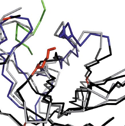
www.nature.com/scientificreports/
# Q R.M.S.D NAlign NResidue Seq. identity [%] Source PDB ID
1 0.85 0.89 220 229 53 Humanized 1L7I
2 0.85 1.00 222 230 54 Human 5ILC
3 0.84 1.01 222 232 50 Human 5I8C
4 0.84 0.99 218 225 54 Human 4KQ3
5 0.83 1.07 222 231 57 unpublished 3NCJ
6 0.83 1.03 220 229 54 Human 3KYM
7 0.83 1.07 218 223 52 Humanized 5TDO
8 0.83 1.08 222 231 57 unpublished 3NAA
9 0.83 1.03 220 229 57 Human 5V7R
10 0.82 1.10 222 231 56 unpublished 3NAB
11 0.82 1.09 222 232 54 Human 4NRY
12 0.82 1.17 221 226 53 Humanized 1T3F
13 0.82 1.07 223 235 60 Human 5ILL
14 0.82 1.13 223 232 54 Human 4NRY
15 0.82 1.07 217 223 51 Human 5TDN
16 0.82 1.18 222 228 57 Human 5IL6
17 0.82 1.07 222 234 56 Human 5IT2
18 0.82 1.12 221 230 50 Humanized 1AD0
19 0.82 1.15 221 229 53 Human 4LLU
20 0.81 1.22 221 226 48 Human 2JIX
Table 3. List of human Fv fragments from the AbDB22 with high structural similarity to the V-regions of
Fab M3/38 based on an alignment with PDBeFold21. This analysis was performed with a coordinate set at a
preliminary stage of refinement (Rcryst = 20.7%/Rfree = 25.1%). Q mean square deviation weighted by the length
of the alignment, NAlign; NResidue total number of amino acid residues in the sequence; R.M.S.D. root mean
square deviation.
(only in 3KYM), Ile(L36), Gly(L46) and Glu(L68) within the so-called vernier r egion28 of the rodent αhGal3-Fab
had to be reconstituted in the human acceptor sequences (Fig. 4). Furthermore, it was known from the cloning
of the V-regions of mAb M3/38 from the hybridoma cell line that the N-termini of both Ig chains influence the
antigen affinity9; therefore, all human acceptor sequences were adapted to the rat Ig via mutation (to the extent
necessary, see Fig. 3) to Glu(H1), Ile(H2), Glu(H3) and Val(L2), Val(L3).
In the human V H sequence the positions H2 and H71 were substituted by the corresponding residues from the
rat mAb according to the following rationale. Residue H2 is situated underneath the CDR-H3, which forms a bent
hairpin loop in the crystal structure of the chimeric αhGal-Fab (see Figs. 2, 4). Especially the interactions with
the residues at the base of this loop are crucial for the orientation of CDR-H3 and thereby relevant for antigen
binding29. Residue H71 is known to influence the conformations of both CDR-H2 and CDR-H3; depending on
the size of its side chain, CDR-H2 gets shifted relative to CDR-H330.
In the human VL sequence altogether five positions were substituted: L2, L3, L36, L46 and L68. The effect of
the N-terminal residues on the antigen affinity was already demonstrated during the cloning of the functional
chimeric αhGal3-Fab from the hybridoma9. In fact, L2 has been described as a critical residue that forms a
platform for CDR-L131. Positions L36 and L46 represent a pair of back-mutations in the vernier zone of the VL
domain. Both are residues at the hydrophobic V L/VH interface. The size of these side chains can influence the
angle between the VH and V L domains, which would change the relative positions of the CDRs from heavy and
light chains and thereby the shape of the paratope32. Finally, residue L68 is located at the tip of a hairpin loop on
the backside of CDR-L1, such that a size difference between the human and rodent side chains would provoke
a shift of this CDR.
The coding regions of the modified light and heavy chain variable domains for each of the four acceptor Fv
portions, with the grafted CDRs from M3/38 (Fig. 3), were obtained by gene synthesis including appropriate
restriction sites and cloned on a derivative of the bacterial expression vector pASK8833,34 carrying coding regions
for the human constant domains and a chloramphenicol resistance gene. The resulting recombinant Fab frag-
ments were produced via functional secretion in E. coli and purified according to published procedures9 by IMAC
from the periplasmic extract via the His6-tag attached to the heavy chain. Following SEC, a yield of approximately
0.5 mg soluble protein per 2 L culture was obtained for the constructs "3KYM", "4KQ3" and "4NRY". Interestingly,
the version "5I8C" showed a significantly lower yield (approximately 0.05 mg). The purified Fabs were tested for
binding of recombinant hGal3 by ELISA (Fig. 5), indicating a KD value of 3.6 ± 0.1 nM for "4NRY", 5.9 ± 1.9 nM
for "4KQ3", 6.9 ± 1.3 nM for "5I8C" but a much lower affinity for "3KYM", with KD = 2.8 ± 0.6 µM. Thus, the
best humanized αhGal3-Fab version, "4NRY", reached an affinity within threefold of the chimeric αhGal3-Fab
(KD = 1.3 ± 0.2 nM) when measured under the same conditions.
Consequently, among the four CDR-grafted constructs, "4NRY" appeared as the most promising humanized
version and was subjected to refinement. In an attempt to reconstitute the full binding activity of the chimeric
αhGal3-Fab, additional point mutations within the vernier zone were devised by rational analysis (see Figs. 3,
Scientific Reports | (2021) 11:7358 | https://doi.org/10.1038/s41598-021-86641-0 6
Vol:.(1234567890)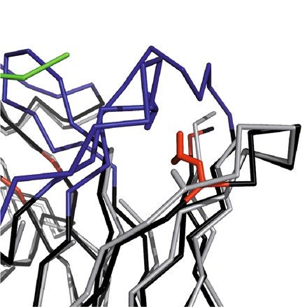
www.nature.com/scientificreports/
VH: 1 10 20 30 40 50 52A 60
. . . . . . . .
GaletuxiFab QIQLVQSGPELKKPGESVKISCKASGYTFTDYAIHWVKQAPGKGLKWMGWINTYTGKPIYADDFKGRFV
3KYM FR EVQ.LE..GG.VQ..G.LRL..A..gftfsiypmf..R.......E.VSwigpsggitkyadsvkg..T
4KQ3 FR EVQ.....A.V....S...V.....ggtfssyais..R....Q..E...siipwfgttnyaqkfqg.VT
5I8C FR .EV.....A.V....A...V..R.Fgytftgnalh..R....Q..E.L.winphsgdtttsqkfqg.VY
4NRY FR .V......A.V......L.....V.gynfasewig..R.M.....E...iiypgdsdtkyspsfqgQVI
GaletuzuFab ........A.V......L.....V.............R.M..........................QVI
CDR-H1 CDR-H2
70 82ABC 90 100A 110
. ... . . .
GaletuxiFab FSLEASASTANLQISNLKNEDTATYLCARGTMMASL------------DYWGQGVMVTVSS
3KYM FR I.RDN.KN.LY..MNS.RA......Y...eghndwyf-----------dl..R.TL.....
4KQ3 FR ITADE.T...YMEL.S.RS....V.Y...dseyyfdh-------------....TL.....
5I8C FR MTRDK.IN..F.DVTR.TSD..GI.Y...dkyygneavgmdv--------....TS.....
4NRY FR I.ADK.IN..Y..W.S..AS...I.Y...qnhygsgsyfyrtayyyamdv....TT.....
GaletuzuFab I..DK.IN..Y..W.S..AS...I.Y..........------------......TT.....
CDR-H3
VL: 1 10 20 27ABCDE 30 40 50 60
. . . ..... . . . .
GaletuxiFab DVVMTQTPVSLSLAIGQPASISCKSSQSLVHSDGKTYLSWILQRPGQSPKGLIYLVSKLDSGVPDRFSG
3KYM FR .IQ...S.GT...SP.ER.TL..rasq-----svssyla.YQ.K...A.R....dasnrat.I.A....
4KQ3 FR .IQ...S.S.V.ASV.DRVT.T.rasq-----gisnwln.YQ.K..KA..L...aasslqs...S....
5I8C FR .IQL..S.SF..ASV.DKVT.T.rasq-----gvrnela.YQ.K..KA.NL...yastlqs...S...A
4NRY FR .IQL..S.S...ASV.DRVT.T.rasq-----sigsyln.YQ.K..KA..L...aastlqs...S....
GaletuzuFab ......S.S...ASV.DRVT.T...................Q.K..KA................S....
CDR-L1 CDR-L2
70 80 90 100
. . . .
GaletuxiFab SGSETEFTLKISRVEAEDLGVYYCWQATHFPLTFGSGTKLEIK
3KYM FR ...G.....T..SLQS..FA....qqydkwplt..G...V...
4KQ3 FR ...G.D...T..SLQP..FAT...qqysddp-t..Q...V...
5I8C FR T..G.H...TV.SLQP..FAT.F.qhmssyplt..G...V...
4NRY FR ...G.D...T..SLQP..SAT...qqsystpft..P...VD.R
GaletuzuFab .....D...T..SLQP..SAT..............P...VD.R
CDR-L3
Figure 3. Amino acid sequence alignment of the V-regions cloned from the M3/38 hybridoma in form of
the chimeric αhGal3-Fab (GaletuxiFab) with suitable human template sequences for CDR grafting as well as
the finally humanized version, GaletuzuFab. Human V-region templates considered in this study were from a
mAb targeting the surface glycoprotein LINGO-1 (PDB ID: 3KYM), an anti-IL-17A antibody (PDB ID 4KQ3),
the HIV-1 neutralizing Fab VRC34.01 (PDB ID: 5I8C) and from the HIV-1 neutralizing antibody m66 (PDB
ID: 4NRY), which appeared to offer the most promising acceptor framework to yield GaletuzuFab. Sequences
are numbered according to K abat20. CDRs of the donor antibody are highlighted in blue whereas those of the
acceptor antibodies are shown in lower case letters. The definition of CDR-L1 also includes positions L26–L30 as
proposed by Chothia et al.31. Initially introduced back-mutations for CDR grafting are highlighted in pink (with
an additional position for 3KYM in green), second generation back-mutations are colored red.
4). As result, back-mutation of the residues Lys(H46) and Met(L4) to the original rat V-sequences showed
positive effects on the antigen affinity. Quantitative ELISA measurements revealed K D values of 1.3 ± 0.1 nM
for the Lys(H46) mutant and of 1.5 ± 0.1 nM for the Met(L4) mutant, compared with a value of 2.3 ± 0.2 nM
measured for "4NRY" side by side, thus approaching the affinity of the chimeric αhGal3-Fab towards hGal3, with
KD = 1.2 ± 0.2 nM in this experiment (Fig. 5C). Both residues are located in close proximity to the antigen-binding
site, suggesting a direct interaction with the presumed full length hGal3 antigen, which is more extended than
the minimal epitope peptide visible in the crystal structure with the chimeric αhGal3-Fab.
Aiming at a synergistic effect, both mutations Lys(H46) and Met(L4) were combined on the "4NRY" back-
ground, which resulted in the final humanized version, dubbed GaletuzuFab. Measured side by side with the chi-
meric αhGal3-Fab (GaletuxiFab), GaletuzuFab showed an indistinguishable KD value of 1.3 ± 0.2 nM in the ELISA
(Fig. 6). Even lower matching values of 0.34 ± 0.02 nM for GaletuzuFab and of 0.29 ± 0.02 nM for GaletuxiFab
were measured in SPR experiments, which further confirmed the functional humanization of the αhGal3-Fab.
Taken together, nine back-mutations to the rodent sequence were introduced into the human acceptor framework
"4NRY": Ile(H2), Lys(H46), Leu(H71), Val(L2), Val(L3), Met(L4), Ile (L36), Gly(L46) and Glu(L68) (see Fig. 3).
Site‑specific labelling of the humanized Fab. Our previous PET imaging study using GaletuxiFab—
in line with studies of some other tumor-targeting F abs10—demonstrated the positive effect of P
ASylation35,36
to moderately increase the plasma half-life and thereby improve tumor-to-blood ratio for TC imaging in an
orthotopic mouse xenograft model9. Therefore, the same approach was followed for GaletuzuFab and a geneti-
cally encoded, structurally disordered 200-residue sequence comprising the small l-amino acids Pro, Ala and
Ser (PAS) was appended to the C-terminus of its light chain. Apart from the enlarged hydrodynamic molecular
Scientific Reports | (2021) 11:7358 | https://doi.org/10.1038/s41598-021-86641-0 7
Vol.:(0123456789)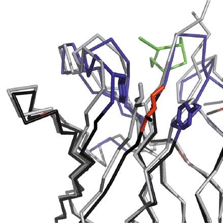
www.nature.com/scientificreports/
H71 H2 L68
ThrH52 ValH2
Leu H71 IleH2
SerL27a
TyrH27
AlaH71 Tyr H102
Val L27c
GluL68
GlyL68
VH VL
H46 L36/L46 L2-L4
AspH62
LysH46 IleL2
Trp L89 Asp L55
ValL2
GluH46 LeuH100a GlnL3
LeuL46 MetL4
TyrL36 LeuL4
GlyL46 ValL3
IleL36
Figure 4. Structural superposition of the chimeric αhGal3-Fab with the crystal structure of mAb m66 (PDB
ID: 4NRY) via Cα positions of the framework regions in both variable domains. The framework residues of the
αhGal3-Fab are shown in black, VH CDRs in dark blue, VL CDRs in light blue, the hGal3 epitope peptide in
green, the m66 framework residues in grey. Side chains of the backmutated framework residues are highlighted
as sticks (αhGal3-Fab: red; m66: grey; structurally interfering residues: blue, see text). Graphics were prepared
using PyMOL version 1.30 (https://www.schrodinger.com).
volume, similar to PEGylation35, the PAS sequence was employed here as a linker to expose a single free Cys
residue at its other end, which subsequently was utilized for the selective chemical coupling with fluorescent or
radioactive probes.
The PAS fusion protein was produced in E. coli and purified in the same manner as GaletuzuFab above, yield-
ing a similar amount of 0.5 mg soluble purified protein per 2 L bacterial shake flask culture. ESI–MS analysis
(Figure S1) revealed a mass of 65,185.01 Da, which is 178 Da higher than the theoretical mass of 65,007.07 Da,
presumably resulting from a disulfide-bridged adduct with a N-acetylcysteine-methylester (177 Da) from the
culture medium or the host cell metabolism. Reconstitution of the free C-terminal Cys residue was achieved via
mild reduction with 1,4-dithiothreitol (DTT), followed by separation from the reducing agent by S EC37.
The resulting GaletuzuFab-PAS200-Cys was individually conjugated with the dye Cy7 and the chelator defer-
oxamine (Dfo) and analysed by ESI–MS, revealing masses of 65,838.37 Da and 65,719.44 Da, respectively, almost
precisely as expected (calculated numbers: 65837.17 Da and 65,718.87 Da; see Figure S1). Further to the specific
labelling of the light chain, the purified and conjugated GaletuzuFab-PAS200-Cys showed quantitative disulfide
bridge formation between light and heavy chains in the same manner as the unfused GaletuzuFab, as demon-
strated by SDS-PAGE (Figure S1D). In addition, ELISA and SPR measurements showed that the antigen affinity
of the conjugated and PASylated GaletuzuFab was fully retained (Figure S2). Finally, the antigen-binding activity
Scientific Reports | (2021) 11:7358 | https://doi.org/10.1038/s41598-021-86641-0 8
Vol:.(1234567890)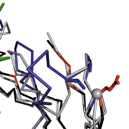
www.nature.com/scientificreports/
15
A B
15
10
∆A405/∆t [10-3 min-1]
∆A405/∆t [10-3 min-1]
10
5
5
0 0
0 20 40 60 80 100 0 1000 2000 3000 4000 5000
Concentration [nM] Concentration [nM]
C
20
∆A405/∆t [10-3 min-1]
15
10
5
0
0 10 20
Concentration [nM]
Figure 5. Investigation of the antigen-binding activity of different humanized Fab versions by ELISA.
Recombinant hGal3 was immobilized on a microtiter plate, then a dilution series of the different Fabs was
applied and, after washing, bound Fab was detected with a goat anti-human-kappa-light-chain-IgG alkaline
phosphatase conjugate. (A) Chimeric αhGal3-Fab (black; KD = 1.3 ± 0.2 nM) and the initial CDR-grafted versions
"4NRY" (red; K D = 3.6 ± 0.1 nM), "4KQ3" (blue; KD = 5.9 ± 1.9 nM) and "5I8C" (green; KD = 6.9 ± 1.3 nM). (B)
The same experiment shown for the humanized Fab version "3KYM" (black; K D = 2.8 ± 0.6 µM). (C) Influence
of single point mutations on the antigen-binding activity of the humanized Fab version "4NRY". The chimeric
αhGal3-Fab (black; KD = 1.2 ± 0.1 nM), the initial CDR-grafted version "4NRY" (dark blue; KD = 2.3 ± 0.2 nM),
the single point mutant "4NRY"(L4M) (red; K D = 1.5 ± 0.1 nM) and the single point mutant "4NRY"(E46K)
(light blue; KD = 1.3 ± 0.1 nM). Ovalbumin (grey) served as a negative control antigen in all three experiments.
Graphics were prepared using Origin(Pro) version 2017 (https://www.originlab.com).
and specificity of GaletuzuFab-PAS200-Cys was confirmed by immuno-histochemical staining of human tissue
sections representative for different TCs in comparison with normal thyroid tissue (Figure S3).
GaletuzuFab radiotracer preparation and characterization. The Dfo conjugate of GaletuzuFab-
PAS200 was labelled with 89Zr(IV) under mild conditions, followed by gel filtration on a PD10 column. The
resulting specific activity was 30 ± 3 GBq µmol−1, and the radiochemical purity, measured via radio-TLC and
SE-radio-HPLC, was > 97%. SDS-PAGE of unlabeled and labeled GaletuzuFab showed a Coomassie-stained
band coinciding with the autoradiography signal, thus confirming the biochemical integrity of the radiotracer
(Figure S4). The GaletuzuFab radiotracer was stable both in 0.25 M Na-acetate, 0.5 g l−1 gentisic acid (formu-
lation buffer) and in human serum for up to 96 h (stable fraction > 95%). In contrast, almost complete trans-
chelation of radioactivity occurred after 24 h incubation in the presence of a 1000-fold concentration of EDTA
(Figure S4B). In vitro binding tests performed on FRO82-1 TC cells (RRID: CVCL_6287) revealed a K D value of
15 ± 7 nM and an immunoreactive fraction of 73 ± 5% (Figure S4C).
PET/CT imaging of orthotopic tumors using GaletuzuFab‑PAS200‑Dfo‑89Zr. After orthotopic
FRO82-1 tumor implantation, mice were monitored weekly by visualising the lesions as hypoechoic areas via US
scanning. Expression of hGal3 in the malignant nodules was confirmed by fluorescence molecular tomography
(FMT) imaging of the neck region 24 h post injection of 130 µg GaletuzuFab-PAS200-Cy7 (Fig. 7). Follow-
ing injection of ~ 80 µCi (3 MBq) of the GaletuzuFab radiotracer, PET/CT images were recorded at different
Scientific Reports | (2021) 11:7358 | https://doi.org/10.1038/s41598-021-86641-0 9
Vol.:(0123456789)www.nature.com/scientificreports/
A
15
∆A405/∆t [10-3 min-1]
10
5
0
0 10 20
Concentration [nM]
B
80
60
∆RU
40
20
0 injection phase injection phase injection phase
injection phase injection phase
0.05 nM 0.15 nM 0.45 nM 1.35 nM 4.05 nM
0 2000 4000 6000 8000
Time [s]
Figure 6. Quantification of the antigen-binding activity of GaletuzuFab. (A) ELISA with the recombinant
hGal3 antigen immobilized on a microtiter plate. Dilution series of the chimeric αhGal3-Fab (black;
KD = 1.3 ± 0.2 nM), the initial CDR-grafted version "4NRY" (grey; KD = 3.6 ± 0.1 nM) and the final humanized
version, GaletuzuFab (blue; K D = 1.3 ± 0.2 nM) were applied. Ovalbumin (grey triangles) served as a negative
control antigen. (B) SPR analysis of the immobilized chimeric αhGal3-Fab (black; KD = 0.29 ± 0.02 nM) and
GaletuzuFab (blue; K D = 0.34 ± 0.02 nM) using hGal3 as analyte at increasing concentrations as indicated (single
cycle kinetics). Measured signals are shown in light shades while the curve fits according to a 1:1 binding model
are depicted in darker colors. Graphics were prepared using Origin(Pro) version 2017 (https://www.originlab.
com).
time points (6, 12 and 24 h), revealing the best tumor-to-background ratio 24 h post injection. The radiotracer
accumulation seen in the PET images correlated well with the dimensions and positions of the tumors found at
necropsy (Figure S5).
GaletuzuFab-PAS200-Dfo-89Zr showed specific uptake in the left thyroid lobe harboring the tumor, thus
allowing the imaging of malignant tissue with high contrast (Fig. 7B). PET images of a group of control animals
with healthy thyroid did not reveal any signal in the neck area. Blocking experiments performed by co-inject-
ing ~ 80 µCi of the radiotracer together with a 1000-fold concentration of unlabeled GaletuzuFab-PAS200 showed
strong decrease in the signal due to the saturation of binding sites, hence confirming the target specificity of the
radiotracer (Fig. 7C). Similar to the previously investigated chimeric αhGal3-Fab9, radioactivity also accumulated
in the kidneys and liver, which is consistent with the known function of these organs in tracer metabolism and
excretion, respectively38.
Quantitative analysis of radiotracer accumulation. The accumulation of GaletuzuFab-PAS200-Dfo-
89
Zr in orthotopic FRO82-1 tumors calculated via image-derived analysis was 3.8 ± 0.9%ID g−1. Biodistribution
analysis of the mice showed similar results, with an uptake of 4.1 ± 0.7%ID g−1 in the left thyroid lobe bearing
the xenograft tumor (Figure S6). This value was around three times higher than the background value meas-
ured for the healthy right thyroid lobe (1.6 ± 0.1%ID g−1) which served as an internal control. The blocking
experiments showed a significant decrease in signal (p < 0.05), comparable with that of the tumor-free thyroid
lobe (2.0 ± 0.3%ID g−1). Elevated accumulation of the tracer was measured in liver (4.6 ± 0.5%ID g−1), spleen
(11 ± 4%ID g−1) and kidneys (85 ± 5%ID g−1), i.e. the main organs for metabolisation of the radiotracer, radio-
metal accumulation and excretion, respectively. In conclusion, the specific accumulation in thyroid tumor and
the tissue distribution measured for GaletuzuFab-PAS200-Dfo-89Zr matched the high contrast tumor images
observed by PET. These functional features of GaletuzuFab-PAS200-Dfo-89Zr were essentially indistinguishable
Scientific Reports | (2021) 11:7358 | https://doi.org/10.1038/s41598-021-86641-0 10
Vol:.(1234567890)www.nature.com/scientificreports/
Figure 7. Imaging of thyroid orthotopic tumors growing in a xenograft mouse model using fluorescence-
labeled and radiolabeled GaletuzuFab-PAS200. (A) Representative FMT imaging of a mouse bearing a FRO82-1
xenograft tumor. The scan performed 24 h after the injection of 130 µg GaletuzuFab-PAS200-Cy7 shows a
hGal3-positive lesion in the neck. (B) PET/CT images acquired 24 h after intravenous injection of GaletuzuFab-
PAS200-Dfo-89Zr (3 MBq) in animals from different groups: (1) co-injected with a 1000-fold excess of non-
radioactive GaletuzuFab-PAS200-Cys to confirm the specificity of tracer binding in vivo (blocking experiment,
left); (2) injected with the radioactive probe only (center); (3) healthy animals (without tumor) injected with
the radioactive probe (control, right). The lower transversal images taken at the level of the neck area reveal the
position of the tumor on the left side of the trachea while the upper images show maximum imaging projection
(MIP) whole-body PET images. (C) Representative PET image of the neck area of an animal from the center
group; the position of the PET signal corresponds to the findings at necropsy. Graphics were prepared using
TrueQuant Imaging Software version 2.0 (https://www.perkinelmer.com) for the fluorescence scans and Inveon
Research Workplace version 4.0 (https://inveon-research-workplace.software.informer.com) for the PET/CT
image analysis.
from those of the PASylated chimeric αhGal3-Fab tracer that was extensively investigated before9, thus providing
proof of success of our antibody humanization endeavor.
Discussion
Despite progress during the last years in improving the preoperative diagnosis of TC as well as the management
of cancer patients, the identification of thyroid carcinoma remains a challenging process that is dominated by
largely outdated methods and involves unnecessary surgery as a prevailing therapeutic a pproach6,7. Hence,
there is an unmet need for a reliable, non-invasive procedure to identify malignant nodules, which can be dis-
tinguished only with difficulty from the highly prevalent benign thyroid proliferations (in particular nodular
hyperplasia) using the currently available diagnostic tools. This is particularly evident for follicular carcinoma,
follicular variants of papillary thyroid carcinoma and for carcinomas that have lost the capacity to accumulate
radioiodine due to reduction or lack of the sodium iodide symporter (NIS), which therefore leads to negative
or equivocal 131I whole-body imaging39. Better methods for TC diagnosis would also be important in the case of
poorly differentiated (PDTC) and anaplastic thyroid (ATC) carcinomas, which are aggressive tumor types with
high metastatic potential. Again, these very rare forms of thyroid carcinoma are characterized by the lack of abil-
ity to accumulate radioiodine due to the absence of NIS and, therefore, are difficult to be diagnosed and cured40.
In recent studies, we demonstrated that immuno-PET imaging by targeting hGal3 can be a powerful diag-
nostic method for TC, with high potential to aid the discrimination between malignant and benign nodules9,41,42.
To enable the translation of hGal3 imaging into clinical application, we now have developed a humanized Fab
fragment derived from the well validated hybridoma mAb M3/388 that shows high affinity and specificity towards
this human TC-associated antigen. In a preceding study, immuno-PET imaging experiments with a chimeric
Fab of the αhGal3 mAb using an orthotopic TC mouse m odel9 revealed advantages for in vivo imaging due to
the faster clearance and higher specific tumor uptake of the PASylated recombinant Fab in comparison with the
full size mAb. To further lower the risk of an ADA response upon repeated administration to human patients,
we have fully humanized the rat variable domains present in the chimeric Fab by CDR grafting onto human
framework regions.
Scientific Reports | (2021) 11:7358 | https://doi.org/10.1038/s41598-021-86641-0 11
Vol.:(0123456789)www.nature.com/scientificreports/
Whereas the concept of CDR grafting between Igs from different species was originally devised on the basis
of structural considerations13,14, common humanization procedures mostly rely on a homology assessment at
the level of the amino acid sequence till today16,43. This is somewhat surprising particularly in cases where three-
dimensional structural information for the donor antibody actually is available44–46. In the early endeavors, accep-
tor framework sequences from structurally characterized antibodies or Bence Jones proteins were chosen13,14,47;
this was followed by the use of human consensus Ig s equences20, including the famous example of the αHER2
antibody trastuzumab15. Today, however, frequently expressed germline Ig sequences from the human genome
serve as preferred templates with presumably low i mmunogenicity43,46,48.
On the other hand, it is well known that the simple replacement of CDRs in the amino acid sequence of
the acceptor Ig by those from the mAb with desired specificity can lead to incomplete preservation of the
antigen-binding activity as a consequence of structural incompatibilities between the donor CDRs and acceptor
frameworks at a finer granular level. Hence, the reconstitution of the original antigen affinity usually requires
back-mutation of certain positions in the acceptor frameworks to the corresponding donor r esidues14,17,28,48. Such
structural incompatibilities can arise from (1) altered packing between CDRs and framework regions in the donor
antibody relative to the situation after grafting on the acceptor frameworks, in particular involving residues of the
vernier zone28, (2) differences in the V
H/VL pairing—i.e. the mutual orientation of the V H and V L domains43—and
(3) differences in key framework residues responsible for the canonical conformations of CDRs31.
To address these potential problems and identify critical residues for CDR function, structural modelling of
the donor antibody was proposed soon after inception of mAb humanization by CDR grafting17. Nevertheless,
published examples of such antibody humanization guided by rational protein design have remained scarce. One
case is the successful humanization of the anti-CEA antibody T84.66 based on crystal structure d ata49; however,
in this example framework regions of the αHER2 antibody 4D5 ver. 8 were chosen as acceptor, which itself is a
humanized mAb based on consensus Ig sequences as explained above and not a natural human antibody.
Today, the situation with regard to the knowledge base has changed: functional fragments (mainly Fab) of
several hundred human mAbs have been structurally elucidated by X-ray crystallography and are easily accessible
from the Protein Data Bank at RCSB (PDB) or from specialized databases such as the A bDb22. Under the plausible
assumption that the likelihood of success of CDR grafting rises with the degree of structural similarity between
the rodent donor and the human acceptor frameworks that support the set of CDRs, we have exploited a strategy
solely based on 3D-structural information. In this concept the choice of a functionally suitable natural human
acceptor framework requires the precise knowledge of the structure of the rodent mAb of interest considering
that modeling approaches, even though useful in the early days of this fi eld17, are associated with substantial
uncertainties. In the present case, the co-crystallization and X-ray structural analysis of the chimeric αhGal3-Fab
with its epitope peptide provided access to that kind of information. Subsequent search in a structural antibody
database led to the identification of four promising Fv regions (PDB IDs: 3KYM, 4KQ3, 5I8C, 4NRY), which
were subsequently employed for experimental CDR grafting.
Not unexpectedly, the different humanized versions resulting from the initial design showed decreased anti-
gen affinity to varying extent, ranging from 2800 nM for "3KYM", over much better values of 6.9 nM for "5I8C"
and 5.9 nM for "4KQ3", to 3.6 nM for the most promising candidate, "4NRY". Remarkably, the latter design,
which made use of the framework regions of the human mAb m6627 for CDR grafting—including some obvi-
ous back-mutations to the rat sequence—showed an affinity already within a factor 3 of the original chimeric
Fab (1.3 nM). Of note, the mAb m66, which was cloned using a phage-display Fab library prepared from frozen
PBMC mRNA of a patient whose serum contained cross-reactive HIV-1-neutralizing antibodies, is particularly
well characterized. Its light and heavy chain sequences closely match the predicted human germ line precursors
IGHV5-51*01/IGHD3-10*01/J6*02 and IGKV1-39*01/IGKJ3*01, r espectively27,50, thus providing a reliable basis
for low immunogenic potential of these acceptor framework regions.
In general, the modest loss in antigen affinity for 3 of the 4 humanization attempts validates our structural
design concept of searching the most closely related light/heavy chain framework with a fixed V H/VL pairing—
compared with the strategy of choosing the most similar light chain and heavy chain frameworks individually,
as it is still common practice in antibody humanization43. Since the likelihood of finding a most similar human
VH/VL pair for a given mAb of animal origin correlates with the size of the growing database of fully human mAb
structures, this approach will become increasingly successful for future CDR grafting campaigns.
Nevertheless, plain grafting of the CDRs alone cannot be expected to guarantee full retention of the antigen-
binding activity of the original rodent mAb. Thus, back-mutations to the donor sequence, in particular in the
vernier region, are typically introduced to account for remaining small structural d eviations15,28. Of course,
the number of such substitutions has to be kept low in order not to compromise the goal of the humanization
procedure, i.e. reducing non-human sequence stretches to minimum. In total seven back-mutations were intro-
duced during our first design for "4NRY" due to obvious structural differences between the donor and acceptor
frameworks.
At the second stage of humanization, after characterization of the initial "4NRY" version as the most promising
candidate, additional back-mutations were evaluated in order to further reduce the difference in affinity com-
pared with the original chimeric Fab. In particular, two positions attracted our attention: H46 and L4 (see Fig. 4).
Residue H46 and its neighbors, H47-H49, create a platform that structurally supports CDR-H229. Thereby, the
side chain of H46 is located at close distance (2.5 Å) to H62 and H63, which form the tip of the CDR-H2 hairpin
loop, and can influence their positions. On the other hand, residue L4 is part of the β-sheet underneath CDR-L3,
in proximity to its C-terminal residue L97, and plays a role for the conformation of this hypervariable l oop29.
The combination of both mutations eventually led to recovery of the full binding affinity, which might be
explained by a more extended interaction between the complete hGal3 antigen and the Fab fragment than it
appeared from the crystal structure with the truncated 9mer epitope peptide. This epitope is located in the ND
of hGal3, which is described as a random c oil51. Thus, additional contacts of the Fab with the highly flexible ND
Scientific Reports | (2021) 11:7358 | https://doi.org/10.1038/s41598-021-86641-0 12
Vol:.(1234567890)www.nature.com/scientificreports/
as part of the full size antigen are possible. As the two residues H46 and L4 are both located within the same
groove at the V L/VH interface, and both have sufficient accessible surface to engage in intermolecular interac-
tions, a synergistic effect seems conceivable. As result, GaletuzuFab, which was developed in just two steps and
carries only nine rat residues in the human framework regions for both Ig chains, shows an antigen affinity that
is indistinguishable from the original chimeric Fab. This demonstrates the efficiency of our structure-based
antibody humanization approach.
Apart from the minimization of potentially immunogenic sequence epitopes, a high degree of molecular
definition of a biopharmaceutical is critical, whereas molecular heterogeneity can have detrimental effects on
pharmacokinetics, in vivo efficacy and t olerability52,53. In this context the posttranslational modification made by
introduction of the chelator Dfo for radionuclide labeling of GaletuzuFab poses a critical factor. Here, the strategy
of site-specific coupling via a single free Cys residue, specifically introduced at the freely accessible C-terminus
of the structurally disordered PAS tail and, thus, prone to reaction with a maleimide group54, was chosen. While
initially incorporated to effect a moderately increased plasma half-life and boosting tumor a ccumulation10, the
PAS sequence at the end of one of the Ig chains conveniently served as a spacer between the functional protein
moiety and the reactive Cys residue. In this way, the need for introducing a potentially critical amino acid
exchange and/or modification of the Ig scaffold, as frequently described for the construction of recombinant
antibody drug conjugates (ADCs)53, was avoided. In fact, PASylation and labeling via the C-terminal Cys residue
led to a precisely defined, monodisperse protein conjugate, as demonstrated by ESI–MS analysis, and had only
marginal effect on the antigen affinity (1.9 ± 0.1 nM for GaletuzuFab-PAS200-Dfo versus 1.3 nM for GaletuzuFab
or 1.6 nM for GaletuzuFab-PAS200-Cy7 as measured by ELISA).
Finally, the GaletuzuFab-PAS200-Dfo-89Zr radiotracer was prepared via labeling with the β-emitter zirco-
nium-89 (89Zr; t1/2 = 78.41 h; Emax β+ = 0.9 meV) under mild conditions (at a temperature ≤ 37 °C) and character-
ized with regard to antigen-binding activity and stability. The radiochemical purity of the humanized Fab was
high and similar to the one previously obtained for the chimeric αhGal3-Fab tracer (> 97%)9 while the specific
activity was even higher (30 ± 3 GBq µmol−1 versus 19.8 ± 1.2 GBq µmol−1), probably due to the placement of the
Dfo chelator at the sterically accessible C-terminus of the PAS chain instead of the random conjugation with Dfo
isothiocyanate in the preceding study. A saturation binding assay with FRO82-1 cells indicated an E C50 value
about one order of magnitude higher than the KD measured via SPR, which can be explained by some hindered
accessibility of the hGal3 antigen within the glycocalyx at the cell surface.
In PET/CT scans of mice carrying orthotopic human thyroid tumors high contrast images were obtained 24 h
post tracer injection, showing a high target-to background ratio in the tumor-bearing left thyroid lobe compared
to the healthy right lobe. No radiotracer uptake in the neck region of healthy animals without xenograft was
visible and blocking tests with an excess of unlabeled GaletuzuFab-PAS200 further confirmed the specificity of
the GaletuzuFab radiotracer, in line with biodistribution experiments. In conclusion, we were able to develop a
highly homogeneous preparation of a PASylated humanized Fab tracer directed against hGal3, thus minimizing
the risk of failure in a future clinical trial. The clinical potential of this novel agent for imaging and, also, therapy
planning of TC has been demonstrated via high and specific uptake by orthotopic tumor xenografts in mice at
early time points after injection.
Materials and methods
Bacterial production and purification of human Gal3. The full length human Gal3 protein (Uni-
ProtKB ID: P17931), with the unpaired thiol residue Cys173 mutated to Thr, was produced as a soluble protein
in the cytoplasm of E. coli as previously described9. An N-terminally truncated fragment starting with Pro113
was prepared by amplifying the corresponding coding region with the primers 5′-GCGCATATGCCATACGGT
GCTC-3′ and 5′-CGCAGTAGCGGTAAACG-3′, thus introducing an NdeI restriction site and an ATG start
codon directly upstream of Pro113. The truncated P113-hGal3 protein, which carried a C-terminal Strep-tag II
like the recombinant full-length hGal355, was expressed as soluble protein in the cytoplasm of E. coli in 2 l shake
flasks, followed by purification via Strep-Tactin affinity chromatography as well as size exclusion chromatogra-
phy (SEC).
Western blotting. Following SDS-PAGE56 of the recombinant full-length hGal3 and its fragment P113-
hGal3, the polypeptides were electro-transferred onto a methanol-activated PVDF membrane (Merck Mil-
lipore, Billerica, MA, USA). After washing in phosphate-buffered saline (115 mM NaCl, 15 mM Na2HPO4,
4 mM KH2PO4; PBS) containing 0.01% v/v Tween 20 (PBS/T0.01) the membrane was incubated with the chimeric
αhGal3-Fab-PAS200-Cy5.5 (2 µg ml−1 in PBS/T0.01)9 for 1 h at room temperature. After washing the membrane
with PBS/T0.01, fluorescence was detected with an Odyssey fluorescence scanner (excitation: 685 nm; emission:
720 nm; LI-COR, Lincoln, NE, USA) and evaluated using Quant version 12.2 software (TotalLab, Newcas-
tle-Upon-Tyne, UK).
Peptide SPOT synthesis and analysis. A set of 8-mer peptides covering residues 1–112 of hGal3, with
2 residue overlap, was directly synthesized with a fully automated MultiPep RS instrument (Intavis, Cologne,
Germany) according to the SPOT method19 on a Gly-PEG500-derivatised cellulose-membrane57 starting from
the C-terminus using Boc-protected amino acids. After N-terminal acetylation and deprotection of the pep-
tides, the membrane was rehydrated with PBS/T0.01 and incubated with the chimeric αhGal3-Fab-PAS200-Cy5.5
(2 µg ml−1 in PBS/T0.01)9 for 1 h at room temperature. After washing with PBS/T0.01, fluorescence signals were
directly detected on the membrane and quantified as described for the western blot above.
Scientific Reports | (2021) 11:7358 | https://doi.org/10.1038/s41598-021-86641-0 13
Vol.:(0123456789)You can also read