Existence of multiple organ aging in animal model of emphysema induced by cigarette smoke extract
←
→
Page content transcription
If your browser does not render page correctly, please read the page content below
Tobacco Induced Diseases
Research Paper

Existence of multiple organ aging in animal model of
emphysema induced by cigarette smoke extract
Guibin Liang1, Zhihui He1, Yan Chen2, Hongbo Zhang3, Huaihuai Peng4, Dandan Zong2, Yingjiao Long2

ABSTRACT
INTRODUCTION   It is commonly considered that COPD or at least emphysema                                                                    AFFILIATION
                                                                                                                                            1 Department of Critical Care
represents accelerated lung aging induced in part by oxidative damage from                                                                  Medicine, The Third Xiangya
cigarette smoke components. However, the issue if there are any aging signs in                                                              Hospital, Central South
                                                                                                                                            University, Changsha, China
other organs in patients with COPD or emphysema remains unclear. The aim of                                                                 2 Department of Respiratory
this study is to explore whether there is multiple organ aging in the animal model                                                          Medicine, The Second Xiangya
of emphysema induced by cigarette smoke extract (CSE), and to ascertain the                                                                 Hospital, Central South
                                                                                                                                            University, Changsha, China
possible mechanisms, if any.                                                                                                                3 Department of Pathology,
METHODS The animal model of emphysema was induced by CSE. Histomorphological                                                                The Second Xiangya Hospital,
                                                                                                                                            Central South University,
changes in lung, heart, liver, kidney and spleen tissues were measured after                                                                Changsha, China
staining with hematoxylin and eosin (H&E). The concentrations of stem cell factor                                                           4 Department of Intensive
                                                                                                                                            Care Unit, The Second
(SCF), CyclinD1 and superoxide dismutase (SOD) in serum were determined by                                                                  Xiangya Hospital, Central
ELISA kit. The expressions of p16 (INK4a), Sca-1, eNOS proteins and mRNA in                                                                 South University, Changsha,
                                                                                                                                            China
lung, heart, liver, kidney and spleen tissues were detected by Western blotting
and quantitative reverse transcriptase polymerase chain reaction (qRT-PCR),                                                                 CORRESPONDENCE TO
respectively. Decitabine (Dec) was applied to examine whether it could alter the                                                            Zhihui He. Department of
                                                                                                                                            Critical Care Medicine, The
changes caused by CSE.                                                                                                                      Third Xiangya Hospital,
RESULTS The histomorphology of lung tissue was significantly changed, while other                                                           Central South University,
                                                                                                                                            Changsha, 410013 Hunan,
organs exhibited normal structure and histomorphology. The concentrations of                                                                China.
SCF, CyclinD1 and SOD in serum were lower in the CSE group than in the                                                                      E-mail: hzh703@csu.edu.cn
                                                                                                                                            ORCID ID: https://orcid.org/
control group. The expression levels of p16(INK4a) protein and mRNA in lung,                                                                0000-0001-5672-9691
heart, liver, kidney and spleen tissues were higher in the CSE group than in the
control group, while the expression levels of Sca-1 and eNOS proteins and mRNA                                                              KEYWORDS
                                                                                                                                            cigarette smoke, decitabine,
were lower in the CSE group than in the control group, in the tissues described                                                             emphysema, multiple organs,
above. Dec could partly alleviate the damages caused by CSE and the degree of                                                               aging

alleviation resulted by Dec varied from organ to organ.                                                                                     Received: 3 March 2021
CONCLUSIONS In addition to the aging of the lung tissue in the emphysema animal                                                             Revised: 26 May 2021
                                                                                                                                            Accepted: 10 November 2021
model induced by CSE, the tissues of the heart, liver, kidney and spleen were
also in the progress of aging, but the sensibility and affinity of lung to CSE were
higher than those of the other organs. Multiple organ aging may also exist in
the animal model of emphysema induced by CSE. DEC can partly alleviate the
multiple organ aging caused by CSE.

ABBREVIATIONS acLDL: acetylated low-density lipoprotein, AI: apoptotic index, COPD: chronic obstructive pulmonary disease,
CS: cigarette smoke, CSE: cigarette smoke extract, Dec: decitabine, DAPI: 4',6-diamidino-2-phenylindole, DI: destructive index, Dil:
1,1’-dioctadecyl-3,3,3’,3-tetramethylindocarbocyanine perchlorate, ECs: endothelial cells, EGM-2: endothelial growth medium-2,
eNOS: endothelial nitric oxide synthase, EPCs: endothelial progenitor cells, FBS: fetal bovine serum, FITC: fluorescein isothiocyanate,
LSCM: laser scanning confocal microscope, PBS: phosphate buffered saline, ROS: reactive oxygen species, SCF: stem cell factor,
SMCs: smooth muscle cells, SOD: superoxide dismutase, UEA-1: ulex europaeus agglutinin-1

Tob. Induc. Dis. 2022;20(January):2                                                                                                          https://doi.org/10.18332/tid/143853

Published by European Publishing. © 2022 Liang G. et al. This is an Open Access article distributed under the terms of the Creative Commons Attribution 4.0 International
License. (https://creativecommons.org/licenses/by/4.0/)

                                                                                          1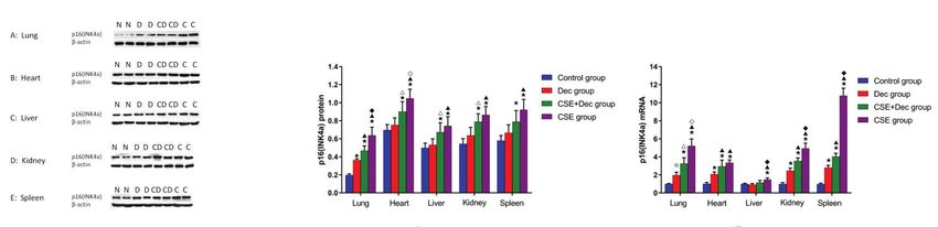
Tobacco Induced Diseases
Research Paper

INTRODUCTION                                                    The mechanisms of their action are mainly through
Aging is characterized by progressive decline in tissue         the release of neutrophilic proteases, including
and organ function and increased risk of mortality,             elastase, cathepsin G and matrix protease that cause
which is associated with a wide range of human                  a chronic mucus hypersecretion state and destroy
disorders, including cancer, diabetes, cardiovascular,          lung parenchyma. In the aging process, the long-
and neurodegenerative diseases. Some studies have               term stimulation of pro-inflammatory cytokines
shown that chronic obstructive pulmonary disease                such as TNF-α, IL-1β and IL-6 in the body can lead
(COPD) or at least emphysema was a disease of                   to a chronic, low-degree and mild inflammatory
accelerated lung aging1. Environmental gases, such              response, thus causing or increasing the occurrence
as cigarette smoke or other pollutants, may accelerate          of age-related degenerative diseases. In this study,
the aging of lungs or worsen aging-related events in            the potential aging of organs in mice was evaluated
lungs by causing defective resolution of inflammation,          by the detection of stem cell factor (SCF), Cyclin D1,
for example, by reducing antiaging molecules, such              superoxide dismutase (SOD), p16(INK4a), stem cell
as histone deacetylases, and this consequently                  antigen-1(Sca-1), and endothelial nitric oxide synthase
induces accelerated progression of COPD. There                  (eNOS), which are linked to oxidation, apoptosis and
are many aging-associated changes in the lungs,                 aging in humans and animals 5. SCF promotes the
including decreased size of thoracic cavity, limiting           proliferation and differentiation of hematopoietic
lung volume. Our previous studies demonstrated that             cells and regulates the growth and development
the expression level of p16(INK4a) was increased,               of hematopoietic cells. Cyclin D1 acts on G1 phase
and the expression level of stem cell antigen-1 (Sca-           and interacts with many proteins to promote cells to
1) was decreased in lung tissue of emphysema 2,3.               enter S phase. SOD could eliminate excess oxygen
However, the issue if there are any aging signs in              free radicals and their derivatives so as to protect
other organs of the body, such as heart, liver, kidney          cells from damage and maintain normal metabolism.
and spleen, with COPD or emphysema, remains                     The antibody p16(INK4a) is one of the best aging
unclear. For ethical reasons, we cannot carry out               biomarkers and is suppressed in early embryogenesis
experiments on organ aging in patients with COPD,               and progressively induced during aging. Sca-1 is
and so we employed an animal model. Smoking is an               usually associated with stem cells or progenitor cells
independent risk factor for cardiovascular and lung             proliferation and self-renewal, and is a marker of
diseases. The animal model of emphysema induced                 youth. eNOS is the key enzyme that catalyzes the
by intraperitoneal injection of cigarette smoke extract         production of NO, an endogenous signal molecule
(CSE) has been verified in previous studies, and it is          which maintains vascular homeostasis, including blood
equivalent to the animal model of emphysema induced             pressure homeostasis, vascular permeability, tension
by cigarette smoke (CS) exposure in lung function               regulation and hypoxic compensatory mechanism.
and histomorphology 4. CSE contains more than                   Decitabine (Dec) is a widely used DNA methylation
4000 chemicals found in CS, including free radicals,            inhibitor, which triggers demethylation in vitro
toxins, and electrophiles. An increasing number                 and in vivo, resulting in continuous reactivation of
of experiments have shown that CS or CSE could                  epigenetic silencing genes. In our previous study3, it
accelerate the senescence of leukocyte, endothelial             was demonstrated that Dec alleviated lung senescence
progenitor cell (EPC) and lung tissue and reduce their          in a CSE-induced emphysema animal model. In the
regeneration ability3. It is commonly considered that           present study, Dec was applied to identify whether it
COPD or at least emphysema represents accelerated               can alleviate the aging of other organs, if any.
lung aging induced in part by oxidative damage from
cigarette smoke components.                                     METHODS
   There are many indicators that can be used to assess         In the present study, we follow the methods of He
the function of the lung, heart, liver and kidney in            et al.3.
humans, but not in mice. The main inflammatory
cells involved in the inflammatory response of COPD             Animals
are neutrophils, macrophages, and T lymphocytes.                Forty male C57BL/6J mice aged 6 weeks were

                                            Tob. Induc. Dis. 2022;20(January):2
                                            https://doi.org/10.18332/tid/143853

                                                            2
Tobacco Induced Diseases
Research Paper

randomly enrolled in this study and fed in a cleaning                                       Measurement of the concentrations of SCF,
unit at 23–25oC, 50–60% humidity, with a 12-hours                                           Cyclin-D1 and SOD in serum
rhythm of night and day. All animals were purchased                                         The mouse was weighed and anesthetized by
from Shanghai laboratory animal center of the Chinese                                       intraperitoneal injection of 10% chloral hydrate (3
Academy of Sciences (SLACCAS, Shanghai, China).                                             mL/kg body weight). Then the blood was sampled
The study was approved by the Institutional Review                                          by cutting the tail and centrifuged within an hour at
Board of Central South University and conformed to                                          700g for 10 min. The supernatant was collected and
the guiding principles for research involving animals                                       stored at -40°C. The concentrations of SCF, Cyclin D1
and human beings6.                                                                          and SOD in serum were measured with ELISA kits
                                                                                            (R&D systems, USA) according to the manufacturer’s
Preparation of CSE and decitabine                                                           instructions.
CSE was prepared according to a previously published
method2. Briefly, one Fu-Rong cigarette (Tar: 13 mg,Slice staining and identification of emphysema
Nicotine: 1.0 mg, carbon monoxide: 14 mg/cigarette; After blood collection, the mice were sacrificed by
China Tobacco Hunan Industrial, Changsha, China)    an overdose of anesthetic. Lung, heart, liver, kidney
was burned and the smoke passed through 4 mL of     and spleen slices were stained with hematoxylin
phosphate-buffered saline (PBS) via a vacuum pump   and eosin (H&E) (Sigma, USA). Emphysema was
at a constant pressure of -0.1 kPa. This product wasquantified based on the measurement of the mean
further filtered through a filter with 0.22 μm poreslinear intercept (MLI) and destructive index (DI).
(Thermo Fisher Scientific, Waltham, MA, USA)        Briefly, MLI was measured by dividing the length
to remove particles and bacteria. The solution was  of a line drawn across the lung section by the total
prepared freshly for each experiment. Five grams of number of intercepts. The DI was calculated by
decitabine (Sigma, MO, USA) was dissolved in 2mL    dividing the defined destructive alveoli by the total
PBS and further diluted to 25 mg/mL, sub-packaged   number of alveoli. Destructive alveolus was defined
and stored under -80oC until the experiments.       as having at least one of the following: alveolar wall
                                                    defects; intraluminal parenchymal rags in alveolar
Experimental protocol                               ducts; obvious abnormal morphology; and typical
The mouse emphysema model was established as emphysematous changes. The analysis was performed
previously described 3. The C57BL/6J mice were using a microscopic point-count technique at ×200
divided into four groups: control, Dec, CSE, and magnification. The histomorphology assessment was
CSE+Dec (10 per group). The total experimental performed in a blinded manner.
period was four weeks, with intraperitoneal
injection of PBS, Dec, or CSE (Table 1). The doses Western blot analysis
of intraperitoneal injection of PBS, Dec, CSE were Briefly, protein was mixed 1:1 with 2 × SDS
0.3 mL/20 g, 0.3 mL/20 g, 2.5 mg/kg (0.3 mL/20 g loading buffer and incubated at 100°C for 4
constant volume), respectively. At Day 28, the mice min. Then, the protein was electrophoresed in
were disposed for the experiments.                  10–12% SDS-polyacrylamide gel and transferred
                                                    electrophoretically onto a polyvinylidene difluoride

Table 1. Experiment schedule (N=10)

 Day                                0                        11                        15                       17                        19                       22
 Control                           PBS                      PBS                       PBS                      PBS                       PBS                      PBS
 Dec                               PBS                      PBS                       Dec                      Dec                       Dec                      PBS
 CSE                               CSE                      CSE                       PBS                      PBS                       PBS                      CSE
 CSE+Dec                           CSE                      CSE                       Dec                      Dec                       Dec                      CSE
All treatments were conducted by intraperitoneal injection. All animals were disposed on Day 28 after the start of treatment. Dec: decitabine. CSE: cigarette smoke extract. PBS:
phosphate buffered saline.

                                                                      Tob. Induc. Dis. 2022;20(January):2
                                                                      https://doi.org/10.18332/tid/143853

                                                                                        3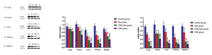
Tobacco Induced Diseases
Research Paper

microporous membrane (Millipore, Billerica, MA,                   Dec, CSE+Dec and CSE groups were 16.60±1.64,
USA). Membranes were incubated with primary                       17.00±1.92, 15.86±1.45 and 16.15±1.80, respectively,
antibody overnight [p16(INK4a): 1:200, Santa Cruz                 and there was no statistical difference between groups
Biotechnology, USA; Sca-1: 1:200, Biolegend, USA;                 (p>0.05). On Day 28, the body weight (g) of mice
eNOS: 1:1000, Proteintech, USA), and then washed                  in the control, Dec, CSE+Dec and CSE groups were
carefully with Tris-buffered-saline with Tween-20                 24.14±0.84, 23.36±0.79, 23.14±1.20 and 24.13±2.27,
(TBST) followed by re-incubation with secondary                   respectively, and there was no significant difference
antibodies (1:3000; 1 h). After washing with TBS-T                between the two groups (p>0.05) (Table 2).
again, immunoreactive bands were detected using
ECL chemiluminescent substrate (Thermo, USA).                     Lung, heart, liver, kidney and spleen
                                                                  histomorphology
Quantitative RT-PCR                                               As shown in Figure 1, the lung tissues of CSE group
Total RNA was extracted from tissues using Trizol                 (Figure 1, Lung C) and CSE+Dec group (Figure 1,
reagent (Invitrogen, USA). First-strand cDNA was                  Lung D) exhibited damaged alveolar walls, thinner
synthesized using the RevertAid™ First Strand cDNA                alveolar septa and enlarged alveolar spaces when
Synthesis Kit (Fermentas, USA) according to the                   compared with the control group (Figure 1, Lung
manufacturer’s instructions, and used as the template             A) and Dec group (Figure 1, Lung B). Quantitative
for quantitative RT-PCR analysis. Gene expression and             analysis showed that MLI and DI were significantly
analysis were performed by RT-PCR using SYBR Green                higher in the CSE group (69.65 ± 10.19 μm, 45.36 ±
qPCR Master Mix (Applied Biosystems, USA) on Bio-rad              6.96%) and CSE+Dec group (53.97 ± 8.86 μm, 35.83
CFX96 Real Time system (Bio-Rad, USA) with β-actin                ± 5.75%) than those in the control group (27.69 ±
used as an internal control. The ΔΔCt method was                  5.37 μm, 9.95 ± 1.19%) and Dec group (29.17 ±
adopted to quantify the fold change of expression levels.         5.31 μm, 10.36 ± 1.40%) (p
Tobacco Induced Diseases
Research Paper
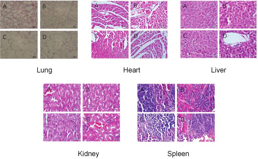
Figure 1. Histomorphological changes of lung, heart, liver, kidney and spleen tissues
Lung tissues of CSE group (C) and CSE+DEC group (D) exhibited destroyed alveolar wall, thinner alveolar septum and enlarged alveolar space when compared with control group
(A) and DEC group (B). Heart tissue of control group (A), DEC group (B), CSE group (C) and CSE+DEC group (D) all exhibited normal structure, no congestion, no bleeding, no
inflammation cells infiltration, cardiomyocytes without degeneration and necrosis. Liver tissue of control group (A), DEC group (B), CSE group (C) and CSE+DEC group (D) all
exhibited normal structure, no congestion, no bleeding, no inflammation cells infiltration, hepatocyte without degeneration and necrosis. Kidney tissue of control group (A),
DEC group (B), CSE group (C) and CSE+DEC group (D) all exhibited normal structure, no congestion, no bleeding, no inflammation cells infiltration, renal tubular cells without
degeneration and necrosis, no glomerular sclerosis. Scale bar represents 50 μm. DEC: decitabine. CSE: cigarette smoke extract.
group (Figure 1, Liver A), Dec group (Figure 1, Liver sclerosis.
B), CSE group (Figure 1, Liver C) and CSE+Dec group As shown in Figure 1, the spleen tissue of control
(Figure 1, Liver D) all exhibited normal structure, group (Figure 1, Spleen A), Dec group (Figure 1,
no congestion, no bleeding, no inflammation cells Spleen B), CSE group (Figure 1, Spleen C) and
infiltration, hepatocytes without degeneration and CSE+Dec group (Figure 1, Spleen D) all exhibited
necrosis. normal structure, splenic nodule presenting, and no
As shown in Figure 1, the kidney tissue of control congestion in splenic sinus.
group (Figure 1, Kidney A), Dec group (Figure 1,
Kidney B), CSE group (Figure 1, Kidney C) and Concentrations of SCF, Cyclin D1 and SOD in
CSE+Dec group (Figure 1, Kidney D) all exhibited serum
normal structure, no congestion, no bleeding, no The concentration of SCF in serum was significantly
inflammation cells infiltration, renal tubular cells lower in the Dec group (41.56 ± 4.04 ng/L), CSE
without degeneration and necrosis, and no glomerular group (34.30 ± 1.67 ng/L) and CSE+Dec group
(36.23 ± 2.56 ng/L) than in the control group (49.75
Table 3. Lung histomorphology (N=10) ± 4.85 ng/L) (pTobacco Induced Diseases
Research Paper
Figure 2. Concentrations of SCF, Cyclin D1 and SOD in serum
Quantitative analyses of concentrations of SCF (A), Cyclin D1 (B) and SOD (C) in serum. Data are represented as mean±SD. Dec: decitabine. CSE: cigarette smoke extract. SCF:
stem cell factor. SOD: superoxide dismutase. pTobacco Induced Diseases Research Paper significantly higher in the CSE+Dec group than those group than in the control group (p
Tobacco Induced Diseases
Research Paper

      Figure 5. Expression levels of eNOS protein and mRNA in lung, heart, liver, kidney and spleen tissues

Left pictures: expressions of eNOS protein in tissues of lung, heart, liver, kidney and spleen, with N: control. D: decitabine. CD: cigarette smoke extract + decitabine: C: cigarette
smoke extract. Right pictures: quantitative analysis regarding expression levels of eNOS protein and mRNA in tissues of lung, heart, liver, kidney and spleen. Data are represented
as mean±SD. Dec: decitabine. CSE: cigarette smoke extract. Sca-1: stem cell antigen-1. p
Tobacco Induced Diseases
Research Paper
is one of the best aging biomarkers; suppressed in protects cells from apoptosis in acute kidney injury18.
early embryogenesis and progressively induced during Decreased cyclin D1 causes apoptotic characteristics
aging. Sca-1 is a glycosylated phosphatidylinositol in the ultrastructure of kidney 19. Sca-1 plays an
anchored protein and a cell surface marker found on important role in renal epithelial cell homeostasis and
hematopoietic stem cells (HSCs), is usually associated in the recovery of renal function damaged by ischemic
with stem cells or progenitor cells proliferation and acute kidney injury20. Upregulating expression level
self-renewal, and is a marker of youth. eNOS is the of eNOS ameliorates kidney fibrosis21. Endothelial
key enzyme that catalyzes the production of NO, an and mesenchymal-like cells secrete SCF to enhance
endogenous signal molecule which maintains vascular erythropoiesis in the spleen of murine embryos22.
homeostasis including blood pressure homeostasis, Reduced SOD level in spleen was associated with the
vascular permeability, tension regulation and the increased risk for cancer in rats23. When 10% of total
hypoxic compensatory mechanism. blood of body is lost, the number of Sca-1(+) cells
Lung aging occurs in lung diseases such as COPD, increased in the spleen of mice, which improved the
idiopathic pulmonary fibrosis (IPF) and acute lung hematopoietic function of the spleen24. Upregulating
injury. Mesenchymal stem cells derived from induced level of eNOS/NO attenuates the inflammatory
pluripotent stem cell may own anti-apoptotic/pro- response in spleen25. Cigarette smoke reduces the
proliferative capacity in vivo and in CS-induced activity of immunoproteasome and histocompatibility
airway cell partly through paracrine secretion complex class I-mediated antigen in spleen, which
of SCF 7. Hyperoxia decreases the expression of damages the immune response in COPD patients
cyclinD1, which induces proliferation restriction and and results in cigarette smoke-induced emphysema
apoptosis of primary type II alveolar epithelial cells in mice26. Patients with COPD have increased risk
in the lung8. Improved SOD expression in mice with for hepatobiliary diseases, asymptomatic elevations
COPD protects lung tissues from oxidative stress and of hepatic transaminases and renal complications27.
inflammation, and inhibits apoptosis of pulmonary Emphysema, an independent common risk factor for
endothelial cells 9. The results of the expression kidney disease, is associated with kidney dysfunction
levels of p16(INK4a), Sca-1 and eNOS in the present in smokers. Intraperitoneal injection of CSE not
study are consistent with our previous studies3,10 only causes emphysema, pulmonary parenchymal
and indicate that the body has damage repair ability apoptosis, but also results in injury of cardiac and
related to senescence. Cardiovascular aging and heart skeletal muscles in mice 28. In the present study,
failure may be due to the limited cardiac regeneration although the histomorphology of heart, liver, kidney
capacity which is mainly caused by excessive oxidative and spleen tissue was unchanged in CSE-induced
stress and chronic low-grade inflammation11. SCF emphysema animal model when compared to the
reduces cardiac myocyte apoptosis in an old heart control, the concentrations of SCF, CyclinD1, SOD in
as well as in a young heart12. Increased expression serum were decreased, and the change trend of the
of Cyclin D1 activates multiple cardiac proliferative expression levels of p16 (INK4a), Sca-1 and eNOS in
pathways, promotes adult cardiomyocytes proliferation heart, liver, kidney and spleen tissues caused by CSE
and preserves cardiac performance after myocardial were consistent with what were seen in lung tissue,
infarction. Genetic deletion of Sca-1 could result suggesting that the capacities of regeneration and
in early-onset of cardiac contractile deficiency repairment of the five organs described above were
as well as age-associated hypertrophy13. In a rat decreased, and not only lung tissue but also heart,
liver injury model induced by thioacetamide, SCF liver, kidney and spleen tissues were in the progress
promotes liver tissue repair14. After liver irradiation, of aging in CSE-induced emphysema animal model.
the level of CyclinD1 in the liver increased in mice, Epigenetic studies are helpful for illuminating some
which promotes the regeneration of hepatocyte15. pathophysiological mechanisms of some diseases.
The increased SOD level could protect mice from Dec is a deoxynucleoside analogue of cytidine, in
acute alcoholic liver injury16. Sca-1+ endothelial which the carbon 5 position of the pyrimidine ring
cells grow aggressively and play an important role is replaced by nitrogen. It is an inhibitor of DNA
in the recovery of severely damaged liver 17. SCF methyltransferase and could trigger demethylation,
Tob. Induc. Dis. 2022;20(January):2
https://doi.org/10.18332/tid/143853
9Tobacco Induced Diseases
Research Paper
leading to gene reactivation. It was verified that Dec 5. Sandhu C, Peehl DM, Slingerland J. p16INK4A mediates
could protect against CSE-induced emphysema in cyclin dependent kinase 4 and 6 inhibition in senescent
animal models3. In the present study, the decreased prostatic epithelial cells. Cancer Res. 2000;60(10):2616-
2622. Accessed May 26, 2021. doi:https://cancerres.
concentration of CyclinD1 and SOD in serum, the aacrjournals.org/content/60/10/2616.full-text.pdf
increased expression of p16(INK4a), the decreased 6. World Medical Association, American Physiological
expression of Sca-1 and eNOS caused by CSE in Society. Guiding principles for research involving
multiple organs could be partially alleviated by animals and human beings. Am J Physiol Regul
Dec, which suggests that the mechanism of DNA Integr Comp Physiol. 2002;283(2):R281-R283.
methylation might be involved in the progress of CSE- doi:10.1152/ajpregu.00279.2002
7. Li X, Zhang Y, Liang Y, et al. iPSC-derived mesenchymal
induced multiple organ senescence. This conclusion
stem cells exert SCF-dependent recovery of cigarette
is consistent with a previous study29. As the degree smoke-induced apoptosis/proliferation imbalance in
of alleviation resulting by Dec described above varies airway cells. J Cell Mol Med. 2017;21(2):265-277.
from organ to organ, the degree of involvement of doi:10.1111/jcmm.12962
methylation mechanism may vary from organ to 8. Wu D, Liang M, Dang H, Fang F, Xu F, Liu C. Hydrogen
organ. The differences between the Dec group and protects against hyperoxia-induced apoptosis in type II
control group may be due to the side effect of Dec on alveolar epithelial cells via activation of PI3K/Akt/Foxo3a
signaling pathway. Biochem Biophys Res Commun.
the organs in normal individuals.
2018;495(2):1620-1627. doi:10.1016/j.bbrc.2017.11.193
9. Xu X, Huang H, Yin X, Fang H, Shen X. Effect of
CONCLUSIONS lentivirus-mediated CFTR overexpression on oxidative
The present study demonstrated that additionally to stress injury and inflammatory response in the lung
the aging of the lung tissue in the emphysema animal tissue of COPD mouse model. Biosci Rep. 2020;40(1).
model induced by CSE, the tissues of the heart, liver, doi:10.1042/bsr20193667
kidney and spleen tissues were also in the progress 10. He Z, Chen Y, Hou C, He W, Chen P. Cigarette Smoke
Extract Changes Expression of Endothelial Nitric Oxide
of aging, although these organs exhibited normal Synthase (eNOS) and p16(INK4a) and is Related to
structure and histomorphology except lung. Dec Endothelial Progenitor Cell Dysfunction. Med Sci Monit.
could partly alleviate the changes caused by CSE 2017;23:3224-3231. doi:10.12659/msm.902746
and the degree of alleviation varies from organ to 11. Triposkiadis F, Xanthopoulos A, Butler J. Cardiovascular
organ. The results of the present study provide novel Aging and Heart Failure: JACC Review Topic of
understanding and perspective in the pathogenesis of the Week. J Am Coll Cardiol. 2019;74(6):804-813.
doi:10.1016/j.jacc.2019.06.053
emphysema or COPD and in the systematic impact of
12. Lehrke S, Mazhari R, Durand DJ, et al. Aging impairs
cigarette smoking on the human body. the beneficial effect of granulocyte colony-stimulating
factor and stem cell factor on post-myocardial
REFERENCES infarction remodeling. Circ Res. 2006;99(5):553-560.
1. Ito K, Barnes PJ. COPD as a disease of accelerated doi:10.1161/01.RES.0000238375.88582.d8
lung aging. Chest. 2009;135(1):173-180. 13. Bailey B, Fransioli J, Gude NA, et al. Sca-1
doi:10.1378/chest.08-1419 knockout impairs myocardial and cardiac progenitor
2. He ZH, Chen Y, Chen P, et al. 5-Aza-2'-deoxycytidine cell function. Circ Res. 2012;111(6):750-760.
protects against emphysema in mice via suppressing doi:10.1161/circresaha.112.274662
p16(Ink4a) expression in lung tissue. Int J 14. Esmaili M, Qujeq D, Yoonesi AA, Feizi F, Ranaee M. Effects
Chron Obstruct Pulmon Dis. 2017;12:3149-3158. of associated SCF and G-CSF on liver injury two weeks
doi:10.2147/copd.S131090 after liver damage: A model induced by thioacetamide
3. He ZH, Chen Y, Chen P, He SD, Ye JR, Liu D. Decitabine administration. Mol Biol Res Commun. 2014;3(2):141-
enhances stem cell antigen-1 expression in cigarette 147. Accessed May 26, 2021. https://www.ncbi.nlm.nih.
smoke extract-induced emphysema in animal model. gov/pmc/articles/PMC6373574/pdf/mbrc-3-141.pdf
Exp Biol Med (Maywood). 2016;241(2):131-139. 15. Liu Y, Shi C, Cui M, Yang Z, Gan D, Wang Y. Different
doi:10.1177/1535370215598402 doses of partial liver irradiation promotes hepatic
4. He ZH, Chen P, Chen Y, et al. Comparison between regeneration in rat. Int J Clin Exp Pathol. 2015;8(6):6554-
cigarette smoke-induced emphysema and cigarette 6559. Accessed May 26, 2021. https://www.ncbi.nlm.nih.
smoke extract-induced emphysema. Tob Induc Dis. gov/pmc/articles/PMC4525869/pdf/ijcep0008-6554.pdf
2015;13(March). doi:10.1186/s12971-015-0033-z 16. Liu X, Hou R, Yan J, et al. Purification and
Tob. Induc. Dis. 2022;20(January):2
https://doi.org/10.18332/tid/143853
10Tobacco Induced Diseases
Research Paper
characterization of Inonotus hispidus exopolysaccharide cigarette smoke extract induced emphysema, and injury of
and its protective effect on acute alcoholic liver cardiac and skeletal muscles in BALB/C mice. Exp Lung Res.
injury in mice. Int J Biol Macromol. 2019;129:41-49. 2013;39(1):18-31. doi:10.3109/01902148.2012.745910
doi:10.1016/j.ijbiomac.2019.02.011 29. Jung M, Pfeifer GP. Aging and DNA methylation. BMC
17. Tsuchiya A, Heike T, Baba S, et al. Sca-1+ endothelial Biol. 2015;13:7. doi:10.1186/s12915-015-0118-4
cells (SPECs) reside in the portal area of the liver and
contribute to rapid recovery from acute liver disease.
Biochem Biophys Res Commun. 2008;365(3):595-601.
doi:10.1016/j.bbrc.2007.10.150
18. Bengatta S, Arnould C, Letavernier E, et al. MMP9
and SCF protect from apoptosis in acute kidney
injury. J Am Soc Nephrol. 2009;20(4):787-797.
doi:10.1681/ASN.2008050515
19. Wan N, Xu Z, Chi Q, et al. microRNA-33-3p involved
in selenium deficiency-induced apoptosis via targeting
ADAM10 in the chicken kidney. J Cell Physiol.
2019;234(8):13693-13704. doi:10.1002/jcp.28050
20. Camarata TD, Weaver GC, Vasilyev A, Arnaout MA.
Negative Regulation of TGFbeta Signaling by Stem
Cell Antigen-1 Protects against Ischemic Acute
Kidney Injury. PLoS One. 2015;10(6):e0129561.
doi:10.1371/journal.pone.0129561
21. Arfian N, Kusuma MH, Anggorowati N, et al. Vitamin D
upregulates endothelin-1, ETBR, eNOS mRNA expression
and attenuates vascular remodelling and ischemia in
kidney fibrosis model in mice. Physiol Res. 2018;67(Suppl
1):S137-S147. doi:10.33549/physiolres.933823
22. Tan KS, Inoue T, Kulkeaw K, Tanaka Y, Lai MI, Sugiyama
D. Localized SCF and IGF-1 secretion enhances CONFLICTS OF INTEREST
erythropoiesis in the spleen of murine embryos. Biol The authors have each completed and submitted an ICMJE form for
disclosure of potential conflicts of interest. The authors declare that
Open. 2015;4(5):596-607. doi:10.1242/bio.201410686 they have no competing interests, financial or otherwise, related to
23. Ferjani H, Draz H, Abid S, Achour A, Bacha H, Boussema- the current work. Z. He reports that since the initial planning of the
Ayed I. Combination of tacrolimus and mycophenolate work, funding was received from Natural Science Foundation of China
mofetil induces oxidative stress and genotoxicity in spleen (81870040) and from Hunan province science and technology project
(2015JC3033).
and bone marrow of Wistar rats. Mutat Res. 2016;810:48-
55. doi:10.1016/j.mrgentox.2016.10.002 FUNDING
24. Kuzmac S, Grcevic D, Sucur A, Ivcevic S, Katavic V. This study was supported by the Natural Science Foundation of China
Acute hematopoietic stress in mice is followed by (81870040) and Hunan province science and technology project
(2015JC3033).
enhanced osteoclast maturation in the bone marrow
microenvironment. Exp Hematol. 2014;42(11):966-975. ETHICAL APPROVAL AND INFORMED CONSENT
doi:10.1016/j.exphem.2014.07.262 The study was approved (No. 056; 2018) by the Institutional Review
25. Zhang Y, Ding Y, Lu T, et al. Biliverdin reductase-A Board of Central South University, Changsha, China. The study
conformed to the guiding principles for research involving animals.
attenuated GMH-induced inflammatory response in the Informed consent was not applicable.
spleen by inhibiting toll-like receptor-4 through eNOS/
NO pathway. J Neuroinflammation. 2018;15(1):118. DATA AVAILABILITY
doi:10.1186/s12974-018-1155-z The data supporting this research are available from the authors on
reasonable request.
26. Kammerl IE, Dann A, Mossina A, et al. Impairment
of Immunoproteasome Function by Cigarette Smoke AUTHORS’ CONTRIBUTIONS
and in Chronic Obstructive Pulmonary Disease. Am GL conceived the study, performed the experiment, data analysis, and
J Respir Crit Care Med. 2016;193(11):1230-1241. drafted the manuscript. ZH conceived the study, its design, performed
the experiment, data analysis, and critically revised the manuscript. YC
doi:10.1164/rccm.201506-1122OC coordinated the study. HZ, HP, DZ and YL helped in operation and data
27. Mapel D. Renal and hepatobiliary dysfunction in chronic collection. All authors read and approved the final manuscript.
obstructive pulmonary disease. Curr Opin Pulm Med.
2014;20(2):186-193. doi:10.1097/mcp.0000000000000024 PROVENANCE AND PEER REVIEW
Not commissioned; externally peer reviewed.
28. Zhang Y, Cao J, Chen Y, et al. Intraperitoneal injection of
Tob. Induc. Dis. 2022;20(January):2
https://doi.org/10.18332/tid/143853
11You can also read