FACT SHEET Spinal Muscular Atrophy - INTRODUCTION - APTA Pediatrics
←
→
Page content transcription
If your browser does not render page correctly, please read the page content below
FACT SHEET
Spinal Muscular Atrophy

INTRODUCTION
This fact sheet is intended to provide an overview of Spinal Muscular Atrophy including common presentations,
diagnosis, prognosis, pharmacological management, and physical therapy management of the condition. The
information provided can help the physical therapist or physical therapist assistant better understand the
complex nature of this condition and plan appropriate examination and intervention activities.

WHAT IS SPINAL MUSCULAR ATROPHY (SMA)?
SMA is a genetic condition that results in degeneration of the anterior horn cells and muscle weakness. SMA is
the leading genetic cause of death among infants and toddlers.

   QUICK FACTS
       •   SMA is inherited in an autosomal recessive pattern, meaning that both parents are typically genetic
           carriers of the condition
       •   Incidence is 1 in 11,000 live births, carrier frequency is 1 in 401-3, and the typical recurrence risk for
           siblings born of the same parents is 1 in 4
       •   Age of symptom onset is related to severity of phenotype:
                o Type I typically presents in infancy (0-3 months)
                o Type II after the onset of sitting (6-18 months)
                o Type III (after 18 months) after the onset of walking

ETIOLOGY OF SMA
In typically developing children, both SMN1 and SMN2 genes are present on chromosome 5q13 and produce
SMN protein.2 SMN protein has been identified as a critical component of the RNA spliceosome functioning in
the nuclear production of a group of other proteins4 and is considered critical to normal development and
postnatal maintenance of the motor unit (see FIGURE 1)5 SMN is critical in axonal transport and
neuromuscular junction function and development, and also plays a role in muscle sarcomeric structure
possessing critical binding partners at the M band and I band in the muscle.6 The SMN protein is found
throughout the body, but is most prevalent in concentration in the spinal cord.

In children with SMA, the SMN 1 gene is mutated and does not produce functional SMN protein2 Only the
SMN2 gene produces SMN protein, therefore it becomes a modulator of condition severity in SMA. While
additional copies of SMN2 may allow for greater ability to compensate for the absence of SMN1, the
relationship does not fully predict prognosis or outcome7,8 Motor neuron degeneration and atypical
development are hallmarks of SMA but the root cause of motor neuron sensitivity to SMN protein depletion is
still unknown.

May 2021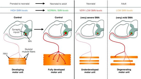
PRESENTATION OF SMA
The natural history of SMA includes presentation at any age, with acute onset of motor weakness and loss of
function. Since the approval of several SMN modulating therapies, a new natural history has begun to emerge.
In some cases, this includes the hallmark characteristic of proximal weakness, while other presentations
include subclinical strength limitations and/or development that appears entirely normal. Newborn screening9
or prenatal genetic testing may allow pre-symptomatic treatment; however, often treatment follows
symptomatic presentation. Those with more copies of SMN2 and earlier treatment have better motor
outcomes.10-12

In the untreated natural history, proximal             FIGURE 1: Survival Motor Neuron (SMN) Protein
extremity and trunk muscles are most                 may be crucial for normal development and postnatal
affected, with the diaphragm being relatively                  maintenance of the motor unit5
spared, even in the most severely affected
children. The initial period of acute progressive
weakness is followed by a prolonged plateau
during which muscle strength and function may
be stable. The degree of severity is related to
age of onset of weakness, which can range
from weakness at birth to adult onset.
Minipolymyoclonus, a fine tremor, is often seen
in the fingers and fasciculations and tremor are
often seen in the tongue. Cognitive function of
individuals with SMA is typically unaffected.
There is often a striking discrepancy between
alertness and cognitive ability and the ability to
                                                       From: D’Ydewall and Sumner, 2015, Neurotherapeutics©
move in the most severe forms of SMA.13

CLASSIFICATION OF SMA
Traditionally childhood onset forms of SMA are classified as one of 3 types with each defined by the maximum
motor skill attained (see FIGURE 2). However, with the advent of SMN modulating therapies, often current
motor function ability (non-sitter, sitter, and walker) is used as a classification.13
   • SMA type I: (Werdnig Hoffman Syndrome, acute SMA, infantile-onset SMA): Most severe type; typically
        presents prior to 6 months; children typically do not attain the ability to sit without assistance. Most
        common type of SMA, occurring in up to 60% of SMA births
   • SMA type II: (Intermediate SMA, juvenile SMA, chronic SMA) Intermediate form; typically presents
        between 6 and 18 months of age; children achieve the ability to sit and may stand with support at an
        early age, but never walk without braces or assistance
   • SMA type III: (Kugelberg-Welander Syndrome): Mildest form; typically presents after onset of
        ambulation and after 18 months of age. Children achieve the ability to walk without bracing or
        assistance at some point; however, loss of motor function (including ambulation) can occur
   • Some also describe a type IV (adult onset) and a type 0 with onset at birth

DIAGNOSIS
Newborn screening for SMA14
  • SMA was added to the federal Recommended Uniform Screening Panel (RUSP) for newborn screening
     in 2018. Screening for SMA is now available on Newborn Screening panels in 36 states, and 74% of
     infants are screened for the disorder in the United States.14
May 2021                                                                         APTA Pediatrics Fact Sheets |   2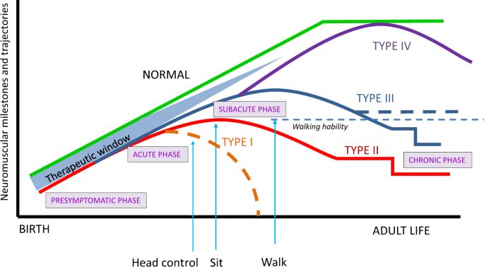
•   Newborn screening allows for early access to treatments which are known to have a greater effect at
       younger ages.15

Diagnostic testing findings:
   • Genetic testing (blood test): Homozygous mutation typically of exon 7 of the survival Motor Neuron 1
      (SMN1) gene on chromosome 5q13, 95% sensitivity with the remaining 5% having undetected
      uncommon mutations identifiable on follow up testing16
   • EMG: Diminished compound muscle action potential (CMAP), fibrillation potentials, normal nerve
      conduction velocity (NCV), diminished motor unit number estimation (MUNE), polyphasic potentials
   • Muscle ultrasound: fasciculations can be visualized within the muscle17
   • Muscle biopsy: Grouped atrophy (not required for diagnosis)

                 FIGURE 2: Neuromuscular Milestones from Birth to Adult Life in SMA

                                                                     From: Serra-Juhe C. & Tizziano e.f. (2019)

CHARACTERISTICS OF SMA
Motor:
Progressive weakness, leading to:
   • A paucity of overall movement in those who are non-sitters (Type I SMA)
   • Decreased active movement, with limited antigravity movement in those who are sitters (Type 2 SMA)
   • Decreased mobility
   • Typical motor development prior to symptom onset followed by motor delay
   • Early hypermobility with contracture development later in disease
   • Poor head control common in non-sitters and some weaker sitters
   • Muscle fatigue2
   • Areflexia or hyporeflexia
   • Axillary slip through
   • Fasciculation of muscles, most common in tongue
   • “Polyminimyoclonus” in the fingers
   • Difficulty swallowing and chewing

May 2021                                                                      APTA Pediatrics Fact Sheets |   3
Postural compensations:18
• Common resting posture: Excessive lower extremity abduction and external rotation with hip and knee
flexion, use of ‘stacking maneuvers’ to maintain head and trunk control, asymmetric weightbearing,
upper extremity pronation with ulnar drift
• Kyphotic sitting posture in those who sit, with scoliosis over time
• Gower’s maneuver in children with Type III SMA who transition from the floor to standing
For a more detailed list of biomechanical and compensatory behaviors please see the
Best Practices Toolkit for PTs and Clinical Evaluators in SMA
Cardiorespiratory:
• Restrictive lung disease and respiratory insufficiency which presents initially as nocturnal
hypoventilation
• Increased risk for cardiac involvement and in the most severe cases congenital heart disease
Cognitive and sensory:
• Cognitive and sensory systems are typically intact,5 though more recently it has become apparent that
cognitive delay may be a characteristic of some individuals with SMA type I.19
At risk for:
• Contractures, osteopenia, scoliosis, kyphosis, hip dislocation, falls and fractures
• Issues with nutrition and weight management
PROGNOSIS
Survival is dependent on severity, age of presentation, management using standard care guidelines, and
treatment with disease modifying therapy13
SMA type I (non-sitters):
• Survival is typically limited to 18 months with rare exceptions without SMN modulating therapies
• With mechanical ventilation (BiPAP, tracheostomy) and gastrostomy feeding this can be extended20
SMA type II (sitters):
• Variable survival without disease modifying therapy. This is dependent on respiratory compromise and
support provided
SMA type III (standers/walkers):
• Typical life expectancy
CHANGING PHENOTYPES WITH PHARMACOTHERAPEUTICS
Decision-making about which SMN modulating therapy is indicated takes place as a part of family-centered
specialized care between the child, their caregivers, and the medical team. As of 2020 there are 3 SMN
modulating therapy options with FDA approval for SMA (see FIGURE 3).
• Spinraza (Nusinersen) approved for all types of 5q SMA; intrathecal administration three times per year
after initial loading doses
• Zolgensma (AVXS101) approved for patientsFIGURE 3: FDA Approval Timeline of SMN Modulating Therapies
December 23, 2016 May 24, 2019 August 7, 2020
Spinraza/Nusinersen Zolgensma/Onasemnogene Evrysdi/Risdiplam
approved abepavovex-xioi approved approved
(Biogen) (Novartis) (Roche/Genentech)
TESTS AND MEASURES
Consideration of an individual and family’s goals, concerns, and needs, as well as SMA type and current
functional level (non-sitter, sitter walker) should guide the selection of outcome measures for those with SMA
across the various levels of the ICF (see FIGURE 4).18,21
Tests of body functions and structure:
• Manual Muscle Test • Goniometry
• Myometry • Pulmonary function tests
Tests of activity and participation:18,22
• Hammersmith Functional Motor Scale- • Egen Klassification-2
Expanded (HFMSE) • Motor-Function Measure (MFM)
• Modified Hammersmith Functional Motor Scale • The 6-minute-walk test (6MWT)
(HFMS) • PedsQL©/PedsQL Neuromuscular Module©
• Revised Hammersmith Scale (RHS) • Assessment of Caregiver Experience with
• The CHOP Infant Test of Neuromuscular Neuromuscular Disease (ACEND)
Disorders (CHOP-Intend) • Pediatric Inventory of Disability Evaluation
• The Test of Infant Motor Performance (PEDI/PEDI-CAT)
Screening Items (TIMPSI) • Fatigue Severity Scale (FSS)
• Hammersmith Infant Neurologic Examination • Modified SMA-Functional Rating Scale
(HINE) Motor Section 2 (Modified SMA-FRS)
• Revised Upper Limb Module (RULM) • Children’s Assessment of Participation and
• Timed tests of function (TFT) (time to walk/run Enjoyment and Preferences for Activities
10m, time to rise to standing from supine on (CAPE-PAC)23
the floor, time to climb 4 steps) • Bayley Scales of Infant Motor Development
• Activlim (III) ©/Bayley-4©
• Adult Test of Neuromuscular Disorders (ATEND) • Peabody Motor Scales-2©
FIGURE 4: Most Commonly Used Outcomes by SMA Functional Level/Phenotype
Non-sitter Sitter Walker Treated presymptomatically
CHOP INTEND/ATEND 6 min walk test (6MWT) PDMS-2
TIMPSI Hammersmith Expanded (HFMSE)/Revised Hammersmith (RHS)
Revised Upper Limb Module (RULM)
Motor Function Measure (MFM)
Egen Klassification-2 (EK2)
Timed Function Tests (TFT)
HINE (Section 2), WHO, BSID-III/Bayley-4, ACEND, FSS, Modified SMAFRS, PEDI-CAT, PedsQL, PROMIS, SMA-HI
May 2021 APTA Pediatrics Fact Sheets | 5PHYSICAL THERAPY INTERVENTIONS24
• Recommended intervention plans should follow SMA standard of care guidelines for stretching,
strengthening, aerobic exercise, standing, bracing, and balance21,20
• Maintain flexibility through range of motion/stretching, positioning, bracing, splinting, standing25
programs, and serial casting21
• Maintain strength and conserve energy through exercise and activity (this includes play, aquatics,
hippotherapy, and developmental exercises)26-32
• Foster good posture, with appropriate seating to optimize alignment33
• Promotion of weight-bearing to promote bone health13,21
• Foster and maintain movement and mobility to allow for independence, and environmental exploration
at younger ages as well as to foster overall health and wellness and participation in activity, adapted
sports, and recreation at all ages
• Educate patient and caregivers on the role of assistive technology to maximize activity and participation
including:
o Manual or power wheelchairs or scooters as early as 12 months to 2 years34
o Standers or long leg braces to initiate standing for patients with SMA who cannot stand on their
own25
o Adapted computer access or switch toys as appropriate
o Equipment for ADLs, including: environmental adaptations, bath equipment and mechanical lifts
• Educate patient and caregivers regarding handling, positioning, optimizing potential and safety and
preparing for changes during growth
REFERENCES
1. Kolb SJ, Kissel JT. Spinal Muscular Atrophy. Neurologic clinics. 2015;33(4):831-846.
2. Lefebvre S, Burglen L, Reboullet S, et al. Identification and characterization of a spinal muscular
atrophy-determining gene. Cell. 1995;80(1):155-165.
3. Sugarman EA, Nagan N, Zhu H, et al. Pan-ethnic carrier screening and prenatal diagnosis for spinal
muscular atrophy: clinical laboratory analysis of >72,400 specimens. Eur J Hum Genet. 2012;20(1):27-
32.
4. Gabanella F, Butchbach ME, Saieva L, Carissimi C, Burghes AH, Pellizzoni L. Ribonucleoprotein
assembly defects correlate with spinal muscular atrophy severity and preferentially affect a subset of
spliceosomal snRNPs. PLoS ONE. 2007;2(9):e921.
5. d'Ydewalle C, Sumner CJ. Spinal Muscular Atrophy Therapeutics: Where do we Stand?
Neurotherapeutics. 2015;12(2):303-316.
6. Berciano MT, Castillo-Iglesias MS, Val-Bernal JF, et al. Mislocalization of SMN from the I-band and M-
band in human skeletal myofibers in spinal muscular atrophy associates with primary structural
alterations of the sarcomere. Cell Tissue Res. 2020;381(3):461-478.
7. Lefebvre S, Burlet P, Liu Q, et al. Correlation between severity and SMN protein level in spinal
muscular atrophy. Nat Genet. 1997;16(3):265-269.
8. Calucho M, Bernal S, Alias L, et al. Correlation between SMA type and SMN2 copy number revisited:
An analysis of 625 unrelated Spanish patients and a compilation of 2834 reported cases. Neuromuscul
Disord. 2018;28(3):208-215.
9. Kellar-Guenther Y, McKasson S, Hale K, Singh S, Sontag MK, Ojodu J. Implementing Statewide
Newborn Screening for New Disorders: U.S. Program Experiences. Int J Neonatal Screen.
2020;6(2):35.
10. Finkel RS, Mercuri E, Darras BT, et al. Nusinersen versus Sham Control in Infantile-Onset Spinal
Muscular Atrophy. N Engl J Med. 2017;377(18):1723-1732.
May 2021 APTA Pediatrics Fact Sheets | 611. Baranello G, Darras BT, Day JW, et al. Risdiplam in Type 1 Spinal Muscular Atrophy. N Engl J Med.
2021;384(10):915-923.
12. Mendell JR, Al-Zaidy S, Shell R, et al. Single-Dose Gene-Replacement Therapy for Spinal Muscular
Atrophy. N Engl J Med. 2017;377(18):1713-1722.
13. Finkel RS, Mercuri E, Meyer OH, et al. Diagnosis and management of spinal muscular atrophy: Part 2:
Pulmonary and acute care; medications, supplements and immunizations; other organ systems; and
ethics. Neuromuscul Disord. 2018;28(3):197-207.
14. Newborn Screening for SMA. 2021. Retrieved from: https://www.curesma.org/newborn-screening-for-
sma/. Accessed May 8, 2021.
15. Dangouloff T, Servais L. Clinical Evidence Supporting Early Treatment Of Patients With Spinal
Muscular Atrophy: Current Perspectives. Ther Clin Risk Manag. 2019;15:1153-1161.
16. Prior TW, Bayrak-Toydemir P, Lynnes TC, et al. Characterization of Reference Materials for Spinal
Muscular Atrophy Genetic Testing: A Genetic Testing Reference Materials Coordination Program
Collaborative Project. J Mol Diagn. 2021;23(1):103-110.
17. Mah JK, van Alfen N. Neuromuscular Ultrasound: Clinical Applications and Diagnostic Values. Can J
Neurol Sci. 2018;45(6):605-619.
18. Krosschell KJ, Dunaway Young S, Cruz R, A. M, Curry M, Peterson I. Best Practices for Physical
Therapists and clinical Evaluators in Spinal Muscular Atrophy (SMA): Recommendations to Support
the effective Conduct of Clinical Trials in SMA. 2019. Retrieved from: https://www.curesma.org/wp-
content/uploads/2019/11/Cure-SMA-Best-Practices-for-PTs-and-CE-in-SMA-Clinical-Trials-Nov-
2019.pdf. Accessed June 29, 2020.
19. Polido GJ, de Miranda MMV, Carvas N, et al. Cognitive performance of children with spinal muscular
atrophy: A systematic review. Dement Neuropsychol. 2019;13(4):436-443.
20. Trenkle J, Brugman J, Peterson A, Roback K, Krosschell KJ. Filling the gaps in knowledge translation:
Physical therapy recommendations for individuals with spinal muscular atrophy compared to standard
of care guidelines. Neuromuscul Disord. 2021;10.1016/j.nmd.2021.02.011.
21. Mercuri E, Finkel RS, Muntoni F, et al. Diagnosis and management of spinal muscular atrophy: Part 1:
Recommendations for diagnosis, rehabilitation, orthopedic and nutritional care. Neuromuscul Disord.
2018;28(2):103-115.
22. Mercuri E, Mazzone E, Montes J, Messina S, Schroth MK. Spinal Muscular Atrophy Motor Functional
Scales and Measures of Pulmonary Function. In: Sumner CJ, Paushkin S, Ko C-P, eds. Spinal
Muscular Atrophy: Academic Press; 2017:371-382.
23. King G, Law M, King S, et al. Children’s Assessment of Participation and Enjoyment (CAPE) and
Preferences for Activities of Children (PAC). San Antonio, TX: Harcourt Assessment Inc; 2004.
24. Mercuri E, Finkel RS, Muntoni F, et al. Diagnosis and management of spinal muscular atrophy: Part 1:
Recommendations for diagnosis, rehabilitation, orthopedic and nutritional care. Neuromuscul Disord.
2017;10.1016/j.nmd.2017.11.005.
25. Townsend EL, Simeone SD, Krosschell KJ, Zhang RZ, Swoboda KJ, Project Cure SMAIsN. Stander
Use in Spinal Muscular Atrophy: Results From a Large Natural History Database. Pediatr Phys Ther.
2020;32(3):235-241.
26. Cunha MC, Oliveira AS, Labronici RH, Gabbai AA. Spinal muscular atrophy type II (intermediary) and
III (Kugelberg-Welander). Evolution of 50 patients with physiotherapy and hydrotherapy in a swimming
pool. Arq Neuropsiquiatr. 1996;54(3):402-406.
27. Lemke D, Rothwell E, Newcomb TM, Swoboda KJ. Perceptions of equine-assisted activities and
therapies by parents and children with spinal muscular atrophy. Pediatr Phys Ther. 2014;26(2):237-
244.
May 2021 APTA Pediatrics Fact Sheets | 728. Lewelt A, Krosschell KJ, Stoddard GJ, et al. Resistance strength training exercise in children with spinal
muscular atrophy. Muscle Nerve. 2015;52(4):559-567.
29. Bartels B, Montes J, van der Pol WL, de Groot JF. Physical exercise training for type 3 spinal muscular
atrophy. Cochrane Database Syst Rev. 2019;3:CD012120.
30. Madsen KL, Hansen RS, Preisler N, Thogersen F, Berthelsen MP, Vissing J. Training improves
oxidative capacity, but not function, in spinal muscular atrophy type III. Muscle Nerve. 2015;52(2):240-
244.
31. Montes J, Garber CE, Kramer SS, et al. Single-Blind, Randomized, Controlled Clinical Trial of Exercise
in Ambulatory Spinal Muscular Atrophy: Why are the Results Negative? J Neuromuscul Dis.
2015;2(4):463-470.
32. Salem Y, Gropack SJ. Aquatic therapy for a child with type III spinal muscular atrophy: a case report.
Phys Occup Ther Pediatr. 2010;30(4):313-324.
33. Peeters LHC, Janssen M, Kingma I, van Dieen JH, de Groot IJM. Patients With Spinal Muscular
Atrophy Use High Percentages of Trunk Muscle Capacity to Perform Seated Tasks. Am J Phys Med
Rehabil. 2019;98(12):1110-1117.
34. Dunaway S, Montes J, O'Hagen J, Sproule DM, Vivo DC, Kaufmann P. Independent mobility after early
introduction of a power wheelchair in spinal muscular atrophy. J Child Neurol. 2013;28(5):576-582.
HELPFUL WEBSITES
• Best Practices Toolkit for Physical Therapists and Clinical Evaluators in SMA. Available at:
https://www.curesma.org/clinical-trial-readiness-toolkits/
• Cure SMA. Available at: www.curesma.org
• SMA Foundation. Available at: www.smafoundation.org
• Muscular Dystrophy Association. Available at: www.mdausa.org
• SMA Outcomes. Available at: www.smaoutcomes.org
• SMA Common Data Elements (NINDS). Available at:
https://www.commondataelements.ninds.nih.gov/Spinal%20Muscular%20Atrophy
• STEP IN SMA: Teaching and Excellence for Physiotherapists. Available at: www.STEPINSMA.org
• TREAT NMD Spinal Muscular Atrophy Core Dataset. Available at: https://treat-nmd.org/patient-
registries/treat-nmd-core-datasets/sma-core-dataset/#1596626402330-3004fec9-c9a6
©2021 by the APTA Academy of Pediatric Physical Therapy, www.pediatricapta.org
Developed by expert contributors Kristin J Krosschell, PT, DPT, MA, Board-Certified Pediatric Clinical
Specialist & Allan Glanzman PT, DPT, Board-Certified Pediatric Clinical Specialist. Special thanks to
Shree Pandya, PT for her contribution to the previous version of this fact sheet. Supported by the Fact
Sheet Committee of APTA Pediatrics.
The APTA Academy of Pediatric Physical Therapy provides access to these member-produced fact
sheets and resources for informational purposes only. They are not intended to represent the position
of APTA Pediatrics or of the American Physical Therapy Association.
May 2021 APTA Pediatrics Fact Sheets | 8You can also read