Gestational Hypoxia and Blood-Brain Barrier Permeability: Early Origins of Cerebrovascular Dysfunction Induced by Epigenetic Mechanisms - Frontiers
←
→
Page content transcription
If your browser does not render page correctly, please read the page content below
MINI REVIEW
                                                                                                                                                published: 19 August 2021
                                                                                                                                          doi: 10.3389/fphys.2021.717550

                                                Gestational Hypoxia and Blood-Brain
                                                Barrier Permeability: Early Origins of
                                                Cerebrovascular Dysfunction
                                                Induced by Epigenetic Mechanisms
                                                Emilio A. Herrera 1 and Alejandro González-Candia 2*
                                                1
                                                 Laboratory of Vascular Function and Reactivity, Pathophysiology Program, ICBM, Faculty of Medicine, University of Chile,
                                                Santiago, Chile, 2 Institute of Health Sciences, University O’Higgins, Rancagua, Chile

                                                Fetal chronic hypoxia leads to intrauterine growth restriction (IUGR), which is likely to
                                                reduce oxygen delivery to the brain and induce long-term neurological impairments.
                                                These indicate a modulatory role for oxygen in cerebrovascular development. During
                                                intrauterine hypoxia, the fetal circulation suffers marked adaptations in the fetal
                                                cardiac output to maintain oxygen and nutrient delivery to vital organs, known as the
                                                “brain-sparing phenotype.” This is a well-characterized response; however, little is known
                                                about the postnatal course and outcomes of this fetal cerebrovascular adaptation. In
                                                addition, several neurodevelopmental disorders have their origins during gestation. Still,
                           Edited by:           few studies have focused on how intrauterine fetal hypoxia modulates the normal brain
                        Zhihong Yang,
   Université de Fribourg, Switzerland
                                                development of the blood-brain barrier (BBB) in the IUGR neonate. The BBB is a cellular
                        Reviewed by:
                                                structure formed by the neurovascular unit (NVU) and is organized by a monolayer of
                         Xiu-Fen Ming,          endothelial and mural cells. The BBB regulates the entry of plasma cells and molecules
   Université de Fribourg, Switzerland
                                                from the systemic circulation to the brain. A highly selective permeability system achieves
            Carmelina Daniela Anfuso,
            University of Catania, Italy        this through integral membrane proteins in brain endothelial cells. BBB breakdown and
                  *Correspondence:              dysfunction in cerebrovascular diseases lead to leakage of blood components into the
           Alejandro González-Candia            brain parenchyma, contributing to neurological deficits. The fetal brain circulation is
           alejandro.gonzalez@uoh.cl
                                                particularly susceptible in IUGR and is proposed to be one of the main pathological
                    Specialty section:          processes deriving BBB disruption. In the last decade, several epigenetic mechanisms
          This article was submitted to         activated by IU hypoxia have been proposed to regulate the postnatal BBB permeability.
                 Integrative Physiology,
                a section of the journal
                                                However, few mechanistic studies about this topic are available, and little evidence shows
                Frontiers in Physiology         controversy. Therefore, in this mini-review, we analyze the BBB permeability-associated
            Received: 31 May 2021               epigenetic mechanisms in the brain exposed to chronic intrauterine hypoxia.
            Accepted: 19 July 2021
          Published: 19 August 2021             Keywords: chronic intrauterine hypoxia, brain endothelial dysfunction, cerebral circulation, fetal growth
                                                restriction, BBB permeability
                             Citation:
   Herrera EA and González-Candia A
      (2021) Gestational Hypoxia and
Blood-Brain Barrier Permeability: Early
                                                INTRODUCTION
           Origins of Cerebrovascular
   Dysfunction Induced by Epigenetic            Fetal growth restriction (FGR) is a severe condition during pregnancy, where the fetus does not
                        Mechanisms.             grow according to its potential as a result of an adverse uterine environment (Kingdom and Smith,
           Front. Physiol. 12:717550.           2000). Placental insufficiency is the predominant cause of FGR, leading to chronic fetal hypoxemia
    doi: 10.3389/fphys.2021.717550              and intrauterine growth restriction (IUGR) (Kesavan and Devaskar, 2019). In addition, several

Frontiers in Physiology | www.frontiersin.org                                         1                                          August 2021 | Volume 12 | Article 717550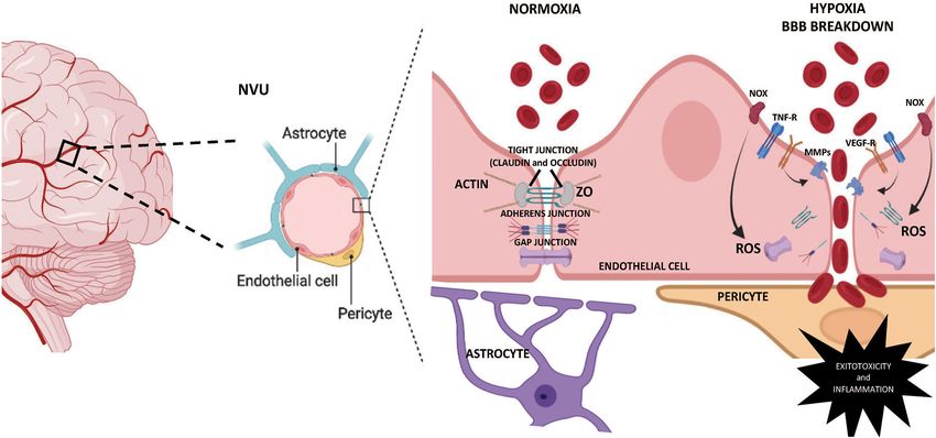
Herrera and González-Candia                                                                     Cerebrovascular Programming by Gestational Hypoxia

babies are exposed to chronic hypoxia and IUGR due to                     barrier (BCSFB), and (iii) the blood-brain barrier (Tietz and
pregnancy at a high altitude (Herrera et al., 2015). Intrauterine         Engelhardt, 2015). While the arachnoid barrier and the BCSFB
hypoxia induces an adaptive fetal redistribution of cardiac               have moderate permeability in the fetal, neonatal, and adult
output, favoring vital organs such as the brain, known as                 period, the BBB is the closest structure to the brain cells and
the brain sparing effect (Giussani, 2016). In this scenario, the          hence, is considered the most important barrier (Benz and
cerebral and heart circulations vasodilate, with a concomitant            Liebner, 2020). The BBB is formed by endothelial cells that
pronounced peripheral vasoconstriction (Villas-Bôas et al., 2008;         separate the capillary blood from the brain interstitial fluid and
Giussani, 2016). However, brain vasodilation does not ensure              parenchyma, limiting transcellular and paracellular transport
normal brain development in growth-restricted fetuses, and the            mechanisms through a differential expression of tight junctions
neurodevelopmental outcomes will depend on the timing of                  (TJ), adherens junctions (AJ), and possibly gap junctions (GJ) in
hypoxia, the severity of IUGR, and the gestational age at delivery        the inter-endothelial cleft (Figure 1). Also, the BBB comprises
(Padilla et al., 2011; Baschat, 2014). Studies in animal models           vascular smooth muscle cells, astrocytes, microglia, pericytes,
have demonstrated that gestational chronic hypoxia reduces                and oligodendrocytes. These cells contribute to the permeability
the neuronal number and vascular and synaptic numbers in                  and integrity of the BBB through their intimate anatomical
the hippocampus, impairing memory function in adult rats                  relationship (Liebner et al., 2018). However, the cross-talk
(Camm et al., 2021). In addition, IUGR may compromise cerebral            between each cell type is partially understood, and our knowledge
vascular homeostasis by increased excitotoxicity, oxidative stress,       of neonatal BBB development remains incomplete.
and neuroinflammation (Miller et al., 2016; Sweeney et al.,                  Every constituent cell of the NVU contributes to the
2019). In structural terms, IUGR is associated with reduced               BBBs integrity, and any dysfunction might result in the
brain and cortical volume, showing a reduced number of cells,             barrier breakdown, with dramatic consequences such as
and myelination shortages. These conditions are evidenced by              neuroinflammation and neurodegeneration (Kempuraj et al.,
less efficient networks with decreased long-range connections             2016; Sweeney et al., 2019). Although there is little or no evidence
(Miller et al., 2016). Even more, some authors have proposed              of the effects of IUGR on vascular permeability in human
an association of fetal hypoxia to later neurodegenerative and            neonates exposed to hypoxia, there is plenty of data obtained
neuropsychiatric disorders (Faa et al., 2014, 2016).                      from different animal models (Clancy et al., 2001, 2007; Kaur and
   However, the principal factor of cerebrovascular diseases is the       Ling, 2008; Disdier and Stonestreet, 2020). Thus, the neonatal
BBB breakdown, characterized by blood component infiltration,             NVU increases the BBB’s permeability by structural changes
aberrant transport, and clearance of molecules into the central           in the seal given by TJ, AJ, or GJ proteins. Brain endothelial
nervous system (CNS) (Yang and Rosenberg, 2011; Zhao et al.,              cells contain low fenestration and selective rates of transcytosis
2015). Structurally, the core of the BBB is a monolayer of brain          mainly due to the high expression of the TJ proteins. TJ are a
endothelial cells; nevertheless, these cells cannot form a barrier        combination of transmembrane proteins (claudins and occludin)
on their own (Gastfriend et al., 2018). Indeed, the development           and cytoplasmic adapter proteins called zonula occludens (ZO)
of integrity characteristics in the cerebrovascular tree requires         that interact with cytoskeleton filaments (Gonzalez-Candia
organized cells interactions from glial cells (i.e., astrocytes,          et al., 2021). Decreases in TJ proteins expression have been
microglia), pericytes, and neurons. Such a complex relationship           reported after hypoxic exposition in neonatal brains. Specifically,
implies the existence of a neurovascular unit (NVU) (Sweeney              hypoxia induces a decrease in claudin 5 and occludin protein
et al., 2019). The NVU represents a structural and functional             levels, which increases the paracellular diffusion of solutes and
multicellular interaction between cerebral parenchyma and brain           ions across the BBB (Andersson et al., 2021). Furthermore,
circulation (Iadecola, 2017), establishing a highly selective BBB         claudins decrease in the long term is associated with BBB
that favors cerebral homeostasis (Bell et al., 2020) (Figure 1).          breakdown and neurovascular disorders in humans (Tietz and
The complexity of this unit opens a wide and interesting field in         Engelhardt, 2015). BBB functions have mainly focused on TJs;
the search for understanding the multiple processes that mediate          however, cadherin/catenin interaction, as AJ proteins, regulate
cerebrovascular health.                                                   cell-cell adhesion between endothelial cells, contributing to
                                                                          the overall junction arrangement and BBB integrity (Li et al.,
                                                                          2018). Vascular endothelial (VE)-cadherin is responsible for
NEUROVASCULAR UNIT IN                                                     the assembly of AJ and is downregulated by BBB breakdown
INTRAUTERINE GROWTH RESTRICTION                                           signaling events (Daneman and Prat, 2015). For instance,
                                                                          under neuroinflammatory conditions, PI3Kα triggers TNFα
The NVU plays various roles within the brain. This unit is                signaling to cause VE-cadherin internalization, reducing the
responsible for the homeostasis and regulation of the cerebral            protein levels at junctions and impairing endothelial barrier
blood flow in response to neuronal activity changes, known as             function (Cain et al., 2010). Gestational or postnatal hypoxia
neurovascular coupling (NVC) (Iadecola, 2017; Hendrikx et al.,            can induce an unbalanced oxidative tone, as described elsewhere
2019). In addition, the same unit is in charge of protecting the          (Herrera et al., 2014; Villamor et al., 2019). The induction
CNS from harmful blood-borne and toxic substances (Blanchette             of the NADPH oxidase (NOX) system by proinflammatory
and Daneman, 2015; Keaney and Campbell, 2015). From a                     mediators can generate BBB permeability by downregulation of
structural view, three layers determine the barrier function in           proteins involved in intercellular junctions such as VE-cadherin,
CNS, (i) the arachnoid barrier, (ii) the blood-cerebrospinal fluid        occludin, and claudin-5 (Rochfort et al., 2014). Another family

Frontiers in Physiology | www.frontiersin.org                         2                                    August 2021 | Volume 12 | Article 717550
Herrera and González-Candia Cerebrovascular Programming by Gestational Hypoxia FIGURE 1 | Schematic representation of the BBB permeability mechanisms. The NVU is the main anatomical unit of the blood-brain barrier (BBB), sheltering the brain from systemic influences by limiting transcellular and paracellular transport. The Brain endothelial cells contain no fenestrae and undergo very low rates of transcytosis. Tight junctions, adherens junctions, and gap junctions formed between adjacent endothelial cells underlie the physical barrier that impedes paracellular diffusion of ions, macromolecules, and other solutes. Fetal chronic hypoxia may determine increased permeability in the BBB through pro-inflammatory and oxidative mechanisms, inducing degradation and damage in the membrane’s integral proteins, leading to BBB breakdown. MMPs, metalloproteinases; NOx, NADPH oxidase; NVU, neurovascular unit; ROS, reactive oxygen species; TNF-R, Tumor necrosis factor receptor; VEGF-R, vascular endothelial growth factor receptor; ZO, zonula occludens. of proteins involved in the permeability of the BBB is the GJ, members of the NVU, are likely to contribute to the permeability constituted by connexins. Connexin hemichannels have been of the BBB observed in cerebral hypoxia through downregulation implicated in the propagation of injury by hypoxia (Kim et al., of paracellular proteins such as Claudin-5 (CLDN5), occludin, 2017). Interestingly, neonatal hypoxia can negatively regulate the and ZO-1 (Obermeier et al., 2013). On the other hand, hypoxia- expression of connexin 43 (Davidson et al., 2013). In addition, induced vascular endothelial growth factor (VEGF) type 2 the blockade of connexin 43 decreased oligodendrocyte death receptor (VEGFR-2) pathway activation, increasing permeability and recovered oligodendrocyte maturation in preterm fetuses in the brain microvascular endothelium by decreasing junctional exposed to perinatal asphyxia (Davidson et al., 2014). proteins claudin-5, occludin, and ZO-1 (Castañeda-Cabral et al., Inflammatory mediators are critical for BBB disruption. 2020). Besides, in postnatal cerebral ischemia, VEGF may Microglia, neurons, astrocytes, and endothelial cells can release affect BBB damage by inducing metalloproteinases (MMP)-2 proinflammatory cytokines and chemokines, modulating expression, increasing the BBB permeability by brain endothelial adhesion molecules and transmigration of activated immune dysfunction (Shen et al., 2018). cells into the brain parenchyma (Jickling et al., 2015; Huang et al., Oxidative stress has a critical role in BBB breakdown in 2016). In endothelial cells, proinflammatory molecules regulate different neurological conditions (Olmec and Ozyurt, 2012). the expression of adhesion molecules such as intercellular Although the hypoxia generated in IUGR is sufficient to generate adhesion molecule 1 (ICAM-1) and vascular cell adhesion a redox imbalance (Myatt and Cui, 2004; Herrera et al., protein 1 (VCAM-1), physiologically expressed at low levels in 2014), direct evidence in human or animal models of BBB the BBB. However, their expression is increased in response to permeability is associated with IUGR remains to be elucidated. hypoxia, increasing the extravasation of molecules into the brain CNS contains several sources of ROS, such as NOX, uncoupling parenchyma (Kong et al., 2018). of the mitochondrial electron transport chain, xanthine oxidase In addition, the increase in cellular levels of TNF-α isoform, and uncoupled nitric oxide synthase (NOS) (Warner and IL-1β has been related to the decrease in occludin et al., 2004). The NOX family seems to be a principal source of expression and ZO-1 and 2 in the hypoxic brain (Rochfort oxidative stress in the hypoxic brain through the generation of and Cummins, 2015; Abdullah et al., 2018). This causes an superoxide (O2•-) radicals (Yang et al., 2019). The predominant increased paracellular permeability, modulation of transcytosis, isoform is the NOX2 in brain endothelial cells, and it has been and endocytotic transport mechanisms, leading to changes in observed that the Nox2- knockout mice induce less MMP- transcellular transport and inflammatory damage in the brain 9 and diminished expression of occludin, a critical protein parenchyma (Sweeney et al., 2019). Besides, reactive glial cells, of the BBB permeability (Liu et al., 2011). In addition, ROS Frontiers in Physiology | www.frontiersin.org 3 August 2021 | Volume 12 | Article 717550
Herrera and González-Candia                                                                      Cerebrovascular Programming by Gestational Hypoxia

generated by NOX can act as activators of MMPs (Li et al.,                 studies or adult models of cerebral ischemia. In models of
2018), thus enhancing their proteolytic degradation to the BBB.            cerebral hypoxia-ischemia, an increase of DNA methylation was
Among MMP family members, MMP-2 and 9 possess a substrate                  described as an increase in global DNA methylation in the
specificity for fibronectin, laminin, collagen fibers, and TJ, all         murine cerebral hemispheres, in the promoter of tissue inhibitors
of them structural components of the BBB. Interestingly, these             of MMP-2 (TIMP2). Increased MMP-2 and MMP-9 expression
proteins can be induced by hypoxia (Rosenberg and Yang, 2007).             and activity can affect BBB permeability by proteolysis of
                                                                           extracellular matrix and structural proteins in brain endothelial
                                                                           cells, increasing the BBB breakdown (Figure 2) (Yang et al.,
PERINATAL PROGRAMMING OF THE NVU:                                          2007; Wang et al., 2012). Late gestational maternal hypoxia in
POTENTIAL EPIGENETIC MECHANISMS                                            rats induce hypomethylation in the fetal brain by a mechanism
                                                                           dependent on HIF-1α expression (Li et al., 2016). This is relevant
Adverse environmental conditions during development, such                  as the HIF-related pathway is recognized as the primary sensor
as prenatal hypoxia, can increase the risk of diseases in                  and effector for hypoxic cellular adaptation in the fetus (Herrera
adulthood, such as vascular and parenchymal brain diseases                 et al., 2014). Hypomethylation induced by maternal hypoxia
(Berson et al., 2018). Basic and translational studies have                increased the vulnerability to subsequent postnatal hypoxia and
demonstrated that epigenetic programming of gene patterns in               worsened neurobehavioral outcomes in rat pups (Chen et al.,
response to gestational stress have a critical function in the             2008; Li et al., 2016). Interestingly, some authors have shown that
fetal origins of neurological cells dysfunction (Ducsay et al.,            HIF-1 expression levels and its transcriptional activity are under
2018). In particular, during gestational hypoxia, the epigenetic           strong epigenetic regulation (Nguyen et al., 2013; Ma et al., 2014)
programming of genes determines the functional outcome of                  and others that HIF-1 itself controls the expression of several
the genome (Ducsay et al., 2018). Epigenetics as heritable                 epigenetic regulators (Bustelo et al., 2020). The role of HIF in BBB
patterns in gene expression which are not associated with                  functional programming during fetal hypoxia is still unknown
DNA sequence alteration (Smith et al., 2016). The epigenetic               and needs further study.
mechanisms include methylation and/or demethylation of DNA,                    Histone modification by acetylation and deacetylation plays a
post-translational modifications of histones, and non-coding               central role in chromatin remodeling and epigenetic regulation.
RNAs such as microRNAs (Casanello et al., 2016; Ducsay                     In particular, histone deacetylases (HDAC) are potential
et al., 2018; Zeng and Chen, 2018). Epigenetic events respond              therapeutic targets in different neurological conditions (Gräff
to endogenous and exogenous signals, having central roles in               and Tsai, 2013). In a recent study, the treatment with
regulating appropriate sets of gene expression (Zeng and Chen,             a HDAC inhibitor in mice subjected to cerebral ischemia
2018). Epigenetic modifications serve as remembrance in early              leads to an enhanced expression of the TJ proteins ZO-1,
life stages, that can induce long-term changes in gene expression,         Occludin, and Claudin-5 in brain endothelial cells, further
which may induce disease in later postnatal life (Ducsay et al.,           decreasing the BBB permeability (Su et al., 2020). Conversely,
2018).                                                                     hypoxia and glucose deprivation in the brain promotes
    Hypoxic stress activates multiple epigenetic mechanisms                HDAC9 expression in endothelial cells, which has been
in the fetal brain that increase the vulnerability for                     associated to decreased expression of ZO-1, claudin-5, and
neurodevelopment disturbances in adult offspring (Ma et al.,               occludin (Shi et al., 2016). These findings demonstrate the
2014; Faa et al., 2016), such as increased vulnerability to ischemic       effect of hypoxia on the post-translational modifications
or hypoxic insults (Li et al., 2012; Gonzalez-Rodriguez et al.,            of histones in the regulation of proteins involved in the
2014), disruption of the normal endocrine axis (Wood et al.,               maintenance of the BBB structure and that these mechanisms
2014), and increased risks for adult cardiovascular disease                may be determining the dysfunction of the BBB induced by
(Ducsay et al., 2018). The mechanisms underlying the effects of            the hypoxia.
chronic fetal hypoxia and IUGR on epigenetic programming of                    Another mechanism of epigenetic regulation is mediated by
the fetal brain endothelial cells or NVU has not been studied.             microRNAs, which cause the degradation of genes involved in
However, the effects induced by hypoxia and oxidative stress in            the development and progression of BBB dysfunction (Figure 2)
the fetal brain suggest the involvement of epigenetic mechanisms           (Ma et al., 2020). Currently, there are no data relating to
(Camm et al., 2021).                                                       IUGR and microRNA regulating BBB structure and function;
    DNA methylation regulates the accessibility of DNA to the              however, evidence in adult pathophysiology may give some
transcription machinery modifying the chromatin state. This                clues about microRNAs and BBB disruption. Hypoxic-ischemic
DNA methylation is generated by a group of enzymes known                   models in adult animals have shown that microRNAs can
as DNA methyltransferases (DNMTs) (Ducsay et al., 2018);                   directly or indirectly degrade BBB proteins. In this sense, it has
composed by three principal isoforms: maintenance DNMTs                    been reported that miR-132 is negatively regulated by hypoxia,
(DNMT1) and de novo DNMTs (DNMT3a and DNMT3b)                              which increases MMP-9 activity, which degrades TJ proteins
(Moore et al., 2013). However, no mechanisms have been                     in brain endothelial cells or extracellular matrix components
proposed to demonstrate the DNA methylation events during                  in the NVU, favoring an increased permeability of the BBB
gestational hypoxia, that may regulate the expression of BBB               (Cichon et al., 2014). There are significant correlations between
structural proteins and permeability. In this sense, the effects           microRNAs and TJs by hypoxia in adult models (Toyama
of hypoxia and IUGR can only be extrapolated in neonatal                   et al., 2017). For instance, miR-125-5p has a critical role in

Frontiers in Physiology | www.frontiersin.org                          4                                    August 2021 | Volume 12 | Article 717550
Herrera and González-Candia Cerebrovascular Programming by Gestational Hypoxia
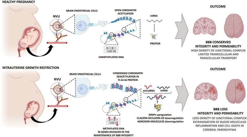
FIGURE 2 | Potential epigenetic mechanisms determining impairment of the BBB permeability by gestational hypoxia. Hypoxic and IUGR injury causes a series of
molecular events, including programming of BBB through genomic DNA methylation signature and an altered expression of microRNAs. These mechanisms
contribute to changes in the expression of molecules related to junctional complexes of the BBB, increasing BBB permeability and further brain damage. AJ, adherens
junction; BBB, blood-brain barrier; GJ, gap junction; MMPs: metalloproteinases; NVU, neurovascular unit; TJ, tight junction; ZO, zonula occludens.
the brain endothelial tightness during an inflammatory response placental and vascular function (Gheorghe et al., 2010;
(Toyama et al., 2017). Part of this response involves specific Herrera et al., 2014; Fajersztajn and Veras, 2017; Soares
mRNA targets of miR-125-5p by down-regulating Claudin- et al., 2017). However, advances in understanding how
1 and Claudin-5, and disrupting adhesion molecules in BBB gestational hypoxia induces variations in the expression of
(Toyama et al., 2017). Furthermore, cerebral endothelial miR- proteins involved in the integrity of the cerebrovascular network
144 downregulates claudin-5, Claudin-12, occludin, and ZO-1, remain widely unexplored. BBB permeability is a major factor
ZO-2, and ZO-3 in a model of BBB permeability associated determining the cause, progression, outcome, and therapeutic
with a blood-tumor barrier (Cai et al., 2017). Cerebral ischemia effectiveness of different neurological impairments in postnatal
triggers an enhanced expression of miR126, which is considered life. Therefore, fetal programming of BBB permeability
endothelial-specific. miR126 is one of the most studied by hypoxia and IUGR pose a unique challenge to the
microRNAs that regulates vascular inflammation. miR126 scientific community in searching for involved mechanisms
downregulates the expression of the ICAM-1 and VCAM- and effective clinical treatment to prevent detrimental
1 molecules and controls inflammatory cells extravasation postnatal outcomes.
into the brain in BBB dysfunction models (Stamatovic et al.,
2016). These data suggest that epigenetic mechanisms define
and regulate the vascular responses to pathological stimuli AUTHOR CONTRIBUTIONS
such as chronic hypoxia (Figure 2). However, evidence from
AG-C and EH drafted and edited the manuscript. Both
fetal exposure to hypoxia leading to epigenetic modifications
authors contributed to the article and approved the
remains elusive.
submitted version.
CONCLUSION
FUNDING
Chronic deprivation of oxygen during gestation dramatically
impacts fetal brain development. Gestational hypoxia can This work was funded by Fondecyt de Inicio grant No 11200798
act through an altered epigenetic fashion to compromise and Fondecyt Regular grant No 1201283.
Frontiers in Physiology | www.frontiersin.org 5 August 2021 | Volume 12 | Article 717550Herrera and González-Candia Cerebrovascular Programming by Gestational Hypoxia
REFERENCES Daneman, R., and Prat, A. (2015). The blood-brain barrier. Cold Spring Harb.
Perspect. Biol. 7:a020412. doi: 10.1101/cshperspect.a020412
Abdullah, W., Tripathi, D., and Ronaldson, P. T. (2018). Blood-brain barrier Davidson, J. O., Drury, P. P., Green, C. R., Nicholson, L. F., Bennet, L., and Gunn,
dysfunction in ischemic stroke: targeting tight junctions and transporters A. J. (2014). Connexin hemichannel blockade is neuroprotective after asphyxia
for vascular protection. Am. J. Physiol. Cell Physiol. 315, C343–C356. in preterm fetal sheep. PLoS ONE 9:e96558. doi: 10.1371/journal.pone.0096558
doi: 10.1152/ajpcell.00095.2018 Davidson, J. O., Green, C. R., Nicholson, L. F., Bennet, L., and Gunn, A. J. (2013).
Andersson, E. A., Mallard, C., and Ek, C. J. (2021). Circulating tight-junction Connexin hemichannel blockade is neuroprotective after, but not during,
proteins are potential biomarkers for blood-brain barrier function in a global cerebral ischemia in near-term fetal sheep. Exp. Neurol. 248, 301–308.
model of neonatal hypoxic/ischemic brain injury. Fluids Barriers CNS 18:7. doi: 10.1016/j.expneurol.2013.06.026
doi: 10.1186/s12987-021-00240-9 Disdier, C., and Stonestreet, B. S. (2020). Hypoxic-ischemicrelated cerebrovascular
Baschat, A. A. (2014). Neurodevelopment after fetal growth restriction. Fetal Diagn changes and potential therapeutic strategies in the neonatal brain. J Neurosci
Ther. 36, 136–142. doi: 10.1159/000353631 Res. 98, 1468–1484. doi: 10.1002/jnr.24590
Bell, A. H., Miller, S. L., Castillo-Melendez, M., and Malhotra, A. (2020). The Ducsay, C. A., Goyal, R., Pearce, W. J., Wilson, S., Hu, X. Q., and Zhang, L. (2018).
neurovascular unit: effects of brain insults during the perinatal period. Front. Gestational hypoxia and developmental plasticity. Physiol. Rev. 98, 1241–1334.
Neurosci. 13:1452. doi: 10.3389/fnins.2019.01452 doi: 10.1152/physrev.00043.2017
Benz, F., and Liebner S. (2020). “Structure and function of the blood-brain Faa, G., Manchia, M., Pintus, R., Gerosa, C., Marcialis, M. A., and Fanos, V. (2016).
barrier (bbb),” in Handbook of Experimental Pharmacology (Berlin; Heidelberg: Fetal programming of neuropsychiatric disorders. Birth Defects Res. C Embryo
Springer). doi: 10.1007/164_2020_404 Today. 108, 207–223. doi: 10.1002/bdrc.21139
Berson, A., Nativio, R., Berger, S. L., and Bonini, N. M. Faa, G., Marcialis, M. A., Ravarino, A., Piras, M., Pintus, M. C., and Fanos,
(2018). Epigenetic regulation in neurodegenerative diseases. V. (2014). Fetal programming of the human brain: is there a link with
Trends Neurosci. 41, 587–598. doi: 10.1016/j.tins.2018. insurgence of neurodegenerative disorders in adulthood?. Curr. Med. Chem.
05.005 21, 3854–3876. doi: 10.2174/0929867321666140601163658
Blanchette, M., and Daneman, R. (2015). Formation and maintenance Fajersztajn, L., and Veras, M. M. (2017). Hypoxia: from placental development to
of the BBB. Mech. Dev. 138, 8–16. doi: 10.1016/j.mod.2015. fetal programming. Birth Defects Res. 109, 1377–1385. doi: 10.1002/bdr2.1142
07.007 Gastfriend, B. D., Palecek, S. P., and Shusta, E. V. (2018). Modelling the blood-
Bustelo, M., Barkhuizen, M., van den Hove, D. L. A., Steinbusch, brain barrier: beyond the endothelial cells. Curr. Opin. Biomed. Eng. 5, 6–12.
H. W. M., Bruno, M. A., Loidl, C. F., et al. (2020). Clinical doi: 10.1016/j.cobme.2017.11.002
implications of epigenetic dysregulation in perinatal hypoxic- Gheorghe, C. P., Goyal, R., Mittal, A., and Longo, L. D. (2010). Gene expression
ischemic brain damage. Front. Neurol. 11:483. doi: 10.3389/fneur.2020. in the placenta: maternal stress and epigenetic responses. Int. J. Dev. Biol. 54,
00483 507–523. doi: 10.1387/ijdb.082770cg
Cai, W., Zhang, K., Li, P., Zhu, L., Xu, J., Yang, B., et al. (2017). Dysfunction Giussani, D. A. (2016). The fetal brain sparing response to hypoxia: physiological
of the neurovascular unit in ischemic stroke and neurodegenerative mechanisms. J. Physiol. 594, 1215–1230. doi: 10.1113/JP271099
diseases: an aging effect. Ageing Res. Rev. 34:77–87. doi: 10.1016/j.arr.2016. Gonzalez-Candia, A., Rogers, N. K., and Castillo, R. L. (2021). “Blood-brain
09.006 barrier dysfunction in the detrimental brain function,” in Connectivity and
Cain, R. J., Vanhaesebroeck, B., and Ridley, A. J. (2010). The PI3K Functional Specialization in the Brain, ed T. Heinbockel (London: Intechope).
p110alpha isoform regulates endothelial adherens junctions via doi: 10.5772/intechopen.94572
Pyk2 and Rac1. J. Cell Biol. 188, 863–876. doi: 10.1083/jcb.2009 Gonzalez-Rodriguez, P. J., Xiong, F., Li, Y., Zhou, J., and Zhang,
07135 L. (2014). Fetal hypoxia increases vulnerability of hypoxic-
Camm, E. J., Cross, C. M., Kane, A. D., Tarry-Adkins, J. L., Ozanne, S. ischemic brain injury in neonatal rats: role of glucocorticoid
E., and Giussani, D. A. (2021). Maternal antioxidant treatment protects receptors. Neurobiol. Dis. 65, 172–179. doi: 10.1016/j.nbd.2014.
adult offspring against memory loss and hippocampal atrophy in a rodent 01.020
model of developmental hypoxia. FASEB J. 35:e21477. doi: 10.1096/fj.20200 Gräff, J., and Tsai, L. H. (2013). The potential of HDAC inhibitors as
2557RR cognitive enhancers. Annu. Rev. Pharmacol. Toxicol. 53, 311–330.
Casanello, P., Herrera, E. A., and Krause, B. J. (2016). Epigenetic programming doi: 10.1146/annurev-pharmtox-011112-140216
of cardiovascular disease by perinatal hypoxia and fetal growth restriction. Hendrikx, D., Smits, A., Lavanga, M., De Wel, O., Thewissen, L., Jansen, K., et al.
Hypoxia Human Dis. 17, 330–347. doi: 10.5772/66740 (2019). Measurement of neurovascular coupling in neonates. Front. Physiol.
Castañeda-Cabral, J. L., Colunga-Durán, A., Ureña-Guerrero, M. E., Beas- 10:65. doi: 10.3389/fphys.2019.00065
Zárate, C., Nuñez-Lumbreras, M. L. A., Orozco-Suárez, S., et al. Herrera, E.A., Krause, B., Ebensperger, G., Reyes, R.V., Casanello, P., Parra-
(2020). Expression of VEGF- and tight junction-related proteins in the Cordero, M., et al. (2014). The placental pursuit for an adequate oxidant
neocortical microvasculature of patients with drug-resistant temporal balance between the mother and the fetus. Front. Pharmacol. 24:149.
lobe epilepsy. Microvasc. Res. 132:104059. doi: 10.1016/j.mvr.2020. doi: 10.3389/fphar.2014.00149
104059 Herrera, E. A., Farías, J. G., Ebensperger, G., Reyes, R. V., Llanos,
Chen, W., Jadhav, V., Tang, J., and Zhang, J. H. (2008). HIF-1 alpha A. J., and Castillo, R. L. (2015). Pharmacological approaches
inhibition ameliorates neonatal brain damage after hypoxic-ischemic in either intermittent or permanent hypoxia: a tale of two
injury. Acta Neurochir. Suppl. 102, 395–399. doi: 10.1007/978-3-211- exposures. Pharmacol Res. 101, 94–101. doi: 10.1016/j.phrs.2015.
85578-2_77 07.011
Cichon, C., Sabharwal, H., Rüter, C., and Schmidt, M. A. (2014). MicroRNAs Huang, T., Gao, D., Hei, Y., Zhang, X., Chen, X., and Fei, Z. (2016).
regulate tight junction proteins and modulate epithelial/endothelial D-allose protects the blood brain barrier through PPARγ-mediated
barrier functions. Tissue Barriers 2:e944446. doi: 10.4161/21688362.2014. anti-inflammatory pathway in the mice model of ischemia reperfusion
944446 injury. Brain Res. 1642, 478–486. doi: 10.1016/j.brainres.2016.
Clancy, B., Darlington, R. B., and Finlay, B. L. (2001). 04.038
Translating developmental time across mammalian species. Iadecola, C. (2017). The neurovascular unit coming of age:a journey
Neuroscience 105, 7–17. doi: 10.1016/S0306-4522(01) through neurovascular coupling in health and disease. Neuron 96, 17–42.
00171-3 doi: 10.1016/j.neuron.2017.07.030
Clancy, B., Finlay, B. L., Darlington, R. B., and Arland, K. J. S. (2007). Extrapolating Jickling, G. C., Liu, D., Ander, B. P., Stamova, B., Zhan, X.,
brain development from experimental species to humans. Neurotoxicology 28, and Sharp, F. R. (2015). Targeting neutrophils in ischemic
931–937. doi: 10.1016/j.neuro.2007.01.014 stroke: translational insights from experimental studies. J.
Frontiers in Physiology | www.frontiersin.org 6 August 2021 | Volume 12 | Article 717550Herrera and González-Candia Cerebrovascular Programming by Gestational Hypoxia
Cereb. Blood Flow Metab. 35, 888–901. doi: 10.1038/jcbfm. Padilla, N., Falcon, C., Sanz-Cortes, M., Figueras, F., Bargallo, N., Crispi, F., et al.
2015.45 (2011). Differential effects of intrauterine growth restriction on brain structure
Kaur, C., and Ling, E. A. (2008). Blood brain barrier in hypoxic-ischemic and development in preterm infants: amagnetic resonance imaging study. Brain
conditions. Curr. Neurovas. Res. 5, 71–81. doi: 10.2174/156720208783565645 Res. 1382, 98–108. doi: 10.1016/j.brainres.2011.01.032
Keaney, J., and Campbell, M. (2015). The dynamic blood- Rochfort, K. D., Collins, L. E., Murphy, R. P., and Cummins, P. M. (2014).
brain barrier. FEBS J. 282, 4067–4079. doi: 10.1111/febs. Downregulation of blood-brain barrier phenotype by proinflammatory
13412 cytokines involves NADPH oxidase-dependent ROS generation: consequences
Kempuraj, D., Thangavel, R., Natteru, P. A., Selvakumar, G. P., Saeed, D., Zahoor, for interendothelial adherens and tight junctions. PLoS ONE 9:e101815.
H., et al. (2016). Neuroinflammation induces neurodegeneration. J. Neurol. doi: 10.1371/journal.pone.0101815
Neurosurg. Spine 1:1003. Rochfort, K. D., and Cummins, P. M. (2015). Cytokine-mediated dysregulation
Kesavan, K., and Devaskar, S. U. (2019). Intrauterine growth of zonula occludens-1 properties in human brain microvascular
restriction: postnatal monitoring and outcomes. Pediatr. endothelium. Microvasc Res. 100, 48–53. doi: 10.1016/j.mvr.2015.
Clin. North Am. 66, 403–423. doi: 10.1016/j.pcl.2018. 04.010
12.009 Rosenberg, G. A., and Yang, Y. (2007). Vasogenic edema due to tight junction
Kim, Y., Davidson, J. O., Green, C. R., Nicholson, L. F. B., O’Carroll, S. J., and disruption by matrix metalloproteinases in cerebral ischemia. Neurosurg. Focus
Zhang, J. (2017). Connexins and Pannexins in cerebral ischemia. Biochim. 22:E4. doi: 10.3171/foc.2007.22.5.5
Biophys. Acta Biomembr. 1860, 224–236. doi: 10.1016/j.bbamem.2017.03.018 Shen, Y., Gu, J., Liu, Z., Xu, C., Qian, S., Zhang, X., et al. (2018). Inhibition of HIF-
Kingdom, J., and Smith, G. (2000). “Diagnosis and management of IUGR,” in 1α reduced blood brain barrier damage by regulating MMP-2 and VEGF during
Intrauterine Growth Restriction A Etiology and Management, eds J. Kingdom acute cerebral ischemia. Front. Cell. Neurosci. 12:288. doi: 10.3389/fncel.2018.
and P. Baker (London: v), 257–273. doi: 10.1007/978-1-4471-0735-4_13 00288
Kong, D. H., Kim, H., Kim, M. R., Jang, J. H., and Lee, S. (2018). Emerging roles Shi, W., Wei, X., Wang, Z., Han, H., Fu, Y., Liu, J., et al. (2016). HDAC9 exacerbates
of Vascular Cell Adhesion Molecule-1 (VCAM-1) in immunological disorders endothelial injury in cerebral ischaemia/reperfusion injury. J. Cell. Mol. Med.
and cancer. Int. J. Mol. Sci. 19:1057. doi: 10.3390/ijms19041057 20, 1139–1149. doi: 10.1111/jcmm.12803
Li, W., Chen, Z., Chin, I., Chen, Z., and Dai, H. (2018). Smith, Z. D., Sindhu, C., and Meissner, A. (2016). Molecular features of cellular
The role of VE-cadherin in blood-brain barrier integrity reprogramming and development. Nat. Rev. Mol. Cell Biol. 17, 139–154.
under central nervous system pathological conditions. Curr. doi: 10.1038/nrm.2016.6
Neuropharmacol. 16, 1375–1384. doi: 10.2174/1570159X166661802 Soares, M. J., Iqbal, K., and Kozai, K. (2017). Hypoxia and placental development.
22164809 Birth Defects Res. 109, 1309–1329. doi: 10.1002/bdr2.1135
Li, Y., Gonzalez, P., and Zhang, L. (2012). Fetal stress and programming Stamatovic, S. M., Johnson, A. M., Keep, R. F., and Andjelkovic, A. V. (2016).
of hypoxic/ischemic-sensitive phenotype in the neonatal brain: Junctional proteins of the blood-brain barrier: new insights into function
mechanisms and possible interventions. Prog Neurobiol. 98, 145–165. and dysfunction. Tissue Barriers 4:e1154641. doi: 10.1080/21688370.2016.
doi: 10.1016/j.pneurobio.2012.05.010 1154641
Li, Y., Ma, Q., Halavi, S., Concepcion, K., Hartman, R. E., Obenaus, A., Su, L., Liang, D., Kuang, S. Y., Dong, Q., Han, X., and Wang, Z.
et al. (2016). Fetal stress-mediated hypomethylation increases the brain (2020). Neuroprotective mechanism of TMP269, a selective class
susceptibility to hypoxic–ischemic injury in neonatal rats. Exp. Neurol. 275, IIA histone deacetylase inhibitor, after cerebral ischemia/reperfusion
1–10. doi: 10.1016/j.expneurol.2015.10.007 injury. Neural. Regen. Res. 15, 277–284. doi: 10.4103/1673-5374.
Liebner, S., Dijkhuizen, R. M., Reiss, Y., Plate, K. H., Agalliu, D., and 265562
Constantin, G. (2018). Functional morphology of the blood-brain barrier in Sweeney, M. D., Zhao, Z., Montagne, A., Nelson, A. R., and Zlokovic,
health and disease. Acta Neuropathol. 135: 311–336. doi: 10.1007/s00401-018- B. V. (2019). Blood-brain barrier: from physiology to disease
1815-1 and back. Physiol. Rev. 99, 21–78. doi: 10.1152/physrev.00050.
Liu, W., Chen, Q., Liu, J., and Liu, K. J. (2011). Normobaric hyperoxia protects 2017
the blood brain barrier through inhibiting Nox2 containing NADPH oxidase Tietz, S., and Engelhardt, B. (2015). Brain barriers: crosstalk between
in ischemic stroke. Med. Gas Res. 1:22. doi: 10.1186/2045-9912-1-22 complex tight junctions and adherens junctions. J. Cell Biol. 209, 493–506.
Ma, F., Zhang, X., and Yin, K. J. (2020). MicroRNAs in central nervous doi: 10.1083/jcb.201412147
system diseases: a prospective role in regulating blood-brain barrier Toyama, K., Spin, J. M., and Tsao, P. S. (2017). Role of microRNAs on blood brain
integrity. Exp. Neurol. 323:113094. doi: 10.1016/j.expneurol.2019. barrier dysfunction in vascular cognitive impairment. Curr. Drug Deliv. 14,
113094 744–757. doi: 10.2174/1567201813666160830124627
Ma, Q., Xiong, F., and Zhang, L. (2014). Gestational hypoxia and epigenetic Villamor, E., Moreno, L., Mohammed, R., Pérez-Vizcaíno, F., and Cogolludo,
programming of brain development disorders. Drug Discov. Today 19, A. (2019). Reactive oxygen species as mediators of oxygen signaling during
1883–1896. doi: 10.1016/j.drudis.2014.09.010 fetal-to-neonatal circulatory transition. Free Radic. Biol. Med. 142, 82–96.
Miller, S. L., Huppi, P. S., and Mallard, C. (2016). The consequences of fetal growth doi: 10.1016/j.freeradbiomed.2019.04.008
restriction on brain structure and neurodevelopmental outcome. J. Physiol. 594, Villas-Bôas, J. M., Maestá, I., and Consonni, M. (2008). Mecanismo de
807–823. doi: 10.1113/JP271402 centralização: da insuficiência placentária à adaptação circulatória fetal [Brain
Moore, L. D., Le, T., and Fan, G. (2013). DNA methylation and its basic function. sparing effect: from placental insufficiency to fetal circulatory adaptation]. Rev.
Neuropsychopharmacology 38, 23–38. doi: 10.1038/npp.2012.112 Bras Ginecol. Obstet. 30, 366–371. doi: 10.1590/S0100-72032008000700008
Myatt, L., and Cui, X. (2004). Oxidative stress in the placenta. Histochem. Cell Biol. Wang, Z., Meng, C. J., Shen, X. M., Shu, Z., Ma, C., Zhu, G. Q., et al.
122, 369–382. doi: 10.1007/s00418-004-0677-x (2012). Potential contribution of hypoxia-inducible factor-1α, aquaporin-4,
Nguyen, M. P., Lee, S., and Lee, Y. M. (2013). Epigenetic regulation of hypoxia and matrix metalloproteinase-9 to blood-brain barrier disruption and brain
inducible factor in diseases and therapeutics. Arch. Pharm. Res. 36, 252–263. edema after experimental subarachnoid hemorrhage. J. Mol. Neurosci. 48,
doi: 10.1007/s12272-013-0058-x 273–280. doi: 10.1007/s12031-012-9769-6
Obermeier, B., Daneman, R., and Ransohoff, R. M. (2013). Development, Warner, D. S., Sheng, H., and Batinić-Haberle, I. (2004). Oxidants, antioxidants
maintenance and disruption of the blood-brain barrier. Nat. Med. 19, and the ischemic brain. J. Exp. Biol. 207, 3221–3231. doi: 10.1242/jeb.01022
1584–1596. doi: 10.1038/nm.3407 Wood, C. E., Rabaglino, M. B., Richards, E., Denslow, N., Zarate, M. A., Chang,
Olmec, I., and Ozyurt, H. (2012). Reactive oxygen species and E. I., et al. (2014). Transcriptomics of the fetal hypothalamic response to
ischemic cerebrovascular disease. Neurochem. Int. 60, 208–212. brachiocephalic occlusion and estradiol treatment. Physiol. Genomics. 46,
doi: 10.1016/j.neuint.2011.11.009 523–532. doi: 10.1152/physiolgenomics.00186.2013
Frontiers in Physiology | www.frontiersin.org 7 August 2021 | Volume 12 | Article 717550Herrera and González-Candia Cerebrovascular Programming by Gestational Hypoxia
Yang, C., Hawkins, K. E., Doré, S., and Candelario-Jalil, E. (2019). Conflict of Interest: The authors declare that the research was conducted in the
Neuroinflammatory mechanisms of blood-brain barrier damage absence of any commercial or financial relationships that could be construed as a
in ischemic stroke. Am. J. Physiol. Cell Physiol. 316, C135–C153. potential conflict of interest.
doi: 10.1152/ajpcell.00136.2018
Yang, Y., Estrada, E. Y., Thompson, J. F., Liu, W., and Rosenberg, G. A. Publisher’s Note: All claims expressed in this article are solely those of the authors
(2007). Matrix metalloproteinase- mediated disruption of tight junction and do not necessarily represent those of their affiliated organizations, or those of
proteins in cerebral vessels is reversed by synthetic matrix metalloproteinase the publisher, the editors and the reviewers. Any product that may be evaluated in
inhibitor in focal ischemia in rat. J. Cereb. Blood Flow Metab. 27, 697–709.
this article, or claim that may be made by its manufacturer, is not guaranteed or
doi: 10.1038/sj.jcbfm.9600375
endorsed by the publisher.
Yang, Y., and Rosenberg, G. A. (2011). Blood-brain barrier breakdown
in acute and chronic cerebrovascular disease. Stroke 42, 3323–3328.
doi: 10.1161/STROKEAHA.110.608257 Copyright © 2021 Herrera and González-Candia. This is an open-access article
Zeng, Y., and Chen, T. (2018). DNA methylation reprogramming during distributed under the terms of the Creative Commons Attribution License (CC BY).
mammalian development. Genes 10:257. doi: 10.3390/genes10040257 The use, distribution or reproduction in other forums is permitted, provided the
Zhao, Z., Nelson, A. R., Betsholtz, C., and Zlokovic, B. original author(s) and the copyright owner(s) are credited and that the original
V. (2015). Establishment and dysfunction of the blood- publication in this journal is cited, in accordance with accepted academic practice.
brain barrier. Cell 163, 1064–1078. doi: 10.1016/j.cell.2015.1 No use, distribution or reproduction is permitted which does not comply with these
0.067 terms.
Frontiers in Physiology | www.frontiersin.org 8 August 2021 | Volume 12 | Article 717550You can also read