Polarization-Sensitive Second Harmonic Generation Microscopy for Investigations of Diseased Collagenous Tissues
←
→
Page content transcription
If your browser does not render page correctly, please read the page content below
MINI REVIEW
                                                                                                                                      published: 30 August 2021
                                                                                                                                 doi: 10.3389/fphy.2021.726996

                                             Polarization-Sensitive Second
                                             Harmonic Generation Microscopy for
                                             Investigations of Diseased
                                             Collagenous Tissues
                                             Richard Cisek, Ariana Joseph, MacAulay Harvey and Danielle Tokarz *
                                             Department of Chemistry, Saint Mary’s University, Halifax, NS, Canada

                                             The advancement of non-invasive quantitative optical diagnosis techniques such as
                                             polarization-sensitive second harmonic generation microscopy (PSHG) for diseases
                                             such as cancer presents opportunities for improving disease understanding and
                                             survival rates. Here, novel and developing techniques in PSHG microscopy applied for
                                             the differentiation of cancerous or diseased tissues are presented, including circular
                                             dichroism, modulation of laser linear polarization, detection of outgoing linear laser
                                             polarization, and double-Stokes Mueller. Typically, initial cancer diagnosis is performed
                          Edited by:         by visual inspection of stained biopsy or surgical resection tissue sections under bright-
                   Nirmal Mazumder,
          Manipal Academy of Higher          field microscopy, however, early diagnosis is challenging due to variability in morphological
                     Education, India        interpretation of the tissues, and because cancer initiation regions can be small and easy to
                        Reviewed by:         miss. Therefore, pathologists could benefit in identifying cancer on biopsy or surgical
                    Stefan G. Stanciu,
  Politehnica University of Bucharest,
                                             resection sections by using unbiased quantitative automated technologies with high
                               Romania       spatial resolution and improved disease specificity that can check the entire slide pixel-
                       Mehdi Alizadeh,       by-pixel. Second harmonic generation microscopy offers the opportunity to measure
           Vilnius University, Lithuania
                       Francisco Avila,      ultrastructural alterations in collagenous scaffolds of organ tissues virtually background
  Universidad de Zaragoza de, Spain          free on submicron-sized tissue regions. The approach is particularly interesting for cancer
                 *Correspondence:            diagnosis applications, because during cancer initiation and progression, the collagen in
                        Danielle Tokarz
               danielle.tokarz@smu.ca
                                             the affected tissue extracellular matrix is often deregulated and becomes disorganized.
                                             This mini-review contains a thorough summary of PSHG techniques that have interrogated
                    Specialty section:       diseased tissues, and discusses their technical variations and successes in disease
         This article was submitted to       discrimination.
                Optics and Photonics,
               a section of the journal      Keywords: cancer, optical pathology, medical imaging, nonlinear optical polarimetry, nonlinear optical microscopy
                    Frontiers in Physics
           Received: 17 June 2021
         Accepted: 10 August 2021
                                             INTRODUCTION
         Published: 30 August 2021
                            Citation:        Second harmonic generation (SHG) or frequency doubling is a nonlinear optical process which
   Cisek R, Joseph A, Harvey M and           occurs efficiently in a microscope when two laser photons of wavelength λ interact with matter to
Tokarz D (2021) Polarization-Sensitive
                                             produce light at λ/2 (Figure 1B). Only non-central symmetric materials at both molecular and
       Second Harmonic Generation
     Microscopy for Investigations of
                                             macromolecular scales can produce SHG and therefore, microcrystalline structures are required
      Diseased Collagenous Tissues.          for SHG. In animals, most SHG occurs from fibrous collagenous connective tissues or myosin in
              Front. Phys. 9:726996.         muscle tissues, both having a non-central microcrystalline structure. Since the SHG is emitted due
     doi: 10.3389/fphy.2021.726996           to the presence and ultrastructure of the muscle or collagen interacting with the laser, no dyes or

Frontiers in Physics | www.frontiersin.org                                         1                                    August 2021 | Volume 9 | Article 726996
Cisek et al. Polarization-Resolved SHG of Diseased Tissues
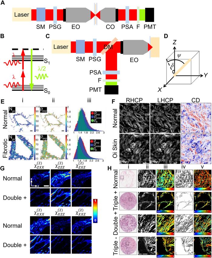
FIGURE 1 | Typical microscope schematics for forward- (A) and epi-detection (C) configurations for polarization-sensitive second harmonic generation (SHG)
microscopy. The energy state diagram of SHG (B) along with the coordinate system, where Z-X is the image plane and the laser propagates along Y, for an arbitrarily
oriented fiber (D) is shown. Abbreviations: SM-scanning mirrors, PSG-polarization state generator, EO-excitation microscope objective, CO-collection microscope
objective, PSA-polarization state analyzer, F-filter, PMT-photomultiplier tube detector and DM-dichroic mirror. Example images obtained using PI-SHG (E), CD-
SHG (F), DSMP-SHG (G) and PIPO-SHG (H). SHG intensity (E-i), fitted ρ (E-ii) and fitted ρ distribution width (E-iii) are shown for normal and fibrotic liver [14]. SHG images
(F) of normal skin and osteogenesis imperfecta (O-i) skin tissues using right-handed circularly polarized light (RHCP), left-handed circularly polarized light (LHCP) and CD-
SHG (CD) where red and blue show positive and negative values, respectively [43]. Laboratory-frame χ (2) values (G) of normal and double + breast tissue using DSMP-
SHG [44]. Brightfield (H-i), SHG (H-ii), ρ (H-iii), δ orientation (H-iv) and DOLP (H-v) images of normal, triple +, double +, ++-, and triple–breast tissue [44]. Images E-H were
adapted with permission from [14, 43, 44] © The Optical Society.
sample modification procedures are needed, and the signal can For biological imaging, lasers with wavelength outside of
be interpreted as a direct indicator of structural sample the tissue absorption spectrum are typically chosen,
changes. therefore reduced heat is deposited into the sample allowing
SHG is energy conserving and consequently does not for long duration functional in vivo studies.
photobleach, differing significantly from non-parametric .
The SHG ..
intensity and polarization can be .written:
processes, such as fluorescence, where absorbed Stokes I SHG ∝ χ 2 : E E , where “:” is the tensor product, E is the
energy often leads to sample damage and photobleaching. intensity and polarization of the laser electric field, and χ (2)
Frontiers in Physics | www.frontiersin.org 2 August 2021 | Volume 9 | Article 726996Cisek et al. Polarization-Resolved SHG of Diseased Tissues
is the second-order nonlinear optical susceptibility tensor, APPLICATIONS OF SHG MICROSCOPY
containing up to 18 unique elements, describing the sample. FOR DETERMINING THE
The equation shows that intensity and polarization of SHG
depends on the sample orientation and ultrastructure,
ULTRASTRUCTURE OF DISEASED
encoded in χ (2) , and on the polarization and intensity of COLLAGENOUS TISSUES
the laser. Kleinman symmetry [1, 2], where
2)
K χ (zxx / χ (xxz
2)
1, as well as cylindrical C6v symmetry are
Polarization-In SHG Microscopy
Pioneered in 1979, polarization-in SHG (PI-SHG) microscopy
typically assumed, resulting in only two unique χ (2) utilizes a polarization state generator (PSG) to rotate the laser
elements whose ratio is historically designated ρ χ zzz (2) /χ (2)
. .
zxx linear polarization state. One method is to use a half-wave plate
[3]. Since E is controlled and I SHG is measured, ρ, and the in- (HWP) in a motorized rotating mount, located before the
plane projection of the orientation angle of collagen, δ, can be excitation objective lens (Figures 1A,C). An SHG intensity
extracted via PSHG measurements (see Figure 1D for image is recorded at each HWP angle, typically with 10–30
coordinate system). This review is focused on polarization- steps in the range 0°–90°. Since SHG is predominantly
sensitive SHG approaches specifically used for discriminating forward-directed, most epi-detected photons require
diseased collagenous tissues, and thus many great papers backscattering, which is less efficient than forward-detection
using SHG microscopy are regretfully not included. [11], and results in depolarization of a fraction of the SHG.
Parameters ρ and δ can be found using this methodology by
performing nonlinear data fitting.
Historical Interpretation of the PSHG PI-SHG microscopy was used to distinguish normal and
Parameters Pertaining to Collagen diseased tissue regions in several tissues (Table 1) with
Researchers, Freund et al. [3–6], pioneered the use of SHG to statistical significance, via the χ (2) zzz values of osteosarcoma,
investigate the structure of collagenous biological tissue by breast cancer and melanoma tissues [12], as well as the ρ and
utilizing different laser polarizations. They focused a θ values of fibrotic and control liver vessels [13, 14]. A significant
Q-switched Nd:YAG laser onto rat tail tendon, collecting SHG increase in the ρ value was also found between breast cancer
in the forward direction, similar to Figure 1A, but without scan biopsy tissues [15, 16], and during thermal denaturation of
mirrors and at different scattering angles. They found an SHG corneal stroma [17]. Several other studies were performed
peak at the 0° scattering angle, indicative of the macroscopic using this technique, including on liver fibrosis [18],
ordered polar structure of collagen, and found that χ (2) of tendon osteoarthritis [19], and keratoconus [20, 21].
exhibited cylindrical symmetry, since rotation about the tendon To avoid delays due to nonlinear data fitting, a fast Fourier
axis did not appreciably change SHG parameters. They attributed transform (FFT) algorithm has been developed to extract ρ values
the signal predominantly to C-N bonds in the amino acids, quickly [22]. It was used in the study of normal and keratoconic
arguing these are the likely dominant polarizable, human corneas [23], human breast carcinoma samples [16, 24]
noncentrosymmetric and non-mobile candidates. They used a and to investigate mechanical load in energy storing versus
polarization state analyzer (PSA) consisting of a polarizer to positional collagen fibrils [25]. The FFT approach has also
directly measure ρ for tendons from rats of different age groups, allowed use of a generic structural model, where rather than
and showed this parameter distinguishes the variation in the assuming a particular symmetry, the second-order FFT
developing biological structure. coefficients (I2 and ϕ2 ), which quantify the modulation depth
Noting the important SHG theoretical work of Dick [7], two of the polarization SHG response (I2 ) and its phase (ϕ2 ), are
groups, Plotnikov et al. [8] and Tiaho et al. [9], attributed ρ to reported and compared. This approach was used for a survey on
the helical pitch (θ) of the protein triple helix via cartilage in osteo-arthritic tissue [26, 27], for a study of ageing via
ρ/(2 + ρ) cos θ. However, it is likely that all the levels of investigating ribose-glycated fibrils isolated from rat tail [28], and
the collagen tissue hierarchy can contribute to the to investigate needle puncture damage in bovine annulus
polarization-dependent SHG parameters [10]. Therefore, the fibrosus [29].
question of which hierarchical levels of collagen in different Several groups have also investigated the excitation anisotropy
tissues are expressed in PSHG microscopy remains open. It (α) for distinguishing diseased tissues, α Iv − IH /Iv + IH , where
also presents an opportunity to investigate which disease states Iv and IH are the SHG intensities when the incident light is
affect the quantitative imaging results. Since the dyes used for vertically and horizontally polarized, respectively. α is related to δ,
standard staining of histopathology samples in hospitals, and was measured in skin exposed to UVB [30, 31], and using a
hematoxylin and eosin Y, seem to be compatible with fast motion-artefact free implementation in cheek and eye corner
PSHG microscopy [10], therefore PSHG capability could be skin at two ages [32].
added to existing histopathology slide scanners enabling the Polarization analysis of the outgoing SHG has also been
new modality without altering standard hospital sample incorporated using a PSA typically located after the collection
preparation procedures. The resulting automated objective in a forward-detection geometry (Figure 1A), or
quantitative analysis could yield pathologists with an after a dichroic mirror that separates SHG in epi-detection
additional quantitative marker for improved diagnosis and (Figure 1C). While variable PSAs are reviewed in Double
evaluation. Stokes Mueller Polarimetric SHG (DSMP-SHG) Microscopy
Frontiers in Physics | www.frontiersin.org 3 August 2021 | Volume 9 | Article 726996Cisek et al. Polarization-Resolved SHG of Diseased Tissues
TABLE 1 | A summary of the PSHG microscopy techniques used for quantifying the differences in collagen structure in diseased tissue and the corresponding parameters
measured with those techniques. The following abbreviation is used NR: Not reported.
Technique Condition Parameters: References
Statistical significance
PI-SHG Osteosarcoma, Breast cancer and Melanoma χ (2)
zzz: Yes, β: NR [12]
PI-SHG Liver fibrosis ρ: Yes, θ: Yes [13, 14]
PI-SHG Mammary dysplasia and breast cancer K: Yes, ρ: Yes, χ (2) (2)
xxx/χ zxx: Yes [15]
PI-SHG Breast ductal carcinoma β: Yes, ρ: Yes [16]
PI-SHG Thermal denaturation θ: Yes [17]
PI-SHG Liver fibrosis ρ: NR, χ (2) (2)
xxz/χ zxx: NR [18]
PI-SHG Osteoarthritis Polarization plots: NR [19]
PI-SHG Keratoconus Polarization plots: NR [20]
PI-SHG Keratoconus δ: No [21]
PI-SHG Keratoconus θ: NR, δ: NR [23]
PI-SHG Breast carcinoma ρ: Yes [24]
PI-SHG Mechanical load ρ: Yes [25]
PI-SHG Osteoarthritis I2 and ϕ2 : NR [27]
PI-SHG Aged I2 and ϕ2 : Yes [28]
PI-SHG Needle puncture damage I2 : No [29]
PI-SHG UVB exposure α : Yes [30, 31]
PI-SHG Aged δ: NR [32]
PI-SHG Skin dysplasia β: NR [33]
PI-SHG Aged β: Yes [34]
PI-SHG Osteogenesis imperfecta β: No [35]
PI-SHG Colorectal dysplasia and cancer β: Yes [36]
PI-SHG Ovarian cancer β: NR [37]
PI-SHG Mechanical load ρ: Yes [38]
PI-SHG Irradiation ρ: Yes [39]
PI-SHG CT26 derived tumor from mice ρ: NR, θ: NR [40]
PI-SHG Implantation of B16 melanoma cells ρ: NR, θ: NR [40]
PI-SHG Implantation of CT26 colon carcinoma cells and 4T1 breast cancer cells ρ: NR, θ: NR [41]
PI-SHG Esophageal squamous cell carcinoma ρ: No [42]
PI/CD-SHG Ovarian cancer θ: Yes, β (0°): Yes, ICD-SHG: Yes [49]
CD-SHG Osteogenesis imperfecta ICD-SHG: Yes [43]
CD-SHG Idiopathic pulmonary fibrosis θ: No, ICD-SHG: Yes [47]
CD-SHG 3D spheroid models of idiopathic pulmonary fibrosis θ: Yes, ICD-SHG: Yes [48]
DSMP-SHG Breast cancer with ER, PgR and HER2 expression K: No, χ (2)
zzz, χ zxx, χ zxz, χ xxx, χ xzz, and χ xxz: NR
(2) (2) (2) (2) (2)
[44]
PIPO-SHG Breast cancer with ER, PgR and HER2 expression ρ: Yes, DOLP: Yes [44]
PIPO-SHG Thyroid carcinomas and diseases ρ: Yes, C: No, DOLP: Yes [55, 58, 59]
PIPO-SHG Non-small cell lung carcinoma ρ: Yes, C: Yes, DOLP: Yes, δ: Yes [56]
PIPO-SHG Non-small cell lung carcinoma ρ: Yes [57, 58]
PIPO-SHG Pancreatic ductal adenocarcinoma ρ: Yes, C: Yes, DOLP: Yes [58, 60]
PIPO-SHG Bone cancer ρ: Yes [61]
PO-SHG Breast cancer θ: No [62]
PO-SHG Atherosclerosis ρ: No [63]
and Polarization-In, Polarization-Out SHG (PIPO-SHG) Stationary PSAs where the collagen axis is aligned to the
Microscopy sections, stationary PSAs are also used. In one laboratory polarization axis have also been used for
implementation, SHG intensity through a polarizer parallel investigating effects of mechanical load [38] and irradiation
(I|| ) and perpendicular (I⊥ ) to the laser linear polarization is with high intensity infra-red light [39].
recorded allowing the SHG anisotropy (β) to be calculated: Interestingly, analysis of the PSHG data has also been
β I|| − I⊥ /I|| + 2I⊥ . A study of dysplastic human skin showed achieved through Fourier projection of the PSHG image
that optimization of the selection of linear laser polarization stacks onto two phasor plots referred to as microscopic
orientations using a Fourier algorithm on the SHG intensity multiparametric analysis by phasor projection of PSHG
obtained with circularly polarized light allowed β values to (µMAPPS). This technique has been used to compare the ρ
distinguish diseased areas more effectively [33], while in rat values of SHG data taken from biopsies of the left flank of mice
skin, β significantly changed during ageing [34]. In another days after implantation of melanoma [40], colon carcinoma
study, the β values of femurs from wild-type mice and oim and breast cancer cells [41].
mice, a model for human osteogenesis imperfecta, were similar PI-SHG microscopy was also used to image submucosa of
[35] while different β values are reported for dysplasia and esophageal squamous cell carcinoma (ESCC). The ρ values from 4
colon cancer [36] as well as ovarian cancer tissue [37]. stages of ESCC showed few differences between one another, and
Frontiers in Physics | www.frontiersin.org 4 August 2021 | Volume 9 | Article 726996Cisek et al. Polarization-Resolved SHG of Diseased Tissues
authors concluded that PI-SHG microscopy cannot be used for (calculated relative to one component), and for each
staging ESCC because the structural changes are likely of component, the real (Figure 1G) and imaginary parts can be
macromolecular rather than micromolecular origin [42]. extracted, for each pixel of the image. Figure 1G can be used to
compare tensor components for each pixel of the image [44].
Additionally, one can obtain the degree of polarization (DOP),
Circular Dichroism Second Harmonic degree of linear polarization (DOLP) and degree of circular
Generation Microscopy polarization (DOCP) at each input polarization. DSMP-SHG
In circular dichroism SHG (CD-SHG) microscopy, two SHG microscopy was used to study microarray slides of human
images are obtained using left-handed (IL) and right-handed (IR) breast cancer types, finding that components of χ (2) are
circularly polarized laser light, with no PSA in the SHG detection predominantly real and validating Kleinman symmetry [44].
path (Figures 1A,C). The CD-SHG intensity (ICD-SHG) is the When the χ (2) components are real and birefringence is
normalized difference of the two quantities, negligible, the polarization of the SHG signal will not have a
ICD−SHG 2(IL − IR )/(IL + IR ). One way to implement CD-SHG circular component, hence a reduced polarimetric measurement
is via a liquid-crystal rotator (LCR) followed by a quarter-wave with only linearly polarized incoming and outgoing states known
plate (QWP) as the PSG [45]. ICD-SHG is a complicated parameter as polarization-in, polarization-out SHG microscopy can be
when expressed in terms of the sample susceptibility, and performed [50–52].
according to equation 12 in [46], only measures structures
with nonzero chirality (χ (2) xyz), nonzero phase between χ
(2)
Polarization-In, Polarization-Out SHG
elements, and nonzero angle to the imaging plane (ψ in
Figure 1D). Nonzero phase between tensor elements can Microscopy
occur due to a resonance transition near the laser wavelength Polarization-in, polarization-out SHG (PIPO-SHG) uses a
and/or the SHG wavelength, due to a molecular magnetic dipole simplified PSA compared to DSMP-SHG as well as the same
or due to an electric quadrupole rather than dipolar PSG as in the PI-SHG technique, and hence is typically
interaction [46]. implemented using a forward-detection geometry (Figure 1A).
CD-SHG microscopy was applied to determine differences The PSA measures the linear polarization state of the SHG signal
between normal and osteogenesis imperfecta skin tissues as well at different angles of the linear polarization of the laser. The
as idiopathic pulmonary fibrosis human lung tissue where simplest PSA consists of a linear polarizer in a motorized rotation
variations in ICD-SHG were statistically significant [43, 47, 48]. mount. SHG images are typically captured with at least 8 PSG
Another study used CD-SHG microscopy to find differences angles and 8 PSA angles, hence at least 64 images are recorded for
between 4 ovarian tissue classifications. The mean ICD-SHG the technique, resulting in ∼20 min acquisition times in
value for normal ovarian tissue was significantly higher than comparison to PI-SHG which takes ∼1.5 min [53]. The
the other tissues [49]. additional dimensionality of the data is thought to produce
higher accuracy fitting, although at the time of writing this
manuscript no study has performed the comparison with the
Double Stokes Mueller Polarimetric SHG other techniques. Fitting of the SHG intensity versus PSA and
Microscopy PSG angles yields δ and ρ. When the more generalized C6
Double Stokes Mueller polarimetric SHG (DSMP-SHG) symmetry is assumed, χ (2) xyz appears, and can be measured as
microscopy, introduced in 2015 [50, 51], is an alternative the chirality ratio (C χ 2) /χ (2) ) [54].
(
xyz zxx
method to performing PSHG microscopy. DSMP-SHG The PIPO-SHG measurements also allow the DOLP of the
microscopy aims to obtain all possible polarization SHG signal to be obtained, where the SHG light is fully linearly
information in the smallest amount of measurements. Stokes polarized when DOLP 1, and increasingly depolarized or
.
vectors are used to describe the laser polarization ( s ) and circularly polarized as it approaches 0. DOLP measurements
.
polarization of the SHG signal ( s ′). Their relation is described were performed at 8 incident laser polarizations
. .
by the function s ′ M s , where M is a double Mueller matrix (θ 0˚, 22.5˚, 45˚, 67.5˚, 90˚, 112.5˚, 135˚, 157.5˚) and for
which can be characterized via measurement. One such each one, two DOLP calculations were averaged, one using
implementation using a single detector required 54 measurements at analyzer angles 0˚, 45˚, 90˚, 135˚ and
measurements. A PSG produced linearly polarized states at another at 22.5˚, 67.5˚, 112.5˚, 157.5˚ [55]. An improved
angles 0°, ±45°, 90°, 157.5°, two elliptical states, and two DOLP is obtained when structures with δ orientations closest
circularly polarized states, while at each PSG state, 6 SHG to the normal of the crystal axis are ignored, since they have the
polarization intensities are measured to obtain a Stokes vector lowest signal in collagen imaging, which can result in a low signal-
using the PSA. Both the PSG and PSA consisted of a polarizer, to-noise ratio [44]. Lower DOLP values can occur due to
HWP and QWP at the laser and SHG wavelengths, respectively, ultrastructural disorder of collagen, or due to fragmentation of
with waveplates in mechanical rotation mounts (Figures collagen within the focal volume, where SHG is emitted from
1A,C) [44]. uncorrelated domains [56].
DSMP-SHG has the advantage that 6 laboratory frame A PIPO-SHG microscopy study of pathology slides of lung
susceptibility components can be extracted from double samples from patients with non-small cell lung carcinoma
Mueller matrix components: χ (xxx 2) (2) , χ (2) , χ (2) , χ (2) , χ (2)
, χ xzz xxz zxx zzz zxz revealed significant differences in the mean ρ values [57]. A
Frontiers in Physics | www.frontiersin.org 5 August 2021 | Volume 9 | Article 726996Cisek et al. Polarization-Resolved SHG of Diseased Tissues
more recent study of that disease showed that the median ρ and another technique, polarimetry analysis can also be performed
the median average distribution (MAD) of δ could differentiate using circularly polarized laser excitation, only requiring a single
stages II and III from normal tissue, while the MAD of C and scan [53, 69]. Fourier techniques could be used for faster data
DOLP could differentiate stage I [56]. Human thyroid tissues analysis [16, 23, 24], while imaging rates can be increased using
were also investigated with PIPO-SHG, and revealed significant higher repetition rate pulsed lasers combined with faster scanners
differences in the average DOLP values between normal thyroid such as spinning mirrors, or by increasing the field of view using a
tissue and follicular variant of papillary thyroid carcinoma, wide-field imaging approach [70]. With these developments, it is
classical papillary thyroid carcinoma, follicular nodular disease, reasonable that PSHG microscopy may be implemented in a
Grave’s disease, and anaplastic or undifferentiated carcinoma. modified hospital pathology slide scanner. Furthermore, since
This was also true for the mean ρ values except for insular thyroid SHG requires no dyes, it can be implemented as an epi-detection
tissues [58, 59]. Pancreatic ductal adenocarcinoma tissue was also setup in an endoscope for in vivo quantitative imaging as a biopsy
investigated with PIPO-SHG and demonstrated significant tool [71–73].
differences between the mean ρ values of tumor tissue and the It is evident that PSHG imaging can quantitatively
periductal, lobular and parenchymal regions of normal pancreatic differentiate certain diseased tissues based on their
tissue. The DOLP values of the pancreatic tumor regions were ultrastructure in pathological sample slides however, care
only significantly different from normal periductal and must be given to PSHG image quality and the image
parenchymal tissues [60] while the C values in diseased analysis methods used. For instance, when assessing the
thyroid and pancreas were not significantly different from quality of PSHG images, it has been found that using SHG
normal tissues [58–60]. intensity as a criterion is only suitable in specific instances [74].
PIPO-SHG microscopy was also applied to study a microarray Furthermore, while statistical discrimination based on many
slide containing 3 pathological human breast cancer types samples has been validated in different tissues, the efficacy of
(Figure 1H) with the overexpression/absence of estrogen diagnosis in individual regions remains unclear and must be
receptor, progesterone receptor, and human epidermal growth addressed. Improved differentiation could be obtained via
factor receptor 2: triple +, double +, and triple -. The mean and additional implementation of complementary SHG intensity
median ρ values of triple + and double + as well as the mean and analysis techniques, such as texture analysis [56, 75–77], the
median DOLP values for double + were significantly different Hough transform [78] and the structure tensor [79, 80]. These
from normal breast tissue [44]. Bone cancer was also investigated analysis techniques could be extended to PSHG ρ or δ images.
and the ρ values of bone adjacent to tumor was found to be Additional information about diseased tissues can be obtained
significantly different to bone adjacent to normal marrow [61]. by coupling complementary techniques, such as polarization-
Polarization-out SHG (PO-SHG) microscopy was also attempted sensitive two-photon excitation fluorescence microscopy [81],
by varying the angle of an analyzer before the detector, as in and polarization-sensitive third harmonic generation
PIPO-SHG, but with only one linear input polarization state, microscopy [82]. The idealized scanning system could find
however, it did not achieve differentiation between normal and potential hotspots and report them to the pathologist who
diseased tissue [62, 63]. could confirm the diagnosis, aiding in the difficult task of
finding small diseased regions in comparatively large
samples, saving pathologists time and lives by pinpointing
DISCUSSION regions of interest.
Automated digital histopathology using PSHG microscopy is a
promising technology for diagnosis of disease in AUTHOR CONTRIBUTIONS
histopathological samples, however, implementation requires
fast imaging and data analysis balanced with maintaining high The authors contributed to manuscript writing (RC, DT) and
measurement accuracy to obtain diagnosis based on the fewest editing (RC, AJ, MH, DT).
amount of analyzed pixels. To reduce imaging time due to the
different laser polarizations needed, liquid crystal or electro-optic
modulators can be used [64–66], or beams with different FUNDING
polarizations can be interleaved. The clever use of fewer input
polarizations could also be used, such as in Stokes-Mueller SHG The research was supported by the Natural Sciences and
[67] which uses four polarizations in a PI-SHG setup, or Engineering Research Council of Canada Discovery Grants
advanced polarimeters with multiple detectors can be used for Program (RGPIN-2018-05444), Canada’s Research Support
simultaneous detection of the SHG polarization states [68]. In Fund, and Saint Mary’s University.
2. Dailey CA, Burke BJ, and Simpson GJ. The General Failure of Kleinman
REFERENCES Symmetry in Practical Nonlinear Optical Applications. Chem Phys Lett (2004)
390:8–13. doi:10.1016/j.cplett.2004.03.109
1. Kleinman DA. Nonlinear Dielectric Polarization in Optical media. Phys Rev 3. Roth S, and Freund I. Second Harmonic Generation in Collagen. J Chem Phys
(1962) 126:1977–9. doi:10.1103/PhysRev.126.1977 (1979) 70:1637–43. doi:10.1063/1.437677
Frontiers in Physics | www.frontiersin.org 6 August 2021 | Volume 9 | Article 726996Cisek et al. Polarization-Resolved SHG of Diseased Tissues
4. Roth S, and Freund I. Optical Second-Harmonic Scattering in Rat-Tail 24. Mercatelli R, Triulzi T, Pavone FS, Orlandi R, and Cicchi R. Collagen
Tendon. Biopolymers (1981) 20:1271–90. doi:10.1002/bip.1981.360200613 Ultrastructural Symmetry and its Malignant Alterations in Human Breast
5. Freund I, and Deutsch M. Second-harmonic Microscopy of Biological Tissue. Cancer Revealed by Polarization-resolved Second-harmonic Generation
Opt Lett (1986) 11:94–6. doi:10.1364/OL.11.000094 Microscopy. J Biophotonics (2020) 13:e202000159. doi:10.1002/jbio.202000159
6. Freund I, Deutsch M, and Sprecher A. Connective Tissue Polarity. Optical 25. Quigley AS, Bancelin S, Deska-Gauthier D, Légaré F, Kreplak L, and Veres SP.
Second-Harmonic Microscopy, Crossed-Beam Summation, and Small-Angle In Tendons, Differing Physiological Requirements lead to Functionally
Scattering in Rat-Tail Tendon. Biophysical J (1986) 50:693–712. doi:10.1016/ Distinct Nanostructures. Sci Rep (2018) 8:4409. doi:10.1038/s41598-018-
S0006-3495(86)83510-X 22741-8
7. Dick B. Irreducible Tensor Analysis of Sum- and Difference-Frequency 26. Duboisset J, Aït-Belkacem D, Roche M, Rigneault H, and Brasselet S. Generic
Generation in Partially Oriented Samples. Chem Phys (1985) 96:199–215. Model of the Molecular Orientational Distribution Probed by Polarization-
doi:10.1016/0301-0104(85)85085-0 Resolved Second-Harmonic Generation. Phys Rev A (2012) 85:043829.
8. Plotnikov SV, Millard AC, Campagnola PJ, and Mohler WA. Characterization doi:10.1103/PhysRevA.85.043829
of the Myosin-Based Source for Second-Harmonic Generation from Muscle 27. Mansfield JC, Mandalia V, Toms A, Winlove CP, and Brasselet S. Collagen
Sarcomeres. Biophysical J (2006) 90:693–703. doi:10.1529/biophysj.105.071555 Reorganization in Cartilage under Strain Probed by Polarization Sensitive
9. Tiaho F, Recher G, and Rouède D. Estimation of Helical Angles of Myosin and Second Harmonic Generation Microscopy. J R Soc Interface (2019) 16:
Collagen by Second Harmonic Generation Imaging Microscopy. Opt Express 20180611. doi:10.1098/rsif.2018.0611
(2007) 15:12286–95. doi:10.1364/OE.15.012286 28. Aït-Belkacem D, Guilbert M, Roche M, Duboisset J, Ferrand P, Sockalingum
10. Tuer AE, Akens MK, Krouglov S, Sandkuijl D, Wilson BC, Whyne CM, et al. G, et al. Microscopic Structural Study of Collagen Aging in Isolated Fibrils
Hierarchical Model of Fibrillar Collagen Organization for Interpreting the Using Polarized Second Harmonic Generation. J Biomed Opt (2012) 17:
Second-Order Susceptibility Tensors in Biological Tissue. Biophysical J (2012) 080506. doi:10.1117/1.JBO.17.8.080506
103:2093–105. doi:10.1016/j.bpj.2012.10.019 29. Wang J-Y, Mansfield JC, Brasselet S, Vergari C, Meakin JR, and Winlove CP.
11. Williams RM, Zipfel WR, and Webb WW. Interpreting Second-Harmonic Micro-mechanical Damage of Needle Puncture on Bovine Annulus Fibrosus
Generation Images of Collagen I Fibrils. Biophysical J (2005) 88:1377–86. Fibrils Studied Using Polarization-Resolved Second Harmonic
doi:10.1529/biophysj.104.047308 Generation(P-SHG) Microscopy. J Mech Behav Biomed Mater (2021) 118:
12. Hompland T, Erikson A, Lindgren M, Lindmo T, and de Lange Davies C. 104458. doi:10.1016/j.jmbbm.2021.104458
Second-harmonic Generation in Collagen as a Potential Cancer 30. Yasui T, Takahashi Y, Fukushima S, Ogura Y, Yamashita T, Kuwahara T, et al.
Diagnostic Parameter. J Biomed Opt (2008) 13:054050. doi:10.1117/ Observation of Dermal Collagen Fiber in Wrinkled Skin Using Polarization-
1.2983664 Resolved Second-Harmonic-Generation Microscopy. Opt Express (2009) 17:
13. Rouède D, Schaub E, Bellanger J-J, Ezan F, Scimeca J-C, Baffet G, et al. 912–23. doi:10.1364/oe.17.000912
Determination of Extracellular Matrix Collagen Fibril Architectures and 31. Fukushima S-i., Yonetsu M, and Yasui T. Polarization-resolved Second-
Pathological Remodeling by Polarization Dependent Second Harmonic Harmonic-Generation Imaging of Dermal Collagen Fiber in Prewrinkled
Microscopy. Sci Rep (2017) 7:12197. doi:10.1038/s41598-017-12398-0 and Wrinkled Skins of Ultraviolet-B-Exposed Mouse. J Biomed Opt (2018)
14. Rouède D, Schaub E, Bellanger J-J, Ezan F, and Tiaho F. Wavy Nature of 24:031006. doi:10.1117/1.JBO.24.3.031006
Collagen Fibrils Deduced from the Dispersion of Their Second-Order 32. Tanaka Y, Hase E, Fukushima S, Ogura Y, Yamashita T, Hirao T, et al. Motion-
Nonlinear Optical Anisotropy Parameters ρ. Opt Express (2020) 28: artifact-robust, Polarization-Resolved Second-Harmonic-Generation
4845–58. doi:10.1364/OE.380089 Microscopy Based on Rapid Polarization Switching with Electro-Optic
15. Ambekar R, Lau T-Y, Walsh M, Bhargava R, and Toussaint KC. Quantifying Pockells Cell and its Application to In Vivo Visualization of Collagen Fiber
Collagen Structure in Breast Biopsies Using Second-Harmonic Generation Orientation in Human Facial Skin. Biomed Opt Express (2014) 5:1099–113.
Imaging. Biomed Opt Express (2012) 3:2021–35. doi:10.1364/BOE.3.002021 doi:10.1364/boe.5.001099
16. Tsafas V, Gavgiotaki E, Tzardi M, Tsafa E, Fotakis C, Athanassakis I, et al. 33. Hristu R, Stanciu SG, Tranca DE, and Stanciu GA. Improved Quantification of
Polarization-dependent Second-harmonic Generation for Collagen-based Collagen Anisotropy with Polarization-Resolved Second Harmonic
Differentiation of Breast Cancer Samples. J Biophotonics (2020) 13: Generation Microscopy. J Biophoton (2017) 10:1171–9. doi:10.1002/
e202000180. doi:10.1002/jbio.202000180 jbio.201600197
17. Matteini P, Cicchi R, Ratto F, Kapsokalyvas D, Rossi F, de Angelis M, et al. 34. Miler I, Rabasovic MD, Aleksic M, Krmpot AJ, Kalezic A, Jankovic A, et al.
Thermal Transitions of Fibrillar Collagen Unveiled by Second-Harmonic Polarization-resolved SHG Imaging as a Fast Screening Method for Collagen
Generation Microscopy of Corneal Stroma. Biophysical J (2012) 103: Alterations during Aging: Comparison with Light and Electron Microscopy.
1179–87. doi:10.1016/j.bpj.2012.07.055 J Biophotonics (2021) 14:e202000362. doi:10.1002/jbio.202000362
18. Lin J, Pan S, Zheng W, and Huang Z. Polarization-resolved Second-Harmonic 35. Nadiarnykh O, Plotnikov S, Mohler WA, Kalajzic I, Redford-Badwal D, and
Generation Imaging for Liver Fibrosis Assessment without Labeling. Appl Phys Campagnola PJ. Second Harmonic Generation Imaging Microscopy Studies of
Lett (2013) 103:173701. doi:10.1063/1.4826516 Osteogenesis Imperfecta. J Biomed Opt (2007) 12:051805. doi:10.1117/
19. Brown CP, Houle M-A, Popov K, Nicklaus M, Couture C-A, Laliberté M, et al. 1.2799538
Imaging and Modeling Collagen Architecture from the Nano to Micro Scale. 36. Birk JW, Tadros M, Moezardalan K, Nadyarnykh O, Forouhar F, Anderson J,
Biomed Opt Express (2014) 5:233–43. doi:10.1364/boe.5.000233 et al. Second Harmonic Generation Imaging Distinguishes Both High-Grade
20. Ávila FJ, Del Barco O, and Bueno JM. Polarimetric Multiphoton Microscopy Dysplasia and Cancer from normal Colonic Mucosa. Dig Dis Sci (2014) 59:
for the Analysis of Ocular Structures. Opt Pura Apl (2019) 52:1–9. doi:10.7149/ 1529–34. doi:10.1007/s10620-014-3121-7
OPA.52.1.51013 37. Nadiarnykh O, LaComb RB, Brewer MA, and Campagnola PJ. Alterations of
21. Raoux C, Schmeltz M, Bied M, Alnawaiseh M, Hansen U, Latour G, et al. the Extracellular Matrix in Ovarian Cancer Studied by Second Harmonic
Quantitative Structural Imaging of Keratoconic Corneas Using Polarization- Generation Imaging Microscopy. BMC Cancer (2010) 10:1471–2407.
Resolved SHG Microscopy. Biomed Opt Express (2021) 12:4163–78. doi:10.1186/1471-2407-10-94
doi:10.1364/boe.426145 38. Gusachenko I, Tran V, Houssen YG, Allain J-M, and Schanne-Klein M-C.
22. Amat-Roldan I, Psilodimitrakopoulos S, Loza-Alvarez P, and Artigas D. Fast Polarization-resolved Second-Harmonic Generation in Tendon upon
Image Analysis in Polarization SHG Microscopy. Opt Express (2010) 18: Mechanical Stretching. Biophysical J (2012) 102:2220–9. doi:10.1016/
17209–19. doi:10.1364/OE.18.017209 j.bpj.2012.03.068
23. Alizadeh M, Merino D, Lombardo G, Lombardo M, Mencucci R, Ghotbi M, 39. Silva DFT, Gomes ASL, De Campos Vidal B, and Ribeiro MS. Birefringence
et al. Identifying Crossing Collagen Fibers in Human Corneal Tissues Using and Second Harmonic Generation on Tendon Collagen Following Red
pSHG Images. Biomed Opt Express (2019) 10:3875–88. doi:10.1364/ Linearly Polarized Laser Irradiation. Ann Biomed Eng (2013) 41:752–62.
boe.10.003875 doi:10.1007/s10439-012-0720-3
Frontiers in Physics | www.frontiersin.org 7 August 2021 | Volume 9 | Article 726996Cisek et al. Polarization-Resolved SHG of Diseased Tissues
40. Radaelli F, D’Alfonso L, Collini M, Mingozzi F, Marongiu L, Granucci F, et al. 58. Tokarz D, Cisek R, Golaraei A, Krouglov S, Navab R, Niu C, et al. Tumor
μMAPPS: a Novel Phasor Approach to Second Harmonic Analysis for In Tissue Characterization Using Polarization-Sensitive Second Harmonic
Vitro-In Vivo Investigation of Collagen Microstructure. Sci Rep (2017) 7: Generation Microscopy. Proceed SPIE (2015) 9531:95310C. doi:10.1117/
17468. doi:10.1038/s41598-017-17726-y 12.2180969
41. Scodellaro R, Bouzin M, Mingozzi F, D’Alfonso L, Granucci F, Collini M, et al. 59. Tokarz D, Cisek R, Joseph A, Asa SL, Wilson BC, and Barzda V.
Whole-Section Tumor Micro-architecture Analysis by a Two-Dimensional Characterization of Pathological Thyroid Tissue Using Polarization-
Phasor-Based Approach Applied to Polarization-dependent Second Harmonic Sensitive Second Harmonic Generation Microscopy. Lab Invest (2020) 100:
Imaging. Front Oncol (2019) 9:527. doi:10.3389/fonc.2019.00527 1280–7. doi:10.1038/s41374-020-0475-7
42. Chen W-C, Chen Y-J, Lin S-T, Hung W-H, Chan M-C, Wu I-C, et al. Label- 60. Tokarz D, Cisek R, Joseph A, Golaraei A, Mirsanaye K, Krouglov S, et al.
free Characterization of Collagen Fibers in Cancerous Esophagus Tissues Characterization of Pancreatic Cancer Tissue Using Multiphoton Excitation
Using Ratiometric Nonlinear Optical Microscopy. Exp Biol Med Fluorescence and Polarization-Sensitive Harmonic Generation Microscopy.
(Maywood) (2020) 245:1213–21. doi:10.1177/1535370220934039 Front Oncol (2019) 9:272. doi:10.3389/fonc.2019.00272
43. Chen X, Raggio C, and Campagnola PJ. Second-harmonic Generation Circular 61. Burke M, Golaraei A, Atkins A, Akens M, Barzda V, and Whyne C.
Dichroism Studies of Osteogenesis Imperfecta. Opt Lett (2012) 37:3837–9. Collagen Fibril Organization within Rat Vertebral Bone Modified with
doi:10.1364/OL.37.003837 Metastatic Involvement. J Struct Biol (2017) 199:153–64. doi:10.1016/
44. Golaraei A, Kontenis L, Cisek R, Tokarz D, Done SJ, Wilson BC, et al. Changes j.jsb.2017.06.008
of Collagen Ultrastructure in Breast Cancer Tissue Determined by Second- 62. Han X, Burke RM, Zettel ML, Tang P, and Brown EB. Second Harmonic
Harmonic Generation Double Stokes-Mueller Polarimetric Microscopy. Properties of Tumor Collagen: Determining the Structural Relationship
Biomed Opt Express (2016) 7:4054–68. doi:10.1364/BOE.7.004054 between Reactive Stroma and Healthy Stroma. Opt Express (2008) 16:
45. Campbell KR, and Campagnola PJ. Wavelength-Dependent Second Harmonic 1846–59. doi:10.1364/oe.16.001846
Generation Circular Dichroism for Differentiation of Col I and Col III 63. Doras C, Taupier G, Barsella A, Mager L, Boeglin A, Bulou H, et al.
Isoforms in Stromal Models of Ovarian Cancer Based on Intrinsic Chirality Polarization State Studies in Second Harmonic Generation Signals to Trace
Differences. J Phys Chem B (2017) 121:1749–57. doi:10.1021/acs.jpcb.6b06822 Atherosclerosis Lesions. Opt Express (2011) 19:15062–8. doi:10.1364/
46. Golaraei A, Kontenis L, Mirsanaye K, Krouglov S, Akens MK, Wilson BC, et al. oe.19.015062
Complex Susceptibilities and Chiroptical Effects of Collagen Measured with 64. Dewalt EL, Sullivan SZ, Schmitt PD, Muir RD, and Simpson GJ. Polarization-
Polarimetric Second-Harmonic Generation Microscopy. Sci Rep (2019) 9: modulated Second Harmonic Generation Ellipsometric Microscopy at Video
12488. doi:10.1038/s41598-019-48636-w Rate. Anal Chem (2014) 86:8448–56. doi:10.1021/ac502124v
47. James DS, Jambor AN, Chang H-Y, Alden Z, Tilbury KB, Sandbo NK, et al. 65. Dow XY, DeWalt EL, Sullivan SZ, Schmitt PD, Ulcickas JRW, and Simpson GJ.
Probing ECM Remodeling in Idiopathic Pulmonary Fibrosis via Second Imaging the Nonlinear Susceptibility Tensor of Collagen by Nonlinear Optical
Harmonic Generation Microscopy Analysis of Macro/supramolecular Stokes Ellipsometry. Biophysical J (2016) 111:1361–74. doi:10.1016/
Collagen Structure. J Biomed Opt (2019) 25:014505. doi:10.1117/ j.bpj.2016.05.055
1.jbo.25.1.014505 66. Reiser K, Stoller P, and Knoesen A. Three-Dimensional Geometry of
48. James DS, Brereton CJ, Davies DE, Jones MG, and Campagnola PJ. Examining Collagenous Tissues by Second Harmonic Polarimetry. Sci Rep (2017) 7:
Lysyl Oxidase-like Modulation of Collagen Architecture in 3D Spheroid 2642. doi:10.1038/s41598-017-02326-7
Models of Idiopathic Pulmonary Fibrosis via Second-Harmonic Generation 67. Ávila FJ, Del Barco O, and Bueno JM. Quantifying External and Internal
Microscopy. J Biomed Opt (2021) 26:066501. doi:10.1117/1.jbo.26.6.066501 Collagen Organization from Stokes-Vector-Based Second Harmonic
49. Campbell KR, Chaudhary R, Handel JM, Patankar MS, and Campagnola PJ. Generation Imaging Polarimetry. J Opt (2017) 19:105301. doi:10.1088/
Polarization-resolved Second Harmonic Generation Imaging of Human 2040-8986/aa825d
Ovarian Cancer. J Biomed Opt (2018) 23:066501. doi:10.1117/ 68. Mazumder N, Qiu J, Foreman MR, Romero CM, Hu C-W, Tsai H-R, et al.
1.JBO.23.6.066501 Polarization-resolved Second Harmonic Generation Microscopy with a Four-
50. Samim M, Krouglov S, and Barzda V. Double Stokes Mueller Polarimetry of Channel Stokes-Polarimeter. Opt Express (2012) 20:14090–9. doi:10.1364/
Second-Harmonic Generation in Ordered Molecular Structures. J Opt Soc Am OE.20.014090
B (2015) 32:451–60. doi:10.1364/JOSAB.32.000451 69. Psilodimitrakopoulos S, Loza-Alvarez P, and Artigas D. Fast Monitoring of
51. Samim M, Krouglov S, and Barzda V. Nonlinear Stokes-Mueller Polarimetry. Iin-Vvivo Conformational Changes in Myosin Using Single Scan Polarization-
Phys Rev A (2016) 93:013847. doi:10.1103/PhysRevA.93.013847 SHG Microscopy. Biomed Opt Express (2014) 5:4362–73. doi:10.1364/
52. Tuer AE, Krouglov S, Prent N, Cisek R, Sandkuijl D, Yasufuku K, et al. BOE.5.004362
Nonlinear Optical Properties of Type I Collagen Fibers Studied by Polarization 70. Zhao H, Cisek R, Karunendiran A, Tokarz D, Stewart BA, and Barzda V. Live
Dependent Second Harmonic Generation Microscopy. J Phys Chem B (2011) Imaging of Contracting Muscles with Wide-Field Second Harmonic
115:12759–69. doi:10.1021/jp206308k Generation Microscopy Using a High Power Laser. Biomed Opt Express
53. Alizadeh M, Ghotbi M, Loza-Alvarez P, and Merino D. Comparison of (2019) 10:5130–5. doi:10.1364/boe.10.005130
Different Polarization Sensitive Second Harmonic Generation Imaging 71. Fu L, and Gu M. Polarization Anisotropy in Fiber-Optic Second Harmonic
Techniques. Methods Protoc (2019) 2:49. doi:10.3390/mps2020049 Generation Microscopy. Opt Express (2008) 16:5000–6. doi:10.1364/
54. Golaraei A, Mirsanaye K, Ro Y, Krouglov S, Akens MK, Wilson BC, et al. oe.16.005000
Collagen Chirality and Three-Dimensional Orientation Studied with 72. Bao H, Boussioutas A, Jeremy R, Russell S, and Gu M. Second Harmonic
Polarimetric Second-Harmonic Generation Microscopy. J Biophotonics Generation Imaging via Nonlinear Endomicroscopy. Opt Express (2010) 18:
(2019) 12:e201800241. doi:10.1002/jbio.201800241 1255–60. doi:10.1364/oe.18.001255
55. Tokarz D, Cisek R, Golaraei A, Asa SL, Barzda V, and Wilson BC. 73. Ducourthial G, Leclerc P, Mansuryan T, Fabert M, Brevier J, Habert R, et al.
Ultrastructural Features of Collagen in Thyroid Carcinoma Tissue Development of a Real-Time Flexible Multiphoton Microendoscope for Label-
Observed by Polarization Second Harmonic Generation Microscopy. free Imaging in a Live Animal. Sci Rep (2015) 5:18303. doi:10.1038/srep18303
Biomed Opt Express (2015) 6:3475–81. doi:10.1364/BOE.6.003475 74. Stanciu SG, Ávila FJ, Hristu R, and Bueno JM. A Study on Image Quality in
56. Golaraei A, Mostaço-Guidolin LB, Raja V, Navab R, Wang T, Sakashita S, et al. Polarization-Resolved Second Harmonic Generation Microscopy. Sci Rep
Polarimetric Second-Harmonic Generation Microscopy of the Hierarchical (2017) 7:15476. doi:10.1038/s41598-017-15257-0
Structure of Collagen in Stage I-III Non-small Cell Lung Carcinoma. Biomed 75. Pouli D, Genega EM, Sullivan TB, Rieger-Christ KM, Wright V, Georgakoudi
Opt Express (2020) 11:1851–63. doi:10.1364/boe.387744 I, et al. Two-photon Images Reveal Unique Texture Features for Label-free
57. Golaraei A, Cisek R, Krouglov S, Navab R, Niu C, Sakashita S, et al. Identification of Ovarian Cancer Peritoneal Metastases. Biomed Opt Express
Characterization of Collagen in Non-small Cell Lung Carcinoma with (2019) 10:4479–88. doi:10.1364/boe.10.004479
Second Harmonic Polarization Microscopy. Biomed Opt Express (2014) 5: 76. Mostaço-Guidolin LB, Osei ET, Ullah J, Hajimohammadi S, Fouadi M, Li X,
3562–7. doi:10.1364/BOE.5.003562 et al. Defective Fibrillar Collagen Organization by Fibroblasts Contributes to
Frontiers in Physics | www.frontiersin.org 8 August 2021 | Volume 9 | Article 726996Cisek et al. Polarization-Resolved SHG of Diseased Tissues
Airway Remodeling in Asthma. Am J Respir Crit Care Med (2019) 200:431–43. 82. Morizet J, Ducourthial G, Supatto W, Boutillon A, Legouis R, Schanne-Klein
doi:10.1164/rccm.201810-1855OC M-C, et al. High-speed Polarization-Resolved Third-Harmonic Microscopy.
77. Gant KL, Jambor AN, Li Z, Rentchler EC, Weisman P, Li L, et al. Evaluation of Optica (2019) 6:385–8. doi:10.1364/OPTICA.6.000385
Collagen Alterations in Early Precursor Lesions of High Grade Serous Ovarian
Cancer by Second Harmonic Generation Microscopy and Mass Spectrometry. Conflict of Interest: The authors declare that the research was conducted in the
Cancers (2021) 13:2794. doi:10.3390/cancers13112794 absence of any commercial or financial relationships that could be construed as a
78. Bueno JM, Ávila FJ, Hristu R, Stanciu SG, Eftimie L, and Stanciu GA. Objective potential conflict of interest.
Analysis of Collagen Organization in Thyroid Nodule Capsules Using Second
Harmonic Generation Microscopy Images and the Hough Transform. Appl Publisher’s Note: All claims expressed in this article are solely those of the authors
Opt (2020) 59:6925–31. doi:10.1364/ao.393721 and do not necessarily represent those of their affiliated organizations, or those of
79. Ávila FJ, and Bueno JM. Analysis and Quantification of Collagen the publisher, the editors and the reviewers. Any product that may be evaluated in
Organization with the Structure Tensor in Second Harmonic Microscopy this article, or claim that may be made by its manufacturer, is not guaranteed or
Images of Ocular Tissues. Appl Opt (2015) 54:9848–54. doi:10.1364/ endorsed by the publisher.
AO.54.009848
80. Bueno JM, Ávila FJ, and Martínez-García MC. Quantitative Analysis of the Copyright © 2021 Cisek, Joseph, Harvey and Tokarz. This is an open-access article
Corneal Collagen Distribution after In Vivo Cross-Linking with Second distributed under the terms of the Creative Commons Attribution License (CC BY).
Harmonic Microscopy. Biomed Res Int (2019) 2019:3860498. doi:10.1155/ The use, distribution or reproduction in other forums is permitted, provided the
2019/3860498 original author(s) and the copyright owner(s) are credited and that the original
81. Artigas D, Merino D, Polzer C, and Loza-Alvarez P. Sub-diffraction publication in this journal is cited, in accordance with accepted academic practice.
Discrimination with Polarization-Resolved Two-Photon Excited No use, distribution or reproduction is permitted which does not comply with
Fluorescence Microscopy. Optica (2017) 4:911–8. doi:10.1364/optica.4.000911 these terms.
Frontiers in Physics | www.frontiersin.org 9 August 2021 | Volume 9 | Article 726996You can also read