Reoccurring bovine anthrax in Germany on the same pasture after 12 years
←
→
Page content transcription
If your browser does not render page correctly, please read the page content below
Preprints (www.preprints.org) | NOT PEER-REVIEWED | Posted: 23 November 2021

                 Reoccurring bovine anthrax in Germany on the same pasture after 12 years
                 Peter Braun1, Wolfgang Beyer2, Matthias Hanczaruk3, Julia M. Riehm3, Markus Antwerpen1, Christian
                 Otterbein4, Jacqueline Oesterheld1 and Gregor Grass1*

                 1
                  Bundeswehr Institute of Microbiology (IMB), Munich, Germany
                 2
                  Department of Livestock Infectiology and Environmental Hygiene, Institute of Animal Science,
                 University of Hohenheim, Stuttgart, Germany
                 3
                  Bavarian Health and Food Safety Authority, Oberschleißheim, Germany
                 4
                  Local Veterinarian Unit, District Rosenheim, Germany
                         *Correspondence: gregorgrass@bundeswehr.org; Tel.: +49-992692-3981

                 Keywords: Bacillus anthracis, anthrax, outbreak, phylogenetics, detection assay

                 Running title: Recurring bovine anthrax after 12 years

                 Abstract
                 The zoonotic disease anthrax caused by the endospore-forming bacterium Bacillus anthracis is very
                 rare in Germany. In the state of Bavaria, the last case occurred in July of 2009 resulting in four dead
                 cows. In August of 2021, the disease reemerged after heavy rains, killing one gestating cow. Notably,
                 both outbreaks affected the same pasture, suggesting a close epidemiological connection. B.
                 anthracis could be grown from blood culture and the presence of both virulence plasmids (pXO1 and
                 pXO2) were confirmed by PCR. Also, recently developed diagnostic tools enabled rapid detection of
                 B. anthracis cells and nucleic acids directly in clinical samples. The complete genome of the strain
                 isolated from blood, designated BF-5, was DNA-sequenced and phylogenetically grouped within the
                 B.Br.CNEVA clade that is typical for European B. anthracis strains. The genome was almost identical
                 to BF-1, the isolate of 2009, separated only by three single nucleotide polymorphisms on the
                 chromosome, one on plasmid pXO2 and three indel-regions. Further, B. anthracis DNA was detected
                 by PCR from soil-samples taken from spots, where the cow had fallen onto the pasture. New tools
                 based on phage receptor binding proteins enabled the microscopic detection and isolation of B.
                 anthracis directly from soil-samples. These environmental isolates were genotyped and found to be
                 SNP-identical to BF-1. Therefore, it seems that the BF-5 genotype is currently the prevalent one at
                 the affected premises. The contaminated area was subsequently disinfected with formaldehyde.

                 Introduction

                 Bacillus anthracis, the causative agent of anthrax, resides dormant in soils as endospores. These
                 spores can resurface after heavy rains [1] or e.g., by disturbances of animal burial-sites [2]. Typically,
                 susceptible grazing mammals become infected by ingesting spore-contaminated soil. The anthrax
                 pathogen is notorious for unexpectedly re-emerging after years or decades of inactivity at previous
                 outbreak -sites [1]. Such instances include outbreaks in Sweden [2], Siberia [3] or Italy [4,5]. In
                 Germany, anthrax is very uncommon. The last human infections in 2012 were associated with illicit
                 drug-consumption of heroin allegedly contaminated with B. anthracis spores [6–8]. Animal cases are
                 equally rare with small-scale bovine outbreaks recorded in 2009 [9], 2012 [10] and 2014 [11]. While
                 these animal cases involved B. anthracis genotypes common for Germany, the human cases raised
                 concern as genotypes involved were distinct from any known German isolate but closely related to
                 strains from the Near and Middle East [12]. Likely, spores of this genotype were introduced via drug-
                 trafficking activities involving contaminated by-products en route [6,12]. Rapid identification and

                                                                                                                          1

           © 2021 by the author(s). Distributed under a Creative Commons CC BY license.
Preprints (www.preprints.org) | NOT PEER-REVIEWED | Posted: 23 November 2021
genotyping of new outbreak isolates is thus of importance to differentiate natural, reoccurring
outbreaks of domestic strains from deliberate release or accidental contamination.
Therefore, occurrence of bovine anthrax in August of 2021 raised initial alarm. However, this
outbreak has affected the same premises as in 2009. Back then, four heifers had succumbed to the
disease and one euthanized [13]. Now, a gestating cow fell with strong suspicion of anthrax.
The genome (BF-1) of the 2009 anthrax-outbreak has been published [9]. This genome is closely
related with other isolates of the B-branch phylogeny of B. anthracis (B.Br. CNEVA) [14]. The B.Br
CNEVA genotype seems to be typical for mountainous areas in central Europe from France [14] to
Slovakia [14] and from Sweden [2] to Switzerland [15]. Also, to this group belongs a historical
genome reconstructed from a microscopy-slide prepared in Germany in 1878 featuring B. anthracis-
infected dried-up cow-blood [14]. In this report, we described and investigated a rare reoccurring
German anthrax-outbreak in southern Bavaria. Because of the very close spatial occurrence of the
2009/2021 outbreaks, the question arose, whether these involved identical or different B. anthracis
strains. We analyzed the genome-sequence of the 2021 outbreak isolate in phylogenetic relation to
closely related strains and offered conclusions on the origin of this B. anthracis strain.
Material and Methods
Bacterial culture
Strain, B. anthracis Sterne (positive control) [16] and B. cereus ATCC10987 (negative control) were
grown on Columbia blood agar (Becton Dickinson, Heidelberg, Germany) or trimethoprim-
sulfamethoxazole-polymyxin blood agar (TSPBA) [17]. B. anthracis was chemically inactivated with
4% (v/v) Terralin PAA (Schülke & Mayr GmbH, Norderstedt, Germany), as in [18]. Blood-samples were
inactivated within a class III biological safety cabinet at the Bundeswehr Institute of Microbiology
BSL-3 facility by adding 50 ml 4% (v/v) Terralin PAA to 0.5 ml blood. After incubation at room
temperature for 30 min, samples were washed twice with phosphate-buffered saline (PBS).
Initial carcass samples, diagnostic polymerase chain reaction for B. anthracis and microscopy
Blood-samples from the left nostril of the cow-carcass were taken and transferred to the federal
state veterinary laboratory for further analysis. Sample-culture was conducted on Columbia blood
agar, and grown overnight at 37°C. A single colony with typical growth morphology was used for DNA
preparation (Qiagen, Hilden, Germany) and polymerase chain reaction (PCR) was performed for
chromosomal and both virulence plasmids markers (pXO1 and pXO2) as described in the
manufacturer’s instructions (RealStar® Anthrax PCR Kit 1.0; Altona, Hamburg, Germany).
For direct PCR-based detection of B. anthracis in blood-samples, 100 µl inactivated blood-sample
were incubated at 95°C for 10 min to lyse cells and centrifuged. Aliquots of 5 µl of the supernatant
were then used as templates for 16S rRNA SNP-PCR or 16S rRNA SNP RT-PCR performed as described
in [19]. Alternatively, total nucleic acid extractions of blood-samples were used as templates.
MasterPure Complete DNA and RNA Purification Kit (Lucigen, Middleton, WI, USA) was used for
extraction of DNA and RNA from blood-samples according to the manufacturer’s instructions for
whole-blood-samples.
For microscopic detection of B. anthracis from blood-samples, receptor binding protein (RBP)
derivative RBPλ031-120 was used. A volume of 0.5 ml blood was inactivated, repeatedly washed with
2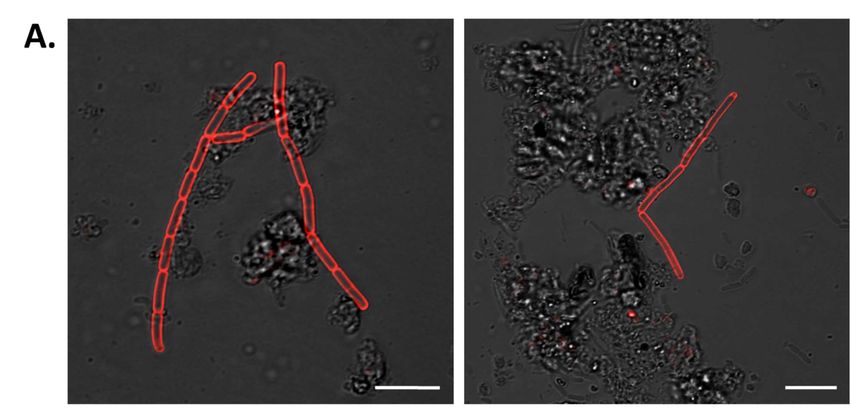
Preprints (www.preprints.org) | NOT PEER-REVIEWED | Posted: 23 November 2021
PBS and mixed with 1 µg mCherry-RBP λ031-120 protein. Fluorescence microscopy was conducted as
described in [18].
Collection of soil-samples
On September 6, 2021, soil-samples were collected from several spots near where the diseased cow
had fallen and subsequently exuded spore-contaminated blood onto the pasture. Because of heavy
rains in the area in the meantime (>50 l/m2), samples were collected from approx. 10 cm below the
surface.
Soil-sample analysis by PCR and culturing of B. anthracis
Soil-samples for PCR analysis were processed as described in [20]. Briefly, three aliquots of soil-
samples were resuspended in sterile water with glass-beads (Ø5 mm) and mixed overnight at room
temperature. Two of the aliquots were spiked beforehand with spores of strain B. anthracis Sterne
34F2 for quantification. The suspensions were filtered through sterile gauze to remove soil particles
and other rough materials. After centrifugation at 4000xg for 15 min, the pellet was washed three
times in sterile water and finally re-suspended in 5 ml aquadest. This suspension was heated to 65-
70°C for 30 min to inactivate vegetative cells. Volumes of 250 µl each were plated onto four semi-
selective agar plates (TSPBA) [21]. Plates were incubated overnight at 37°C. Then, the bacterial lawn
from each plate was scraped off and re-suspended in 4 ml of 0.9 % (w/v) NaCl-solution. An aliquot
(ca. 1 ml) of this suspension was boiled for 20 min in a heating block to release DNA from cells,
centrifuged at 12,000xg for 15 min and the supernatant filtered through a 0.45 µm luer-lock filter.
Aliquots of 5 µl of the filtered supernatant were used for PCR analysis [20]. If PCR-positive, dilutions
of the original suspension were plated and grown on TSPBA [17] for isolation and verification of
suspicious B. anthracis colonies [20]. DNA from a picked colony was tested by PCR for anthrax
markers as described in [1]. Additional enrichment of B. anthracis from soil-samples was achieved by
culturing on semi-selective CEFOMA agar “Bacillus CEreus sensu lato group-specific antibiotics,
FOsfomycin, Macrolides Agar” according to [22].
Enrichment of B. anthracis from soil-samples by magnetic separation and culturing
For enriching B. anthracis from possibly spore-contaminated soil-samples, a newly developed
magnetic bead-assisted magnetic separation-method was applied. In this approach RBP λ031-120 [18]
was re-purposed to capture B. anthracis from soil. In short, Strep-Tactin XT protein (IBA GmbH,
Göttingen, Germany) was coupled to magnetic beads (Dynabeads™ M-280 Tosylactivated,
ThermoFisher, Dreieich, Germany). Then RBPλ031-120 protein was attached to this Strep-Tactin XT via
the Twin Strep-tag epitope. Soil was processed the same as described in [17], i.e., a soil-sample was
shaken in PBS buffer with 0.5 % (v/v) Tween 20 to solubilize spores. The sample was mildly
centrifuged to remove solid material and the crudely cleared supernatant incubated at 62°C for 20
min to inactivate vegetative cells. The supernatant was mixed with Brain Heart Infusion broth
(Merck, Darmstadt, Germany) with fetal calf serum (Merck) and incubated to allow spores to
germinate and develop into vegetative cells. This germination culture was mixed and incubated with
the RBP-loaded magnetic beads to separate B. anthracis spores from the liquid. Separation was
accomplished using a magnetic stand (ThermoFisher). Beads were washed and finally plated onto
3
Preprints (www.preprints.org) | NOT PEER-REVIEWED | Posted: 23 November 2021
TSPBA agar or Columbia blood agar plates (Becton Dickinson). Colonies were evaluated after
incubating over night at 37°C. Full details on the method will be published elsewhere.
Rapid prescreening of candidate B. anthracis colonies
Blood-samples from the carcass or colonies suspicious for B. anthracis obtained after enrichment
from soil-samples, were subjected to colorimetric Enzyme-Linked Phage Receptor Binding Protein
Assay (ELPRA) as described in [23]. In short, the one-step assay version was applied that utilizes
recombinant HRP-coupled RBPλ031-120. Candidate colony material or blood was inactivated, washed
twice with PBS and incubated with 0.1 µg of HRP-RBPλ031-120 protein. Samples were repeatedly
washed with PBS and the pellet resuspended in 50 µL SeramunBlau® slow (containing 3,3′,5,5′-
tetramethylbenzidin) peroxidase substrate (Seramun Diagnostica, Heidesee, Germany). Blue color
development was monitored for several minutes and photo-documented.
DNA isolation from B. anthracis colony material and confirmative PCR
Single bacterial colonies grown on semi-selective agar (TSPBA) were chemically inactivated with 4%
Terralin PAA and DNA isolated using the MasterPure™ Gram Positive DNA Purification kit (Lucigen)
with minor modifications as described in [24]. DNA concentrations were quantified using the Qubit
dsDNA HS Assay Kit (Thermo Fisher Scientific), according to the supplier’s protocol. For confirmation
of B. anthracis DNA via PCR, the chromosomal marker dhp61 was used as described previously [25].
DNA preparations were stored at −20 °C un l further use.
Whole Genome Sequencing
Nanopore sequencing was performed using SQK-LSK109 chemistry on a R10.3 SpotON Flow Cell on
the GridION system (Oxford Nanopore Technologies, Oxford, UK) running system software MinKNOW
21.05.8. A total of 350,000 reads were generated using the implemented “super-accurate base
calling” model. For increasing the assembly-efficacy the amount of reads were down-sampled to
104,110 reads (N50 of 10.01 kb; mean raw quality score of Q13.5). After processing using Flye
assembler V2.9 [26] three circularized high-quality replicons, corresponding to the chromosome
(5,213,322 bp; coverage 174-fold) as well as both plasmids pXO1 (181,920 bp; coverage 614-fold) and
pXO2 (94,735 bp; coverage 491-fold) were obtained. The scaffolds were manually checked for
contaminant reads and annotated automatically by the NCBI Prokaryotic Genome Annotation
Pipeline [27] after submission. All data generated or analyzed during this study are included in this
published article, and its supplementary information files are publically available in the NCBI
Sequence Read Archive (SRA) repository (Bioproject PRJNA309927). CanSNPer (v1.0.10) [28] was
used to classify and subsequently assign the corresponding canSNP-group B.Br.CNEVA to this
genome.
Analysis of whole genome sequencing data and SNP-calling
For rapid core chromosome multiple-alignment, the Parsnp tool from the Harvest Suite was used
[29]. For this, a chromosome-dataset, representing genomes from public databases (Table S1) and
the newly sequenced strains of B. anthracis, were aligned against the chromosome of B. anthracis
‘Ames ancestor’ (NC_007530) as a phylogenetic outgroup using Parsnp (parameters -c -e -u -C 1000).
4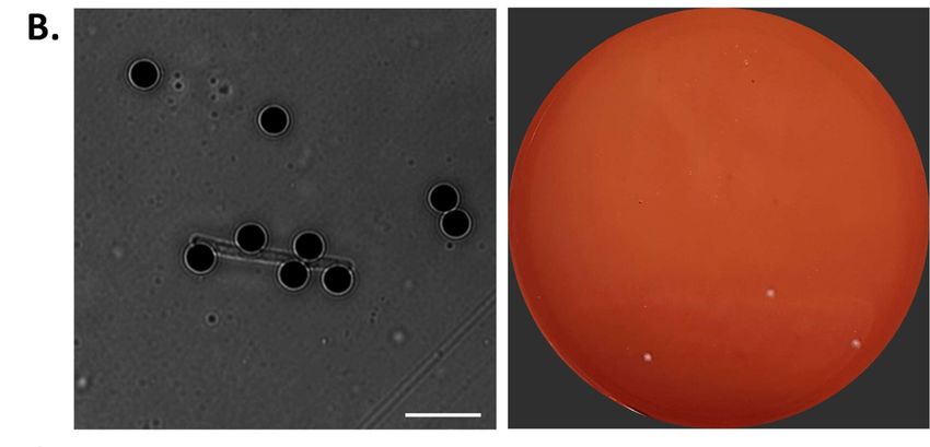
Preprints (www.preprints.org) | NOT PEER-REVIEWED | Posted: 23 November 2021

                 To export the identified SNP-positions, HarvestTools (version 1.0) from the same software suite was
                 used to create a vcf-(Variant Calling File) listing all SNP-positions. In order to enhance data quality,
                 chromosome regions with closely adjacent SNPs (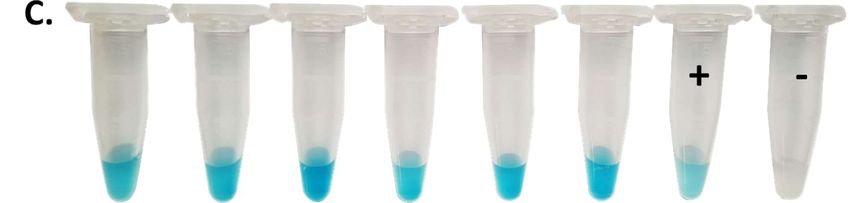
Preprints (www.preprints.org) | NOT PEER-REVIEWED | Posted: 23 November 2021

                 A.

                 B.

                 Figure 1: In situ and molecular PCR diagnostics of a cow diseased of anthrax. A two year old
                 gestating cow fallen to anthrax on a pasture in southern Bavaria (Germany) in August of 2021 (A and
                 B). Close up of the head with bloody discharge out of eyes and left nostril (A) and rear view with
                 bloody anus and vagina (B).

                 Detection of B. anthracis directly in blood-samples by phage RBP-based reporter and 16S rRNA SNP
                 (RT)-PCR

                 Independent to initial diagnostic PCR analysis performed by state health authorities, blood taken
                 from the left nostril of the carcass (Figure 1A) was subjected to recently developed ultrasensitive 16S
                 rRNA SNP (RT)-PCR [19] and phage RBP reporter-based rapid detection assays [18]. Results confirmed
                                                                                                                       6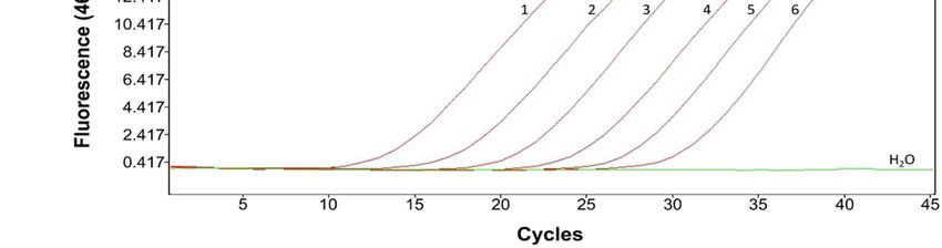
Preprints (www.preprints.org) | NOT PEER-REVIEWED | Posted: 23 November 2021
the previous PCR tests as phage RBP λ031-120 reporter based ELPRA gave positive results when
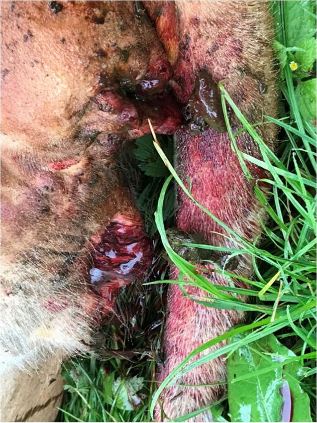
inactivated blood-samples from the carcass were tested (Figure 2A). Using fluorescence microscopy,
mCherry-RBPλ031-120 reporter was found to specifically bind to bacterial chains in blood-sample as
evidenced by red fluorescence (Figure 2B). This indicated that the detected cells were indeed very
likely B. anthracis. Of note, these phage RBP-based tests can be performed in just a few minutes.
Using 16S rRNA SNP-PCR, specific detection of B. anthracis nucleic acids directly in the blood-samples
derived from the carcass as well as from nucleic acid extractions thereof, was also accomplished
(Figure 2C). Dilutions (1:10 to 1:1000) of the inactivated blood-sample (without prior nucleic acid
extraction) yielded Ct values from 24.9 to 31.7. Conversely, dilutions of total nucleic acid extracted
from the same blood-sample yielded Ct values from 13.9 to 21.5 when testing for DNA only (Table
S3). When these total nucleic acid preparations (containing DNA and RNA) were subjected to 16S
rRNA SNP RT-PCR, the same samples (dilutions 1:10 to 1:1000) yielded even lower Ct values (9.7 to
17.8; Table S3). This is because the ultrasensitive RT version of the PCR not only detects 16S rRNA
genes of B. anthracis but also their transcripts, which are more abundant in growing cells compared
to their respective gene copies.
Figure 2: Direct detection of B. anthracis cells in blood from a diseased cow. A: Horseradish
peroxidas (HRP) conjugated RBPλ031-120 was added directly to inactivated blood (taken from the
carcass’ left nostril) (right reaction tube) as well as to inactivated sheep blood which served as a
negative control (left reaction tube). After washing, chromogenic HRP substrate was added and color
development photo-documented after 1 min B: Recombinant fusion protein mCherry-RBPλ031-120 was
added to 100 µl of blood and directly subjected to fluorescence microscopy. Shown are merged
7Preprints (www.preprints.org) | NOT PEER-REVIEWED | Posted: 23 November 2021
images of transmission and fluorescent light (wavelengths: excitation 594 nm, emission: 610 nm).
Scale bar: 5 µm. C: Dilutions of the inactivated cow blood (1 – 1:10, 2 – 1:100, 3 – 1:1000) as well as
dilutions of isolated DNA (4 – 1:10, 5 – 1:100, 6 – 1:1000) from the blood-samples were subjected to
16S rRNA SNP-PCR. Shown are representative realtime PCR amplification-curves.
B. anthracis strains BF-1 and BF-5 are clonal, very closely related outbreak strains
Genomic DNA of B. anthracis strains BF-5 was subjected to sequencing resulting in three contigs
(chromosome, plasmid pXO1 and pXO2) (accession # SRR16572036). Comparison of the genomes of
B. anthracis strains BF-1 and BF-5 revealed that both strains were exceptionally similar (Table 1). The
chromosome of BF-5 featured only three SNPs and two single nucleotide repeat (SNR) differences
(both SNRs in non-coding regions with deletions of a single “T”). While plasmid pXO1 was identical,
pXO2 harbored a single additional SNP- and SNR-insertion (“T”) in three identical repeat regions,
respectively. This clonality of the two outbreak strains clearly supported the hypothesis that a
hitherto non-localized source of unknown origin of contamination exists on-site. This source is very
likely the cause of repeated infection of grassing cows on this pasture.
Table 1: DNA sequence differences between genomes of B. anthracis BF-1 and BF-5.
Reference (BF-1) Position BF-1 nucleotide BF-5 nucleotide Kind of
sequence sequence change
CP047131.1 519877 C T SNP (SNP1)
(chromosome)
CP047131.1 1434950 CTTTTTTTTTTTTTTGTAAA CTTTTTTTTTTTTTGTAAA Deletion
TAA TAA
CP047131.1 1625072 A C SNP (SNP2)
CP047131.1 1878269 GTTTTTTTTTTTTTTTGTAA GTTTTTTTTTTTTTTGTAA Deletion
AATTAA AATTAA
CP047131.1 2472315 T C SNP (SNP3)
CP047133.1 29759 CTTTTTTTAT CTTTTTTTTAT Insertion
(plasmid pX02) 31759
30759
CP047133.1 62640 A G SNP (SNP4)
(plasmid pX02)
Phylogenetically strains B. anthracis BF-5 and -1 group with strains from the Austrian state of Tyrol
The canSNP-type of B. anthracis BF-5 was determined, assigning the strain to the B.Br.CNEVA clade
[32]. Chromosomal sequence analysis inferred the phylogenetic placement of strain BF-5 to a cluster
of central European B. anthracis strains within the B.Br.CNEVA clade. As expected from Table 1, the
closest relative was strain BF-1 (Figure 3). Other close relatives were Tyrol 4675 and Tyrol 6282, from
the Austrian state of Tyrol from 1988 and 1979, respectively. Strains from a large French B.Br.CNEVA
cluster (only three representatives shown in Figure 3) as well as strains from Switzerland, Slovakia,
Germany and Italy were more distantly related. Not shown are additional B.Br.CNEVA genomes
phylogenetically more loosely related to the focus strain, BF-5. Notably, there is a polytomy at the
base of the French cluster, the clade comprising strains A016/17OD930 and Tyrol 3520 and the clade
featuring BF-1, BF-5 and Tyrol 4674 and Tyrol 6282 (Figure 3). This clearly suggests a common
ancestor of all the strains.
8Preprints (www.preprints.org) | NOT PEER-REVIEWED | Posted: 23 November 2021
A.
17OD930 (Switzerland)
A016 (Switzerland)
Tyrol 3520 (Austria)
(France)
French
(France) Cluster
(France)
BF-1 (Germany)
BF-5 (Germany)
Tyrol 4675 (Austria)
Tyrol 6282 (Austria)
A024 (Slovakia)
BA0188 (Italy)
IMB 3011 (Italy)
3011
A046 (Germany)
Ames Ancestor
B. ANSES
CNEVA 11-11
9066
38
13
13
17OD930
ANSES
A016
105 BA0188
Tyrol 21
21
23
3520 26
58
8 IMB 3011
89 9
1 98
7
1
BF-1 11
33
962
962
49 86 Ames
19
2 Ancestor
1 30
A046
1 A024
BF-5 66
Tyrol Tyrol 6282
4675
9Preprints (www.preprints.org) | NOT PEER-REVIEWED | Posted: 23 November 2021
Figure 3: Phylogeny of new B. anthracis isolate BF-5 among its close relatives of the B.Br.CNEVA
canonical SNP-clade. A rooted phylogenetic tree of representatives of the B.Br.CNEVA canonical
single-nucleotide polymorphisms (canSNP) clade of B. anthracis is shown (A). The tree is based on
1558 chromosomal SNPs used to construct a Maximum Likelihood tree (bootstrap confidence from
500 permutations were generated and the tree with the highest likelihood is shown). Isolate names
and countries of origin are indicated at branch termini (red: sequenced in this study; black:
sequences from public databases, Table S1). A Minimum-spanning tree of close relatives of strain BF-
5 within the B.Br.CNEVA canSNP-clade of B. anthracis derived from chromosomal SNPs is shown (B).
Indicated are numerical SNP-differences (logarithmic scale) between chromosomes. Both trees are
rooted to the reference chromosome, B. anthracis strain Ames ‘Ancestor’ that belongs to the
A.Br.Ames canSNP-clade.
Both “classical”, established methods and novel phage RBP reporter fusions enable direct detection
and isolation of B. anthracis from soil-samples
Soil was retrieved from the site of the carcass as well as from surrounding areas up to 80 m away.
(Figure 1A, B). The established analysis methods yielded positive PCR results after cultivation of
original soil materials. Isolated colonies with typical morphology of B. anthracis were positive in PCR
for pagA, capC and saspB (data not shown). The novel, phage protein-based magnetic enrichment
approach fared equally well, yet, can be completed in much shorter time: To screen the possibly
contaminated soil-samples for B. anthracis spores, mCherry-RBPλ031-120 was just added to soil
supernatants pre-incubated with germination medium and the samples were subjected to
fluorescence microscopy. With this method, cells of B. anthracis could be detected directly in soil-
samples as cell chains emitted strong red fluorescence derived from the attached RBP reporter
(Figure 4A). While presence of B. anthracis was indicated by fluorescence microscopy, isolation of B.
anthracis from soil-samples was achieved using magnetic beads coupled with RBP λ031-120. After
binding of the cells to the RBP-loaded magnetic beads, the buffer-washed cell-bead-complexes
(Figure 4B, left panel) were agar-plated and cultured. A representative result is shown in Figure 4B
(right panel). While occasionally hemolytic, non-B. anthracis colonies also grew on the plates, suspect
B. anthracis colonies showing no hemolysis were chemically inactivated and confirmed by ELPRA
(Figure 4C). Genomic DNA from six of these additional isolates was prepared for further analysis.
10Preprints (www.preprints.org) | NOT PEER-REVIEWED | Posted: 23 November 2021
Figure 4
Figure 4: Direct detection and isolation of B. anthracis from contaminated soil-samples associated
with a diseased cow. Soil-samples were shaken in PBST buffer to solubilize spores, centrifuged and
the supernatant mixed with BHI broth containing fetal calf serum and incubated to allow spores to
germinate. A: Recombinant fusion protein mCherry-RBPλ031-120 was added to pre-incubated soil
supernatants and directly subjected to fluorescence microscopy. Shown are two merged images of
transmission and fluorescent light (wavelengths: excitation 594 nm, emission: 610 nm). Scale bar: 5
µm. B: Magnetic beads coupled with RBPλ031-120 were added to pre-incubated soil supernatants to
11Preprints (www.preprints.org) | NOT PEER-REVIEWED | Posted: 23 November 2021
capture B. anthracis cells. A sample was taken for brightfield microscopy (left panel, Scale bar: 5 µm)
and the remainder of the bead suspension buffer-washed, plated on blood agar plates and incubated
at 37°C overnight (right panel). C: Rapid RBP reporter-based assay on inactivated suspicious colony
material from enrichment plates. Inactivated colony material was incubated with RBP λ031-120
covalently linked to horseradish peroxidase for colorimetric identification with chromogenic
substrate. Positive control (+) was B. anthracis Sterne and negative control (-) B. cereus ATCC10987.
Results were scored after about 1 min as positive (blue color development) or negative (no color
development).
Four SNPs found between B. anthracis strains BF-1 and BF-5 were interrogated in additional isolates
derived from contaminated soil
In order to determine the distribution and relative abundance of the four SNPs separating B.
anthracis strains BF-1 and BF-5 (Table1; Table S2), PCRs of the identified four SNP-regions were
conducted on DNA from six soil isolates and the PCR amplicons Sanger-sequenced. We did not
identify any SNP-differences in these six soil isolates relative to BF-5 (data not shown). Thus, these
results indicate that the BF-5 genotype is the prevalent genotype at the affected pasture in 2021.
Discussion
Regarding risk-assessment, re-occurrence of an anthrax-outbreak after 12 years [9] at the same
pasture diminished the suspicion of intentional release of the pathogen as underlying cause.
Conversely, the outbreak strongly indicated that an old anthrax focus was still active. This is
reminiscent to similar situations in other regions of Europe. For instance, in Sweden an outbreak in
cattle occurred in a nature-reserve in 2011. Notably, records positioned an old anthrax burial-site
(mid-1940s) in that area [2,33]. Remarkably, only two years later, an additional cow deceased closely
to this area that had been seen cattle-vaccination after the 2011 outbreak [34]. Similar to the case at
hand, genome sequencing of the two Swedish outbreak isolates from 2011/2013 indicated these
were clonal [2]. The authors offered as plausible explanation for this genomic identity among
spatially and temporally separated outbreaks the spreading of spores by birds or wildlife. Though
these Swedish outbreaks have caused public alarm for the risk of environmental contamination [2],
no more cases were reported in that region since (as of November 2021). More active is the re-
emerging situation in Italy where anthrax resurfaces repeatedly in the southern region of Basilicata
[35,36] and soils at outbreak -sites remained contaminated with viable spores for many years [4,5].
The genomes of strains BF-1 and BF-5 differ by only three chromosomal SNPs (Table 1). A recent
genomic study on an anthrax-outbreak in Italy found strains differing by up to five SNPs [37].
Genome analysis for epidemiologic investigation of strains associated with injectional anthrax have
led the authors to the conclusion that genetic variation is possibly generated as a result of infection
of a single host but some phylogenetic patterns might be best explained by diversity introduced
through several infection-cycles of B. anthracis in several hosts [8]. The 2021 outbreak in Bavaria
seems to follows this pattern with only very few SNP-differences between strains from the same
outbreak-site separated by 12 years. Notably, all six isolates retrieved from soil surrounding the
carcass-site and from 80 m away at a ditch featured the same unique SNP-positions as isolate BF-5
directly grown from the dead cow’s blood. In contrast, it is very unlikely that isolate BF-1 is a direct
ancestor of BF-5. Chromosomal SNP 1 differs from the ancestor-state only in BF-5 but not in BF-1.
Vice versa, however, chromosomal SNP 2 and SNP 3 showed an evolved state in BF-1, while ancestral
in BF-5 (Table 1).
12Preprints (www.preprints.org) | NOT PEER-REVIEWED | Posted: 23 November 2021
In order to acutely diminish the local risk of surface-near spore contamination on-site, the affected
pasture-site where the animal fell (Figure 1A and B), was disinfected with 10 l/m2 10% (v/v)
formaldehyde as similarly advised by [1]. Obviously, this measure will neither be able disinfect
deeper soil horizons nor eliminate the unidentified original contamination-site presumably located
somewhere on the premises. Longer term monitoring of surface-near soil on-site may be able to alert
authorities in case B. anthracis spores can again be detected after favorable weather conditions, e.g.,
heavy rains followed by mild temperatures [38]. Further developments related to sensitive detection
of B. anthracis in soil could facilitate the identification and elimination of the original source of spore
contamination at the affected premises.
In any case, this rare outbreak provided an ideal opportunity for real-life testing of assays developed
beforehand for detection and identification of B. anthracis. Direct microscopy of B. anthracis-
infected blood (Figure 2A) or germinated cells in B. anthracis spore-contaminated soil (Figure 4A) and
rapid testing of inactivated blood (Figure 4B) or suspect colonies [23] yielded similar results with this
authentic materials to previously tested spiked-in materials (unpublished).
Acknowledgements
The authors thank Malena Bestehorn-Willmann for help with genome sequencing, Rahime Terzioglu,
Gabriele Echle, Linda Dobrzykowski, and Laura Madeddu for technical assistance.
Funding
This research work was funded by the Medical Biological Defense Research Program of the
Bundeswehr Joint Medical Service (to GG and MA) and was partially funded through Bundeswehr
Medical Service [SoFo 56Z1-S-43 1922] to MA and the German Federal Ministry of Education and
Research (BMBF) [ZooSeq FKZ 01KI1905A] to MA.
Conflicts of Interest
The authors declare no conflicts of interest. Opinions, interpretations, conclusions, and
recommendations are those of the authors and are not necessarily endorsed by any governmental
agency, department, or other institutions. The funders had no role in the design of the study; in the
collection, analyses, or interpretation of data; in the writing of the manuscript, or in the decision to
publish the results.
Data Availability Statement
All data produced in the present study are available upon reasonable request to the authors.
13Preprints (www.preprints.org) | NOT PEER-REVIEWED | Posted: 23 November 2021
References
1. Turnbull PC. World Health Organization. Anthrax in humans and animals. Geneva (CH): WHO
Press, 2008.
2. Ågren J, Finn M, Bengtsson B, Segerman B. Microevolution during an anthrax outbreak leading
to clonal heterogeneity and penicillin resistance. PLoS One 2014; 9:e89112.
3. Revich BA, Podolnaya MA. Thawing of permafrost may disturb historic cattle burial grounds in
East Siberia. Global Health Action 2011; 4.
4. Fasanella A, Di Taranto P, Battisti A, et al. Old animal anthrax outbreaks discovered through the
analysis of soil. Giornale Italiano die Medicina Tropicale 2011; 16:1–4.
5. Braun P, Grass G, Aceti A, et al. Microevolution of anthrax from a young ancestor (M.A.Y.A.)
suggests a soil-borne life cycle of Bacillus anthracis. PLoS ONE 2015; 10:e0135346.
6. Hanczaruk M, Reischl U, Holzmann T, et al. Injectional anthrax in heroin users, Europe, 2000-
2012. Emerg Infect Dis 2014; 20:322–3.
7. Ringertz SH, Hoiby EA, Jensenius M, et al. Injectional anthrax in a heroin skin-popper. Lancet
2000; 356:1574–5.
8. Keim P, Grunow R, Vipond R, et al. Whole genome analysis of injectional anthrax identifies two
disease clusters spanning more than 13 years. EBioMedicine 2015; 2:1613–1618.
9. Antwerpen M, Proenca DN, Ruckert C, et al. Draft genome sequence of Bacillus anthracis BF-1,
isolated from Bavarian cattle. J Bacteriol 2012; 194:6360–1.
10. Antwerpen M, Elschner M, Gaede W, Schliephake A, Grass G, Tomaso H. Genome sequence of
Bacillus anthracis strain Stendal, isolated from an anthrax outbreak in cattle in Germany.
Genome Announc 2016; 4:e00219-16.
11. Elschner MC, Busch A, Schliephake A, Gaede W, Zuchantke E, Tomaso H. High-quality genome
sequence of Bacillus anthracis strain 14RA5914 isolated during an outbreak in Germany in
2014. Genome Announc 2017; 5.
12. Price EP, Seymour ML, Sarovich DS, et al. Molecular epidemiologic investigation of an anthrax
outbreak among heroin users, Europe. Emerging Infectious Diseases 2012; 18:1307–1313.
13. BAYERISCHE, LANDESTIERÄRZTEKAMMER. Aktueller Fall von Milzbrand bei Weiderindern in
Bayern. BLTK Newsletter 2009; 3:1.
14. Braun P, Knupfer M, Antwerpen M, Triebel D, Grass G. A rare glimpse into the past of the
anthrax pathogen Bacillus anthracis. Microorganisms 2020; 8.
15. Derzelle S, Aguilar-Bultet L, Frey J. Whole genome SNP analysis of bovine B. anthracis strains
from Switzerland reflects strict regional separation of Simmental and Swiss Brown breeds in the
past. Vet Microbiol 2016; 196:1–8.
16. Sterne M. Anthrax. In: Stableforth AW, Galloway IA, eds. Infectious Diseases of Animals, Disease
due to Bacteria. London: Butterworth, 1959: 16–52.
14Preprints (www.preprints.org) | NOT PEER-REVIEWED | Posted: 23 November 2021
17. Fasanella A, Di Taranto P, Garofolo G, et al. Ground Anthrax Bacillus Refined Isolation (GABRI)
method for analyzing environmental samples with low levels of Bacillus anthracis
contamination. BMC Microbiol 2013; 13:167.
18. Braun P, Wolfschläger I, Reetz L, et al. Rapid microscopic detection of Bacillus anthracis by
fluorescent receptor binding proteins of bacteriophages. Microorganisms 2020; 8:934.
19. Braun P, Nguyen MD-T, Walter MC, Grass G. Ultrasensitive detection of Bacillus anthracis by
real time PCR targeting a polymorphism in multi-copy 16S rRNA genes and their transcripts.
medRxiv 2021; [preprint], 2021.09.20 [cited 2021 Oct 26], 2021.09.20.21263746.
20. Beyer W, Bellan S, Eberle G, et al. Distribution and molecular evolution of Bacillus anthracis
genotypes in Namibia. PLoS Negl Trop Dis 2012; 6:e1534.
21. Turnbull, Peter C. Anthrax in Humans and Animals. 4th ed. Geneva: World Health Organization,
2008. Available at: http://www.ncbi.nlm.nih.gov/books/NBK310486/. Accessed 5 October
2021.
22. Rohde A, Papp S, Feige P, Grunow R, Kaspari O. Development of a novel selective agar for the
isolation and detection of Bacillus anthracis. J Appl Microbiol 2020;
23. Braun P, Rupprich N, Neif D, Grass G. Enzyme-Linked Phage Receptor Binding Protein Assays
(ELPRA) Enable Identification of Bacillus anthracis Colonies. Viruses 2021; 13:1462.
24. Knüpfer M, Braun P, Baumann K, et al. Evaluation of a highly efficient DNA extraction method
for Bacillus anthracis endospores. Microorganisms 2020; 8.
25. Antwerpen MH, Zimmermann P, Bewley K, Frangoulidis D, Meyer H. Real-time PCR system
targeting a chromosomal marker specific for Bacillus anthracis. Mol Cell Probes 2008; 22:313–5.
26. Kolmogorov M, Yuan J, Lin Y, Pevzner PA. Assembly of long, error-prone reads using repeat
graphs. Nat Biotechnol 2019; 37:540–546.
27. Angiuoli SV, Gussman A, Klimke W, et al. Toward an online repository of Standard Operating
Procedures (SOPs) for (meta)genomic annotation. OMICS 2008; 12:137–141.
28. Lärkeryd A, Myrtennäs K, Karlsson E, et al. CanSNPer: a hierarchical genotype classifier of clonal
pathogens. Bioinformatics 2014; 30:1762–4.
29. Treangen TJ, Ondov BD, Koren S, Phillippy AM. The Harvest suite for rapid core-genome
alignment and visualization of thousands of intraspecific microbial genomes. Genome Biol
2014; 15:524.
30. Tamura K, Nei M. Estimation of the number of nucleotide substitutions in the control region of
mitochondrial DNA in humans and chimpanzees. Mol Biol Evol 1993; 10:512–26.
31. Kumar S, Stecher G, Li M, Knyaz C, Tamura K. MEGA X: Molecular evolutionary genetics analysis
across computing platforms. Mol Biol Evol 2018; 35:1547–1549.
32. Van Ert MN, Easterday WR, Huynh LY, et al. Global genetic population structure of Bacillus
anthracis. PLoS One 2007; 2:e461.
33. Lewerin SS, Elvander M, Westermark T, et al. Anthrax outbreak in a Swedish beef cattle herd--
1st case in 27 years: Case report. Acta Vet Scand 2010; 52:7.
15Preprints (www.preprints.org) | NOT PEER-REVIEWED | Posted: 23 November 2021
34. Elvander M, Persson B, Sternberg Lewerin S. Historical cases of anthrax in Sweden 1916-1961.
Transbound Emerg Dis 2017; 64:892–898.
35. Fasanella A. Bacillus anthracis, virulence factors, PCR, and interpretation of results. Virulence
2013; 4.
36. Fasanella A, Garofolo G, Galante D, et al. Severe anthrax outbreaks in Italy in 2004:
considerations on factors involved in the spread of infection. New Microbiol 2010; 33:83–6.
37. Abdel-Glil MY, Chiaverini A, Garofolo G, et al. A whole-genome-based gene-by-gene typing
system for standardized high-resolution strain typing of Bacillus anthracis. J Clin Microbiol
2021; 59:e0288920.
38. Van Ness GB. Ecology of anthrax. Science 1971; 172:1303–7.
16You can also read